This document summarizes a study on the combustion route synthesis of monoclinic phase gadolinium oxide phosphors. Gadolinium oxide nanoparticles were synthesized using a low temperature solution combustion method with urea as a fuel. Characterization using XRD, SEM, TEM, Raman spectroscopy, and XPS confirmed the monoclinic phase and average particle size of around 10 nm. XRD showed high crystallinity with no impurities. SEM showed spherical morphology. TEM showed individual nanocrystals around 8 nm. Raman spectroscopy showed characteristic peaks for gadolinium oxide. XPS showed the gadolinium 3d peak was consistent with previous studies. The combustion synthesis method produced phase pure nano-crystalline gadolinium oxide
Preparation, Structure, and Characterization of Nd2mo2o9 fast Oxide Ion Condu...IJARIIT
The structure and ion conductivity of Nd2Mo2O9powders were synthesized by using Nd(NO2)3, MoO3, and aspartic acid (fuel) in assisted combustion method with heating at 550˚C for 6 hours. The thermal decomposition, phase identification, morphology, ionic conductivity of the samples were studied by TGA/DTA, XRD and SEM four probe D.C. method respectively. The formation of Nd2Mo2O9 was confirmed by FTIR studies. The synthesis and crystallization were followed by thermochemical techniques (TGA/DTA) studies. The synthesized materials showed reasonable ionic conductivity. These results indicate that assisted combustion method is a promising method to prepare nanocrystalline Nd2Mo2O9 for solid oxide fuel cell.
Synthesis of Cobalt ferrite by Solid Reaction Methodsank_sanjay
Cobalt ferrite nano-crystalline powder was synthesized from the powder mixture of cobalt carbonate and iron oxide by mixed oxide ceramic method. The effects of temperature of calcination as well as molar ratio of CoCO3/Fe2O3 on the phase structure, morphology and magnetic properties of the products were studied using X-ray diffraction (XRD), field emission scanning electron microscopy (FESEM) and vibrating sample magnetometer (VSM) techniques, respectively. The samples calcined at 800 and 900˚C consisted of cobalt ferrite, iron oxide and cobalt oxide. In the sample calcined at 1000˚C, the reaction was completed and single phase CoFe2O4 with a mean crystallite and particle sizes of 49 and 300 nm, respectively was obtained.
Microstructural and Magnetic Properties of Cobalt Ferrite Nanoparticles Synth...ijtsrd
Cobalt ferrite (CoFe2O4), an inverse spinal ferrite has high permeability, good saturation 1magnetization and no preferred direction of magnetization, high Curie temperature, and high electromagnetic performance. In the present work 0.2M cobalt nitrate 0.3M ferric nitrate and 0.4 M citric acid is used to synthesis cobalt ferrite nanoparticle by sol-gel technique. As the magnetic property depends on the grain size of the synthesized nanoparticle, metal nitrate to citric acid ratio is varied from 0.8, 0.6 and 0.4 and the structural, functional morphological and magnetic characteristics are analyzed. The structural analysis shows the decrease in the average crystallite from 37 to 27nm when CAMN ratio decreases from 0.8 to 0.4. The strain is directly proportional to dislocation density and it reflects the growth of the average grain size, and in the present study, it reflects the same. The calculated lattice parameter is found to be close to 8.373 Ã… and the volume of the cell is found to be 5.63x10⁻²⁸ m is close to the standard value for the cobalt ferrite nanoparticles. From the EDS spectrum, the presence of Co, Fe, and O in the synthesized nanoparticles are noted. Functional groups analysis by FTIR shows the presence of organic sources. Surface morphology by Scanning electron microscope shows the distribution of spherical sized nanoparticles agglomerated in different sizes and the grain size calculated by image J software are close to the calculated value by Scherrer formula from XRD. Chitra | T Raguram | K S Rajni"Microstructural and Magnetic Properties of Cobalt Ferrite Nanoparticles Synthesized by Sol-Gel Technique" Published in International Journal of Trend in Scientific Research and Development (ijtsrd), ISSN: 2456-6470, Volume-2 | Issue-5 , August 2018, URL: http://www.ijtsrd.com/papers/ijtsrd15871.pdf http://www.ijtsrd.com/physics/other/15871/microstructural-and-magnetic-properties-of-cobalt-ferrite-nanoparticles-synthesized-by-sol-gel-technique/chitra
International Journal of Engineering Research and DevelopmentIJERD Editor
Electrical, Electronics and Computer Engineering,
Information Engineering and Technology,
Mechanical, Industrial and Manufacturing Engineering,
Automation and Mechatronics Engineering,
Material and Chemical Engineering,
Civil and Architecture Engineering,
Biotechnology and Bio Engineering,
Environmental Engineering,
Petroleum and Mining Engineering,
Marine and Agriculture engineering,
Aerospace Engineering.
Porosity and the Magnetic Properties of Aluminium Doped Nickel Ferriteijtsrd
The nanocrystalline particles of Aluminium Al doped nickel Ni ferrites with general formula NiAlxFe2 xO4 x = 0.0, 0.2, 0.4, 0.6, 0.8 and 1.0 were synthesized by sol gel auto combustion technique. The formation of single phase cubic spinel was confirmed by X ray diffraction analyses. Morphological features of the samples are studied by Scanning Electron Microscopy SEM to examine the particle size, shape and homogeneity of sample. The magnetic hysteresis graphs were obtained to understand their magnetic behaviours. The relative permeability µr of AlNi ferrite samples shows a decrease for all samples as Al content increases. Sandar Oo | Ye Wint Tun | Shwe Zin Oo "Porosity and the Magnetic Properties of Aluminium Doped Nickel Ferrite" Published in International Journal of Trend in Scientific Research and Development (ijtsrd), ISSN: 2456-6470, Volume-3 | Issue-5 , August 2019, URL: https://www.ijtsrd.com/papers/ijtsrd25240.pdfPaper URL: https://www.ijtsrd.com/physics/other/25240/porosity-and-the-magnetic-properties-of-aluminium-doped-nickel-ferrite/sandar-oo
Photo-induced reduction of CO2 using a magnetically separable Ru-CoPc@TiO2@Si...Pawan Kumar
An efficient photo-induced reduction of CO2 using magnetically separable Ru-CoPc@TiO2@SiO2@Fe3O4
as a heterogeneous catalyst in which CoPc and Ru(bpy)2phene complexes were attached to a solid
support via covalent attachment under visible light is described. The as-synthesized catalyst was characterized
by a series of techniques including FTIR, UV-Vis, XRD, SEM, TEM, etc. and subsequently tested for
the photocatalytic reduction of carbon dioxide using triethylamine as a sacrificial donor and water as a
reaction medium. The developed photocatalyst exhibited a significantly higher catalytic activity to give a
methanol yield of 2570.78 μmol per g cat after 48 h.
International Journal of Engineering Research and Applications (IJERA) is an open access online peer reviewed international journal that publishes research and review articles in the fields of Computer Science, Neural Networks, Electrical Engineering, Software Engineering, Information Technology, Mechanical Engineering, Chemical Engineering, Plastic Engineering, Food Technology, Textile Engineering, Nano Technology & science, Power Electronics, Electronics & Communication Engineering, Computational mathematics, Image processing, Civil Engineering, Structural Engineering, Environmental Engineering, VLSI Testing & Low Power VLSI Design etc.
Removal of lead ions by nife2 o4 nanoparticleseSAT Journals
Abstract Nickel ferrite nanoparticles have been prepared by polymeric precursor method. The nanoparticles application in the removal of lead ion was investigated. The results showed that the adsorptive properties were dependent on pH, duration and temperature. Highest percentage (99%) lead adsorption was observed under basic condition at room temperature during 1 h stirring. Analysis of lead adsorbed nickel ferrite by powder XRD, FTIR and XPS techniques revealed the adsorption took place based on hydroxide mechanism. The lead salt formed on the surface of the ferrite powder was identified to be lead carbonate and lead hydroxy carbonate at pH 7 and 9 respectively. Keywords: Nickel ferrite; Nanoparticles; Lead; Adsorption; Hydroxide
Structural and photoluminescence study of SrAl2O4:Eu3+ phosphors synthesized ...AI Publications
The combustion synthesis method was employed for the synthesis of red-emitting monoclinic SrAl2O4:Eu3+ phosphors. Structural characterization of annealed samples was carried out via X-ray Diffraction (XRD). XRD patterns reveal that strontium aluminate samples were cubic spinel nanoparticles and the grain size determined by the Debye-Scherrer formula is 35.34 nm. The vibrational stretching frequencies corresponding to the composites were confirmed by FT-IR spectroscopy. The PL spectra show the strongest emission at 612 nm corresponds to the 5D0 → 7F2 transition of Eu3+, which results in bright red color emitting phosphor used for display devices and lamp industries.
Facile Synthesis and Characterization of Pyrolusite, β-MnO2, Nano Crystal wit...Editor IJCATR
MnO2 nanoparticles have been synthesized by a simple combustion method using MnSO4.4H2O. The crystalline phase, morphology, optical property and magnetic property of the as prepared nanoparticle were characterized using XRD, FT-IR, FT-Raman, SEM, UV-Vis, PL and VSM respectively. Structural studies by XRD indicate that the synthesized material as tetragonal rutile crystal structure. FT-IR and FT-Raman analysis revealed the stretching vibrations of metal ions in tetrahedral co-ordination confirming the crystal structure. The PL and UV analysis having an emission band at 390 nm, showed a prominent blue peak at 453 nm as well as a green emission lines at 553 nm with band gap energy of 3.2eV. Magnetic measurements indicate that the Néel temperature of the β-MnO2 structures is 92.5K for Hc = 100 Oe which showed antiferromagnetic behaviour.
International Journal of Engineering Research and Applications (IJERA) is an open access online peer reviewed international journal that publishes research and review articles in the fields of Computer Science, Neural Networks, Electrical Engineering, Software Engineering, Information Technology, Mechanical Engineering, Chemical Engineering, Plastic Engineering, Food Technology, Textile Engineering, Nano Technology & science, Power Electronics, Electronics & Communication Engineering, Computational mathematics, Image processing, Civil Engineering, Structural Engineering, Environmental Engineering, VLSI Testing & Low Power VLSI Design etc.
Optical and Electrical Properties of TiO2 Doped Fe2O3 Thin Film Prepared by S...IJAEMSJORNAL
In this work, iron oxide (Fe2O3) and titanium oxide (TiO2) doping (Fe2O3) thin films have been prepared by spray pyrolysis technique (SPT) on a glass substrate. We have studied the effects of various doping percentage of (TiO2) on (Fe2O3) optical and electrical properties. The optical parameters of the prepared films as absorbance, absorption coefficient, optical energy gap and refractive index have been calculated for all prepared films. D.C conductivity and Hall effect for all films are measured. The results showed that absorbance of prepared films decreases with increasing percentage of (TiO2) this lead to increase the energy gap and conductivity decreased with increasing of percentage of (TiO2) as well as the results showed throughout the study that all films have tow activation energy, its value increase with increasing of percentage of (TiO2). Hall effect measurement showed all films have n-type conductivity.
International Journal of Engineering Research and Applications (IJERA) is an open access online peer reviewed international journal that publishes research and review articles in the fields of Computer Science, Neural Networks, Electrical Engineering, Software Engineering, Information Technology, Mechanical Engineering, Chemical Engineering, Plastic Engineering, Food Technology, Textile Engineering, Nano Technology & science, Power Electronics, Electronics & Communication Engineering, Computational mathematics, Image processing, Civil Engineering, Structural Engineering, Environmental Engineering, VLSI Testing & Low Power VLSI Design etc.
Synthesis and Characterisation of Copper Oxide nanoparticlesIOSR Journals
Cupric oxide (CuO) nanoparticles were prepared by the chemical route by calcinations at a higher temperature from 300oC to 400 oC. For the comparison transmission electron microscopy (TEM) and x-ray diffraction (XRD) measurements were made through JCPDS. There is good agreement between data produced by spectroscopy and the microscopic measurements.
International Journal of Engineering Research and Applications (IJERA) is an open access online peer reviewed international journal that publishes research and review articles in the fields of Computer Science, Neural Networks, Electrical Engineering, Software Engineering, Information Technology, Mechanical Engineering, Chemical Engineering, Plastic Engineering, Food Technology, Textile Engineering, Nano Technology & science, Power Electronics, Electronics & Communication Engineering, Computational mathematics, Image processing, Civil Engineering, Structural Engineering, Environmental Engineering, VLSI Testing & Low Power VLSI Design etc.
Crystal Growth and Studies of Dihydrogen Phosphates of Potassium and Ammonium...IJERA Editor
A nonlinear optical (NLO) material Potassium dihydrogen phosphates (KDP) and Ammonium dihydrogen
phosphates (ADP) are grown by slow evaporation solution growth technique. The FTIR studies confirm the
presence of the functional group in the grown crystal. The optical transmittance studies show that the crystal has
transparence in the entire visible and IR region. The thermal stability of the materials was assessed by TG/DTA
analysis. The mechanical stability of the grown crystals was analyzed by Vicker’s microhardness test. The
dielectric behavior of the crystals was tested by dielectric analysis. The second harmonic generation (SHG) of
KDP is confirmed by Kurtz and Perry powder technique using Nd: YAG laser.
Spectral studies of praseodymium doped heavy metal borate glass systemsinventy
Praseodymium doped HMO glasses are fabricated with the following compositions using conventional melt quenching technique. The compositions of the glass systems are 12 ZnO + 33 B2O3 + (50-x) PbO + (x+10) CaO + 4 Al2O3 + 1 Pr6O11 where (x = 0,10,20,30 and 40 mol %.). Certain physical properties of these systems have been evaluated and reported. Spectral data for all these systems were recorded for X-ray diffraction, Optical absorption and Fluorescence properties. The Judd-Ofelt intensity parameters Ωλ ( λ = 2,4,6) were evaluated from the spectral data and in turn employed to evaluate the lasing parameters of Pr3+ HMO glass systems such as radiative transition probabilities (A), radiative life-times (τR), branching ratios (βR) absorption cross-sections (σa) and Stimulated emission cross-sections (σe). The experimental and calculated branching ratios (βR) for the lasing transitions 3P0 3H4, 3P0 3H6, and 3P0 3F2 are found to be in good agreement in the present work.
Spectroscopic studies on Mn2+ ions doped Cadmium Aluminum Fluoro Lead Borate ...IJERA Editor
This article reports on optical properties of Mn2+ (0.5 mol%): 49.5 B2O3 - 10PbO - 30CdO -10AlF3 glasses.
Optical absorption spectrum of the manganese ions doped glass exhibits broad absorption band at 473 nm.
Photoluminescence spectrum of Mn2+ (0.5 mol %): 49.5B2O3- 30CdO – 10PbO - 10AlF3 glass has revealed a red
emission at 618 nm with an excitation wavelength 467 nm.
Synthesis, Characterization of ZnS nanoparticles by Coprecipitation method us...IOSR Journals
ZnS nanoparticles are prepared by coprecipitation method using various capping agents like PVP (polyvinylpyrrolidone), PVA (polyvinylalcohol) and PEG-4000 (polyethyleneglycol). These are characterized by UV-Visible spectra, X-ray diffraction (XRD) studies, Fourier Transform Infra-red spectra (FTIR) and Transmission electron microscopy (TEM). UV-Visible absorption spectra are used to find the optical band gap and the values obtained have been found to be in the range of 3.80-4.00eV. The particle size of nanoparticles calculated from XRD pattern has been in the range of 2-4 nm. It is also observed that the particle size of nanoparticle is affected by the nature of capping agent. Photo catalytic degradation of xylenol orange (XO) by the nanoparticles shows that these act as photo catalysts under sunlight irradiation. The XO dye was degraded more than 87.24, 83.42 and 73.05% in the presence of PEG-4000, PVA and PVP capped ZnS nanoparticles in 120, 150 and 180 min. respectively. The kinetics of catalyzed by synthesized ZnS nanoparticles with XO dye follows pseudo-first order kinetics with reasonable apparent rate constants.
A facile method to prepare CdO-Mn3O4 nanocompositeIOSR Journals
CdO-Mn3O4 nanocomposite has been prepared by a simple solvothermal method using a domestic microwave oven. Cadmium acetate, manganese acetate and urea were used as the precursors and ethylene glycol as the solvent. The as-prepared sample was annealed for 1 hour in each case at different temperatures, viz. 100, 200 and 300°C. The as-prepared and annealed samples were characterized by X-ray diffraction and scanning electron microscopic analyses. Results indicate that annealing at 300°C is required to get the sample with high phase purity and homogeneity. The present study indicates that the method adopted can be considered as an economical and scalable one to prepare the proposed nanocomposite with reduced size, phase purity and homogeneity.
Synthesis and Structural Characterization of Cu Substituted Ni-Zn Nano-Ferrit...IJERA Editor
The ferrite nano particles having chemical formula Ni0.2CuxZn0.8-xFe2O4 (where x=0.0 to 0.8 with step of 0.2) were synthesized by Citrate-Gel Auto Combustion method at low temperature. The synthesized powders were sintered at 500oC for 4 hours in air and characterised by XRD, SEM with EDS. XRD analysis of prepared samples were confirmed the single phase cubic spinel Structure. The crystallite size (D) of prepared ferrites were in the range of 24-73nm. The values of lattice parameter (a) decreased and X-ray density (dx) were increased with the increasing of Cu substitution. The surface morphology of the prepared samples was investigated by Scanning Electron Microscope(SEM). An elemental composition of the samples was studied by Energy Dispersive Spectroscopy(EDS). The observed results can be explained on the basis of composition and crystal size.
Effect of calcination on the electrical properties and quantum confinement of...eSAT Publishing House
IJRET : International Journal of Research in Engineering and Technology is an international peer reviewed, online journal published by eSAT Publishing House for the enhancement of research in various disciplines of Engineering and Technology. The aim and scope of the journal is to provide an academic medium and an important reference for the advancement and dissemination of research results that support high-level learning, teaching and research in the fields of Engineering and Technology. We bring together Scientists, Academician, Field Engineers, Scholars and Students of related fields of Engineering and Technology
Effect of calcination on the electrical properties and quantum confinement of...eSAT Journals
Abstract Fe2O3 nanoparticles have been prepared by a simple solvothermal method using a domestic microwave oven. X-ray powder diffraction measurement indicates the amorphous nature of the as-prepared sample. Calcined samples were obtained by annealing the as-prepared sample at different temperatures, viz. 400, 500, 600 and 700oC. Transmission electron microscopic images indicate that all the five samples are spherical in shape. AC electrical measurements were carried out on pelletized samples by the parallel plate capacitor method at various temperatures ranging from 40-130oC and frequencies ranging from 100 Hz -1 MHz. Results indicate low AC electrical conductivities and consequently show the occurrence of nano confined states. The exciton Bohr radii obtained from the dielectric constant values at 40oC temperature and 1 kHz frequency are 41.8, 54.8, 55.3, 56.3 and 27.0 respectively for the as-prepared sample and samples calcined at 400, 500, 600 and 700oC which indicate a strong quantum confinement effect. The impedance spectra observed exhibit non-ideal behavior. Keywords: Semiconductors, Magnetic materials, Nanoparticles, Electrical properties
Consistently High Voc Values in p-i-n Type Perovskite Solar Cells Using Ni3+-...Pawan Kumar
Leading edge p-i-n type halide perovskite solar cells (PSCs) severely underperform n-i-p PSCs. p-i-n type PSCs that use PEDOT:PSS hole transport layers (HTLs) struggle to generate open-circuit photovoltage values higher than 1 V. NiO HTLs have shown greater promise in achieving high Voc values albeit inconsistently. In this report, a NiO nanomesh with Ni3+ defect grown by the hydrothermal method was used to obtain PSCs with Voc values that consistently exceeded 1.10 V (champion Voc = 1.14 V). A champion device photoconversion efficiency of 17.75% was observed. Density functional theory modeling was used to understand the interfacial properties of the NiO/perovskite interface. The PCE of PSCs constructed using the Ni3+-doped NiO nanomesh HTL was ∼34% higher than that of conventional compact NiO-based perovskite solar cells. A suite of characterization techniques such as transmission electron microscopy, field emission scanning electron microscopy, intensity-modulated photocurrent spectroscopy, intensity-modulated photovoltage spectroscopy, time-resolved photoluminescence, steady-state photoluminescence, and Kelvin probe force microscopy provided evidence of better film quality, enhanced charge transfer, and suppressed charge recombination in PSCs based on hydrothermally grown NiO nanostructures.
1. Advance Physics Letter
________________________________________________________________________________
ISSN (Print) : 2349-1094, ISSN (Online) : 2349-1108, Vol_1, Issue_1, 2014
1
Raman and XPS studies of Combustion Route Synthesized Monoclinic
Phase Gadolinium Oxide phosphors
1
Raunak Kumar Tamrakar, 2
D. P. Bisen, 3
Ishwer Prasad Sahu, 4
Nameeta Brahme
1
Department of Applied Physics, Bhilai Institute of Technology (Seth Balkrishna Memorial), Bhilai House, Durg (C.G.)
Pin-401001,India
2,3,4
School of studies in Physics and Astrophysics, Pt. Ravishankar Shukla University, Raipur (C.G.) Pin-492010, India
Abstract:- Gd2O3 nanoparticles have been synthesized by the
low temperature solution combustion method using urea as
fuels in a short time. The structural, morphology and
luminescence properties have been carried out using powder
X-ray diffraction (PXRD), Scanning electron microscopy
(SEM), Transmission electron microscopy (TEM), Raman
and XPS. For synthesis gadolinium nitrate and Urea were
mixed and kept stirring for 30 min, resultant was transferred
to crucible and fired in a furnace for Few seconds at 6000
C
soon the water got evaporated and a vigorous redox reaction
occurred and Gd2O3 Nano crystals were achieved. The
combustion synthesis method which is reported here is
advantageous from the perspectives of small size of the
nanoparticle.
I. INTRODUCTION:-
In recent years, low dimensional structures having
remarkable research interest owing to their novel
electrical, optical and magnetic properties, elongated as
compared to bulk and other It is reported that at nano
scale, the differences in electrical and optical
characteristics of very small particles are caused by
quantum effects due to their high surface to volume
ratio, which increases the band gap by reduction of the
number of allowable quantum states in the small
particles, and improves surface and interfacial effects[1-
3]
Lanthanide hydroxides and oxides have actively been
investigated because of their wide range of applications
including dielectric materials for multilayered
capacitors, luminescent lamps and displays, solid-laser
devices, optoelectronic data storages, waveguides, and
heterogeneous catalysts. Recently, lanthanide-doped
oxide nanoparticles are of special interests as potential
materials for an important new class of nanophosphors.
When applied for a fluorescent labeling, they present
several advantages such as sharp emission spectra, long
life times, and resistance against photobleaching in
comparison with conventional organic fluorophores and
quantum dots [4-9].
Gadolinium oxide based nanophosphors are found to be
promising candidates in the field of high performance
luminescent devices, catalysis and other functional
devices based on their excellent electronic, optical and
physico-chemical responses arising from 4f electrons.
Not surprisingly, all these properties could be largely
influenced by their chemical composition, crystal
structure, shape and dimensionality. Thus, high surface
area nanomaterial which has a larger fraction of defect
sites per unit area should be of interest as adsorbents in
environmental remediation processes. Cost of synthesis,
simplicity and morphological characteristics of prepared
phosphor are important parameters for their use in the
commercial applications, it is imperative that a self-
propagating combustion route offers the best choice for
the synthesis of Gd2O3 powder[10-13].
Nanoparticles prepared by this combustion route, have
size of ~10 nm such approaches involve the use of fuel
like urea, glycine, alanine, hydrazide etc. to initiate
decomposition reaction of precursor metal salt at high
temperature. The combustion synthesis and low
temperature processing minimize the size of the
materials, which is very important for the most
applications in the electronic, optoelectronic, chemical
industries etc. The higher reactivity of smaller size
Gd2O3 particles is not only because of the large specific
surface area but also due to the high concentration of
low coordinated sites and structural defects on their
surface. Due to these merits, these are in high demand
for various technological applications including
optoelectronic devices, high definition televisions,
biological imaging and tagging, MRI, luminescent
paints and inks for security codes etc. [14-17].
The Present paper deals with synthesis of Gd2O3 by
combustion method. The particle size of the prepared
phosphor was determined by using X-ray
diffraction(XRD) analysis, the morphology of the
prepared phosphor was determined by scanning electron
microscopy(SEM) and transmission electron
microscopy(TEM). The Raman and X-ray photoelectron
spectroscopic studies of the prepared phosphor were also
carried out.
2. Advance Physics Letter
________________________________________________________________________________
ISSN (Print) : 2349-1094, ISSN (Online) : 2349-1108, Vol_1, Issue_1, 2014
2
II. MATERIALS AND METHODS
2.1. Chemicals
Chemicals used in this study are of analytical grade.
Gadolinium nitrate and urea were purchased from
Sigma-Aldrich Chemicals Limited. All reagents were
used without further purification.
2.2 Synthesis: - In Combustion synthesis method
aqueous solutions of Gd(NO3)3.6H2O is prepared by
addition of a suitable amount to prepare the precursor
solution and the mixture was stirred for 4 hour at 60˚C,
the resultant solution converted in to a transparent gel
form. Then the above sample was heated in a furnace at
600˚C for 2 min, the sample changed into its anhydrous
form followed by a vigorous redox reaction to form
Gd2O3 powder[10]. The resulting brownish powder was
heated until a controlled explosion took place yielding a
very fine, white powder. Since the reaction is so rapid,
the crystal growth will be highly restrained.
2Gd(NO3)3 + CH4N2O→Gd2O3+ CO2 + 2H2O +4N2
Figure 1. Show the flow chart of synthesis of Gd2O3
2.3. Material characterization: - The crystallinity as
well as the particle size of the phosphor were monitored
X-ray diffraction measurement. The X-ray powder
diffraction data was collected by using Bruker D8
Advanced X-ray diffractometer using Cu Kα radiation.
The X-rays were produced using a sealed tube and the
wave length of X-ray was 0.154 nm. The X-rays were
detected using a fast counting detector based on Silicon
strip technology (Bruker Lynx Eye detector). The
surface morphology of the prepared phosphor was
determined by field emission scanning electron
microscopy (FESEM) JSM-7600F. Energy dispersive X-
ray analysis (EDX) was used for elemental analysis of
the phosphor. Particle diameter and surface morphology
of prepared phosphor were determined by Transmission
Electron Microscopy (TEM) using Philips CM-200.
Raman studies were carried out on Jobin-Yvon, France,
Ramnor Hg-2s Spectrometer with Ar-Laser with 4 W
power having resolution of 0.5 cm-1
and wave number
accuracy 1 cm-1
over 5000 cm-1
. XPS analysis was
performed in a VG instrument with a CLAM2 analyzer
and a twin Mg/Al anode. The pressure in the analysis
chamber was approximately 9x10⁻¹⁰
mbar. The
measurements were carried out with unmonochromated
Al Ka photons (1486.6 eV). The power of the X-ray
source was kept constant at 300 W.
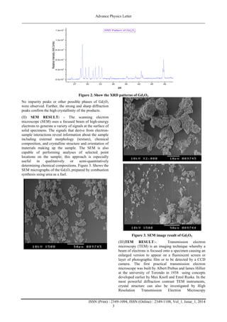
(I) XRD RESULT:-The XRD patterns of the Gd2O3
sample is shown in Figure 2. The diffraction patterns are
well matched with standard JCPDS card no. 43-1015
[18]. The particle size were calculated by the Scherer
formula[19]
where Dv=volume weighted crystallite size, k=shape
factor (0.9),=wavelength of Cu Kα1 radiation,
βhkl=instrumental corrected integral breadth of the
reflection (in radians) located at 2θ, and θ=angle of
reflection (in degrees) was utilized to relate the
crystallite size to the line broadening. The average
crystallite size of Gd2O3 nanoparticles was found to be
in the range of ~10nm.
3. Advance Physics Letter
________________________________________________________________________________
ISSN (Print) : 2349-1094, ISSN (Online) : 2349-1108, Vol_1, Issue_1, 2014
3
27 30 33 36 39 42 45 48
2.0x10
3
4.0x10
3
6.0x10
3
8.0x10
3
1.0x10
4
1.2x10
4
RelativeIntensity(ArbUnits)
2
XRD Pattern of Gd2
O3
Figure 2. Show the XRD patterns of Gd2O3
No impurity peaks or other possible phases of Gd2O3
were observed. Further, the strong and sharp diffraction
peaks confirm the high crystallinity of the products.
(II) SEM RESULT: - The scanning electron
microscope (SEM) uses a focused beam of high-energy
electrons to generate a variety of signals at the surface of
solid specimens. The signals that derive from electron-
sample interactions reveal information about the sample
including external morphology (texture), chemical
composition, and crystalline structure and orientation of
materials making up the sample. The SEM is also
capable of performing analyses of selected point
locations on the sample; this approach is especially
useful in qualitatively or semi-quantitatively
determining chemical compositions. Figure 3. Shows the
SEM micrographs of the Gd2O3 prepared by combustion
synthesis using urea as a fuel.
Figure 3. SEM image result of Gd2O3
(III)TEM RESULT:-. Transmission electron
microscopy (TEM) is an imaging technique whereby a
beam of electrons is focused onto a specimen causing an
enlarged version to appear on a fluorescent screen or
layer of photographic film or to be detected by a CCD
camera. The first practical transmission electron
microscope was built by Albert Prebus and lames Hillier
at the university of Torondo in 1938 using concepts
developed earlier by Max Knoll and Emsl Ruska. In the
most powerful diffraction contrast TEM instruments,
crystal structure can also be investigated by High
Resolution Transmission Electron Microscopy
4. Advance Physics Letter
________________________________________________________________________________
ISSN (Print) : 2349-1094, ISSN (Online) : 2349-1108, Vol_1, Issue_1, 2014
4
(HRTEM), also known as phase contrast imaging as the
images are fonned due to differences in phase of
electron waves scattered through a thin specimen. In
Figure 4 HRTEM micrograph shows a Gd2O3
nanocrystal with a diameter of ~8 nm are seen
throughout the particle[20].
Figure 4. HRTEM images of Gd2O3 prepared by the
combustion method
(IV) X-ray photoelectron spectroscopy (XPS):- XPS
is a surface chemical analysis technique that can be used
to analyze the surface chemistry of a material in its as-
received state, or after some treatment, for example:
fracturing, cutting or scraping in air or UHV to expose
the bulk chemistry, ion beam etching to clean off some
or all of the surface contamination (with mild ion
etching) or to intentionally expose deeper layers of the
sample (with more extensive ion etching) in depth-
profiling XPS, exposure to heat to study the changes due
to heating, exposure to reactive gases or solutions,
exposure to ion beam implant, exposure to ultraviolet
light.The chemical composition of Gd2O3 nanoparticles
was studied with X-ray Photoelectron Spectroscopy
(XPS) and the experimental data was analyzed using
curve fitting. The Gd(3d) level consists of a spin orbit
split, with the Gd(3d)5/2 peak is found at 1186.74 eV
(Fig. 5). The line shape and peak positions are in good
agreement with earlier published data on Gd2O3 powder
pressed into an in sheet[13,21-25].
1197 1194 1191 1188 1185 1182 1179
1.06
1.07
1.08
1.09
1.10
1.11
1.12
Intensity(Arb.Units)
Binding Energy (eV)
Gd(3d)5/2
(1186.74 eV )
Figure 5. The Gd (3d) XPS spectrum of Gd2O3
nanocrystals
(V) Raman spectroscopy: - The Raman Effect has been
an important technique for the elucidation of molecular
structure. Similar information is obtained from the
infrared spectra. Since the infrared and Raman spectra
are governed by different selection rules, the
information obtained from Raman Spectroscopy
supplements the information obtained from infrared
spectra. Samples which cannot be handled in the
infrared (e.g. Aqueous solutions, biological samples
etc.) can easily be studied through Raman Spectroscopy.
Raman spectroscopy is highly informative to elucidate
the structure of the synthesized sample. It is a
nondestructive tool to explore vibrational, rotational and
other low frequency modes in the systems under
study[26]. Figure 6 shows the Raman spectra of Gd2O3
prepared by combustion syntheis using urea as a fuel,
recorded at room temperature with an excitation
wavelength of 633 nm He–Cd laser. A broad and intense
Raman peak at 340 cm-1
along with less intense peaks
were observed at 375, 395, 424 and 451 cm-1
. The
results are good agreement with the previously
published Raman spectroscopic studies on Gd2O3
nanoparticles[,14,17,25,27].
500 475 450 425 400 375 350 325 300
(451)
(424)
(395)
(374)
Ramanintensity(ArbUnits)
Wavenumber(cm
-1
)
Raman spectra of Gd2
O3
(340)
Figure 6. Raman spectra of Gd2O3 nanoparticles
III. CONCLUSION:
Formation of stable gel is an important criterion for a
successful solution combustion synthesis process. Urea
form stable gel with mixed nitrate solution of
gadolinium and the combustion of the gel produces
phase pure nano-crystalline powder without any residual
reactant. In this study, Lattice parameter (a) for cubic
Gd2O3 was found to be 10.7602. The particle sizes are
confined by powder X-ray diffraction studies. The
particle size estimated from Debye–Scherrer’s was well
comparable to TEM results. The advantages of the
phosphors prepared by this combustion process are the
easy availability of homogeneous spherical morphology
in different size, and its wide practicality for other
phosphor materials. X-ray Photoelectron Spectroscopy
(XPS) show the Gd(3d) level consists of a spin orbit
split doublet, with the Gd(3d)5/2 peak is found at 1186.74
eV. Raman spectra with excitation of 633 nm
5. Advance Physics Letter
________________________________________________________________________________
ISSN (Print) : 2349-1094, ISSN (Online) : 2349-1108, Vol_1, Issue_1, 2014
5
wavelength, we found a broad and intense Raman peak
at 340 cm-1
along with less intense peaks were observed
at 375, 395, 424 and 451 cm-1
.
IV. ACKNOWLEDGEMENT
We are very grateful to IUC Indore for XRD
characterization and also thankful to Dr. Mukul Gupta
for his cooperation. I am very thankful to SAIF, IIT,
Bombay for other characterization such as SEM, TEM,
Raman and XPS.
REFERENCES:
[1] Du G and Tendeloo G V 2005 Nanotechnology
16 595.
[2] Xu Z, Yang J, Hou Z, Li C, Zhang C, Huang S
and Lin J 2009 Mater. Res. Bull. 44 1850.
[3] Bockrath M, Liang W, Bozovic D, Hafner J.H,
Lieber C.M, Tinkham M and Park H 2001
Science 291 283.
[4] N. Dhananjaya, H. Nagabhushana, B.M.
Nagabhushana, B. Rudraswamy, C.
Shivakumara, R.P.S. Chakradhar, J. Alloys
Compd. 509(5), 2368–2374 (2011)
[5] G.Z. Li, M. Yu, Z.L. Wang, J. Lin, R.S. Wang,
J. Fang, J. Nanosci. Nanotechnol. 6(5), 1416–
1422, (2006).
[6] Yanhong Li and Guangyan Hong, , Journal of
Luminescence 124 (2007) 297–301.
[7] T. Kim Anh, L. Quoc Minh, N. Vu, T. Thu
Huong, N. Thanh Huong, C. Barthou, W. Strek,
Journal of Luminescence, 102-103. (2003) 391-
394.
[8] E. Downing, L. Hesselink, J. Ralston, and R.
Macfarlane, “A Three-Color, Solid-State, Three
Dimensional. Display,” Science 273, 1185-89
(1996).
[9] M. L. Pang, J. Lin, J. Fu, R.B. Xing, C.X. Luo,
Y.C. Han, Opt. Mater. 23, 547–558 (2003).
[10] Raunak Kumar Tamrakar, D. P. Bisen and
Nameeta Brahme, Research on Chemical
Intermediates May 2014, Volume 40, Issue
5, pp 1771-1779.
[11] M.A. McDonald, K.L. Watkin, Invest. Radiol.
38 (2003) 305.
[12] A. Borel, J.F. Bean, R.B. Clarkson, L. Helm, L.
Moriggi, A.D. Sherry, M. Woods, Chem.–Eur.
J. 14 (2008) 2658.
[13] A.T.M. Anishur Rahman, Krasimir Vasilev,
Peter Majewski, Journal of Colloid and
Interface Science 354 (2011) 592–596
[14] Yan-li Li, Nuo-fu Chen, Jian-ping Zhou, Shu-
lin Song, Li-feng liu, Zhi-gang Yina, Chun-lin
Cai, Journal of Crystal Growth 265 (2004)
548–552
[15] A Brenier, G. Boulon, J. Lumin. 82 (1999) 285
[16] Ytterbium Doped Gadolinium Oxide
(Gd2O3:Yb3+
) Phosphor: Topology,
Morphology, and Luminescence Behaviour in
Hindawi Publishing Corporation Indian Journal
of Materials Science Volume 2014, Article ID
396147, 7 pages, Accepted 4 February 2014
[17] H. Guo, Y. Li, D. Wang, W. Zhang, M. Yin, L.
Lou, S. Xia, J. Alloy. Compd. 376(2004) pp.
23–27
[18] P. Klug, L.E. Alexander, X-ray Diffraction
Procedure (Wiley, New York, 1954).
[19] D. Grier, G. McCarthy, North Dakota State
University, Fargo, North Dakota, USA, ICDD
Grant-in- Aid (1991).
[20] P. E J. Flewit and R. K. Wild, Physical methods
for material characterization, second edition,
lOP publishing, L.9ndon (2003).
[21] Y. Mizokawa, H. Iwasaki, R. Nishitani, J.
Electron Spectrosc. Relat. Phenom. 14 (1978)
129.
[22] F. Soderlind, H. Pedersen, R.M. Petoral Jr, P.
Kall, K. Uvdal, J. Colloid Interface Sci. 288
(2005) 140.
[23] D. Raiser, J.P. Deville, J. Electron Spectrosc.
57 (1991) 91.
[24] J. Zhou, C. Chai, S. Yang, Z. Liu, S. Song, Y.
Li, N. Chen, J. Cryst. Growth 270 (2004) 21.
[25] N. Dhananjaya, H. Nagabhushana, B.M.
Nagabhushana d, B. Rudraswamy, S.C.
Sharma, D.V. Sunitha, C. Shivakumara, R.P.S.
Chakradhar, Spectrochimica Acta Part A:
Molecular and Biomolecular Spectroscopy 96
(2012) 532–540
[26] Szymanski H. A., `Raman Spectroscopy',
Plenum Press, New York(1967).
[27] Ningthoujam R S, Shukl R, Vatsa R K,Duppel
V, Kienle L and Tyagi A K 2009 J. Appl. Phys.
105, 084304