081 carotid artery plaque
•Download as PPT, PDF•
1 like•92 views

This document contains images and descriptions of various plaques and lesions found in arteries and the heart from MRI studies. It shows a plaque in the left internal carotid artery with a resolution of 450μm, a 4.5mm plaque in the aorta identified as a type Va fibroatheroma, and images of a plaque and wall of the left anterior descending coronary artery comparing MRI to X-ray angiogram. The sources listed are papers and studies from 2000 from Imaging Science Laboratories at Mount Sinai School of Medicine.

More Related Content
Viewers also liked
Viewers also liked (12)
More from Society for Heart Attack Prevention and Eradication
More from Society for Heart Attack Prevention and Eradication (20)
Recently uploaded
Recently uploaded (20)
081 carotid artery plaque
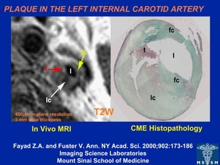
- 1. PLAQUE IN THE LEFT INTERNAL CAROTID ARTERY T2W CME HistopathologyIn Vivo MRI 450µm in-plane resolution 3 mm slice thickness Fayad Z.A. and Fuster V. Ann. NY Acad. Sci. 2000;902:173-186 Imaging Science Laboratories Mount Sinai School of Medicine
- 2. TEE T2W 4.5 mm plaque AHA TYPE Va (fibroatheroma) AORTIC PLAQUE Fayad ZA et al. Circ 2000;101;2503-2509 Imaging Science Laboratories Mount Sinai School of Medicine Plaque Fibrous Cap
- 3. Fayad ZA et al. Circ. 2000;102;506-510 Imaging Science Laboratories Mount Sinai School of Medicine X-ray Angiogram high grade stenosis LAD LAD Wall MR CORONARY WALL IMAGING
Editor's Notes
- In vivo transverse T2-weighted fast SE MR imaging of a left internal carotid artery. Plaque characterization was based on information obtained from T1-, intermediate-, and T2-weighted MR images. Left: T2-weighted MR image (repetition time, two R-R intervals; echo time, 55 msec; 3-mm section thickness; 450-µm in-plane resolution) shows low-signal-intensity lipid core (lc), high-signal-intensity fibrous cap (fc), and very high–signal-intensity thrombus (t). l = arterial lumen. Right: Corresponding histopathologic section. (Mason-eosin stain; original magnification, X10). Fayad ZA, Fuster V. Characterization of atherosclerotic plaques by magnetic resonance imaging. Ann N Y Acad Sci. 2000;902:173-86.
- • In vivo magnetic resonance images of a 4.5 mm thick plaque in the descending thoracic aorta: A) T1-weighted; B) Proton density-weighted; C) T2-weighted; with the corresponding transesophageal echocardiography (TEE) image (panel D). The MR images show an example of an AHA type Va plaque with a dark area in the center (arrow) identified on the T2-weighted image as a lipid rich core (panel C). The lipid rich core is separated from the lumen by a fibrous cap. Plaque characterization was based on the information obtained from T1-, proton-density-, and T2- weighted MR images. Image resolution is 0.8 mm.
- .