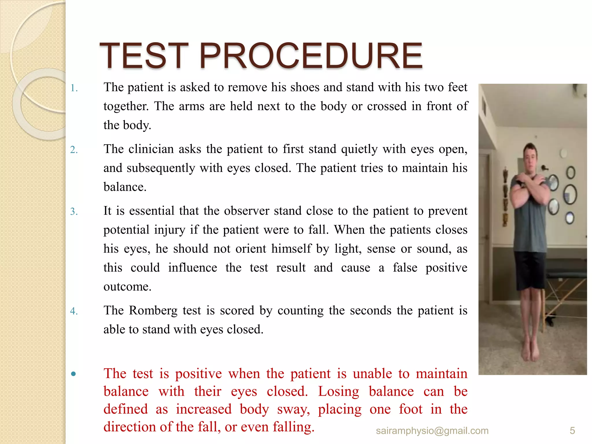
this power point presentation is a detailed description about some of neurological special test and signs. that helps the students and practitioners in diagnosing and quick reviewing for students during examinations.
2. List Of Special Tests
Romberg’s
Kernig’s sign
Brudenzki sign
Tinels’s sign
Slump test
Lhermitte's sign
Bell’s Phenomenon
Gower’s sign
Sun set sign
Battle’s sign
Glabellar tap sign
2
sairamphysio@gmail.com
3. Romberg’s Test
The Romberg test is a test that measures a persons sense of balance.
Specifically, the test assesses the function of the dorsal column of the
spinal cord (the dorsal column is responsible for proprioception).
THE ROMBERG TEST IS AN APPROPRIATE TOOL TO DIAGNOSE
sensory ataxia (a gait disturbance caused by abnormal proprioception
involving information about the location of the joints).
Sub-acute combined degeneration of spinal cord (Vitamin B12
deficiency);
Posterior cord syndrome (Posterior spinal artery infarction).
Hemisection of spinal cord (Brown Sequard syndrome)
It is also proven to be sensitive and accurate means of measuring the
degree of disequilibrium caused by central vertigo, peripheral vertigo
and head trauma.
It has been used in clinic for 150 years.
3
sairamphysio@gmail.com
4. The Romberg test, tests the function of: Dorsal Column
Medial Lemniscal Pathway, the neural pathways by
which sensory information from the peripheral nerves is
transmitted to the cerebral cortex.
When the person is standing with the eyes open, visual,
proprioceptive, and vestibular information is used to
maintain postural stability.
When the eyes are closed, the patient must rely on
proprioception and vestibular function.
4
sairamphysio@gmail.com
5. TEST PROCEDURE
1. The patient is asked to remove his shoes and stand with his two feet
together. The arms are held next to the body or crossed in front of
the body.
2. The clinician asks the patient to first stand quietly with eyes open,
and subsequently with eyes closed. The patient tries to maintain his
balance.
3. It is essential that the observer stand close to the patient to prevent
potential injury if the patient were to fall. When the patients closes
his eyes, he should not orient himself by light, sense or sound, as
this could influence the test result and cause a false positive
outcome.
4. The Romberg test is scored by counting the seconds the patient is
able to stand with eyes closed.
The test is positive when the patient is unable to maintain
balance with their eyes closed. Losing balance can be
defined as increased body sway, placing one foot in the
direction of the fall, or even falling. 5
sairamphysio@gmail.com
6. THE SHARPENED OR TANDEM
ROMBERG TEST
The Sharpened or Tandem Romberg test is a variation of the original
test. The implementation is mostly the same.
For this second test, the patient has to place his feet in heel-to-toe
position, with one foot directly in front of the other.
As with the original Romberg test, the assessment is performed first
with eyes open and then with eyes closed.
The patient crosses his arms over his chest, and the open palm of the
hand lies on the opposite shoulder. The patient also distributes his
weight over both his feet and holds his chin parallel with the floor.
6
sairamphysio@gmail.com
7. KERNIG’S SIGN
Kernig's sign is one of the physically
demonstrable symptoms of meningitis. Severe
stiffness of the hamstrings causes an inability to
straighten the leg when the hip is flexed to 90
degrees.
Kernig's sign is used to diagnose meningitis
7
sairamphysio@gmail.com
8. Step 1. The patient is positioned in supine with hip and
knee flexed to 90 degrees
Step 2. The knee is then slowly extended by the
examiner (Repeat on both legs)
Step 3. Resistance or pain and the inability to extend the
patient's knee beyond 135 degrees, because of pain,
bilaterally indicates a positive Kernig's sign
8
sairamphysio@gmail.com
9. BRUDENZKI SIGN
Brudzinski's sign is one of the physically
demonstrable symptoms of meningitis. Severe
neck stiffness causes a patient's hips and knees
to flex when the neck is flexed.
Brudzinski's sign is used to diagnose meningitis
9
sairamphysio@gmail.com
10. TESTING PROCEDURE
Step 1. Patient in supine position
Step 2. Gently grasp the patient's head from behind and
place the other hand on the patient's chest
Step 3. Gently flex the neck, bringing chin to chest
Positive sign is involuntary flexing of hips and knees (an
involuntary reaction to lessen the stretch on the inflamed
meninges)
10
sairamphysio@gmail.com
11. TINEL'S SIGN
Tinel's test is used to test for compression
neuropathy, commonly in diagnosing carpal
tunnel syndrome.
Technique
It is performed by lightly tapping (percussing)
over the nerve to elicit a sensation of tingling
or "pins and needles" in the distribution of the
nerve.
The Tinel sign is the tingling or prickling
sensation elicited by the percussion of an
injured nerve trunk at or distal to the site of the
lesion. The sign also indicates nerve
regeneration.
Positive test-The test is positive when a tingling
or prickling sensation is felt in the distribution
of the nerve.
11
sairamphysio@gmail.com
12. SLUMP TEST
The Slump Test is a neural tension test used to detect altered
neurodynamics or neural tissue sensitivity.
Slump Test is a neural physical examination that is used for detecting
disc bulging / herniation of the lumbar spine or irritation of the dura of
the spinal cord. It is a progressive series of maneuvers designed to
place the sciatic nerve roots under increasing tension.
The test can cause impingement or irritation of the dura and/ or nerve
roots, with pain radiating down into the areas supplied by the sciatic
nerve.
If the patient is unable to extend the knee because of pain, the
examiner reduces the pressure on the cervical spine and asks the
patient to slowly raise the head.
The Slump test is considered to be positive if the patient is then able to
extend the knee further without pain or with less pain, indicating that
neural structures are affected.
12
sairamphysio@gmail.com
13. The examination is performed step by step as
following:
1. First the patient is asked to “slump” the back into
thoracic and lumbar flexion while the examiner
supports the head to keep it in a neutral position.
2. With one arm , the examiner then applies pressure
across the shoulders to maintain increased flexion
in the thoracic and lumbar spine.
3. At the same time, the patient is asked to actively
flex the cervical spine and head as far as possible.
4. Using the same hand, the examiner then applies
pressure to maintain flexion in all three parts of the
spine.
5. With the other hand the examiner holds the
patient’s foot in maximum dorsi flexion. In this
position the patient is asked to actively straighten
the knee as much as possible.
6. The test is then repeated with the other leg and, if
possible, with both legs at the same time.
13
sairamphysio@gmail.com
14. LHERMITTE'S SIGN
It was first introduced by Pierre Marie and Chatelin in
1917. Then it was named after Jean Lhermitte who was
a French neurologist.
Lhermitte Sign (also called Lhermitte’s phenomenon)
is an electric shock like sensation that occurs on flexion
of the neck.
The sensation radiates down the spine, often into the
legs, arms, and sometimes to the trunk.
It differentiates between spinal cord lesions and
peripheral nerve root lesions, suggests dural or
meningeal irritation of the spinal cord (root irritation) or
possibly cervical myelopathy.
14
sairamphysio@gmail.com
15. TEST PROCEDURE
1. The patient sits with outstretched legs on the examination table.
2. The examiner grasps the patient’s foot with one hand and places
the other on the back of the patient’s head.
3. The examiner then simultaneously flexes the outstretched leg at
the hip and increasingly flexes the cervical spine.
The Lhermitte test is positive if an acute pain or electric shock like
sensation occurs that radiates into the upper or lower extremity,
A positive Lhermitte sign can indicate stenosis of the cervical spinal
canal, where the patient describes a sudden, generalized electric
shock in the arms and trunk, especially when inclining the head.
15
sairamphysio@gmail.com
16. BELL’S PHENOMENON
In 1823 charles Bell a great British anatomist observed
this when trying to close the eyelids of a patient with
facial palsy.
The Bell’s phenomenon, also called the Palpebral-
oculogyric reflex,
It refers to the movement of the eyeballs in an upward
direction when the eyelids are forcefully closed Both the
facial nerve paralysis and the eyeball deviation observed
in this condition are named after him.
The facial nerve carries the afferent fibers for this reflex,
while the efferent fibers travel via the oculomotor nerve
to the superior rectus muscle that controls upper eyelid
movement.
16
sairamphysio@gmail.com
18. GOWER’S SIGN
Gower’s Sign is a very common physical finding in
individuals with Duchenne’s Muscular Dystrophy (DMD).
It involves using their hands to ‘climb’ up their legs in
order to stand up. It is due to weakness in the child’s
proximal hip muscles and thigh muscles.
It is named after William Richard Gowers a British
neurologist.
The child arises from a prone position on the floor. The
child uses the hands-and-knees position and then
gradually go up to a stand by “walking” his hands
progressively up his shins, knees, and thighs. This
maneuver, known as Gowers’ sign, has been seen
exclusively in Duchenne’s muscular dystrophy (DMD).
18
sairamphysio@gmail.com
19. Gowers’s sign is mainly seen in DMD (Duchenne’s muscular
dystrophy) where it is mostly appear at 4–6 years, but also
presents in centronuclear myopathy, myotonic dystrophy and
various other conditions associated with proximal muscle
weakness, such as Becker muscular dystrophy (BMD),
dermatomyositis and Pompe disease.
19
sairamphysio@gmail.com
20. SUNSET SIGN
The “setting sun” sign is an ophthalmologic phenomenon
where the eyes appear driven downward bilaterally. The
inferior border of the pupil is often covered by the lower
eyelid, creating the “sunset” appearance. This finding is
classically associated with hydrocephalus in infants and
children.
The setting sun sign (also known as the sunset eye
sign or setting sun phenomenon) is a clinical phenomenon
encountered in infants and young children with
raised intracranial pressure.
Seen in up to 40% of children with obstructive hydrocephalus and
13% of children with shunt dysfunction.
20
sairamphysio@gmail.com
22. BATTLE’S SIGN
• Battle’s sign is a crescent-shaped bruise that
appears behind one or both ears. It was named
after an English surgeon, Dr. William Henry
Battle, and can be an indication of a serious
head injury.
• When one of the basilar bones is broken,
blood may pool behind the ear, creating
the Battle’s sign bruise.
• While Battle’s sign may look like an
ordinary bruise, it is not a result of direct
injury behind the ear. Instead, it is a sign
that one or more of the skull’s bones have
been broken.
• The size of Battle’s sign can vary but may
also extend down the back of the neck.
22
sairamphysio@gmail.com
23. GLABELLAR TAP SIGN
The glabellar reflex, also known as the "glabellar tap
sign", is a primitive reflex elicited by repetitive tapping of
the glabella—the smooth part of the forehead above the
nose and between the eyebrows.
The glabellar reflex is a primitive reflex - normally
present in infants and absent in adults. In patients with
frontal lobe damage or Parkinsonism, 'frontal release'
signs such as glabellar, grasp, suck, snout and
palmomental reflexes will be present.
23
sairamphysio@gmail.com
24. Interpretation
Increased - ongoing blinking without habitualization.
Parkinsonism or frontal lobe pathology
Normal - up to five blinks and then no further blinking
following habitualization
Decreased - no blinking in response to stimulus.
Upper or lower motor neuron weakness.
24
sairamphysio@gmail.com