
Recommended
More Related Content
What's hot
What's hot (20)
Similar to ELISA.pptx
Similar to ELISA.pptx (20)
Recently uploaded
Recently uploaded (20)
ELISA.pptx
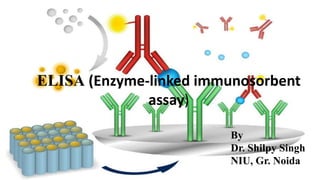
- 1. ELISA (Enzyme-linked immunosorbent assay) By Dr. Shilpy Singh NIU, Gr. Noida
- 2. What is an ELISA? ELISA (which stands for enzyme-linked immunosorbent assay) is a technique to detect the presence of antigens in biological samples. An ELISA, like other types of immunoassays, relies on antibodies to detect a target antigen using highly specific antibody-antigen interactions. Originally described by Engvall and Perlmann (1971), the method enables analysis of protein samples immobilized in microplate wells using specific antibodies.
- 3. Principle of ELISA In an ELISA assay, the antigen is immobilized to a solid surface. This is done either directly or via the use of a capture antibody itself immobilized on the surface. The antigen is then complexed to a detection antibody conjugated with a molecule amenable for detection such as an enzyme or a fluorophore. An ELISA assay is typically performed in a multi- well plate (96- or 384-wells), which provides the solid surface to immobilize the antigen. Immobilization of the analytes facilitates the separation of the antigen from the rest of the components in the sample. This characteristic makes ELISA one of the easiest assays to perform on multiple samples simultaneously. Fig. The basic setup of an ELISA assay. A capture antibody on a multi-well plate will immobilize the antigen of interest. This antigen will be recognized and bound by a detection antibody conjugated to biotin and streptavidin-HRP.
- 4. ELISA depend on the same basic elements: Coating/capture–direct or indirect immobilization of antigens to the surface of polystyrene microplate wells. Plate blocking–addition of irrelevant protein or other molecule to cover all unsaturated surface-binding sites of the microplate wells. Probing/detection–incubation with antigen-specific antibodies that affinity-bind to the antigens. Signal measurement–detection of the signal generated via the direct or secondary tag on the specific antibody. The most commonly used enzyme labels are horseradish peroxidase (HRP) and alkaline phosphatase (AP). Other enzymes have been used as well; these include β-galactosidase, acetylcholinesterase, and catalase. A large selection of substrates is available commercially for performing ELISA with an HRP or AP conjugate. The choice of substrate depends upon the required assay sensitivity and the instrumentation available for signal-detection (spectrophotometer, fluorometer, or luminometer).
- 5. Types of ELISA There are four main types of ELISA: direct ELISA, indirect ELISA, sandwich ELISA and competitive ELISA. Direct ELISA In a direct ELISA, the antigen is immobilized to the surface of the multi-well plate and detected with an antibody specific for the antigen The antibody is directly conjugated to HRP or other detection molecules.
- 6. Indirect ELISA Indirect ELISA is a technique that uses a two- step process for detection, whereby a primary antibody specific for the antigen binds to the target, and a labeled secondary antibody against the host species of the primary antibody binds to the primary antibody for detection. As for direct ELISA assays, the antigen is immobilized to the surface of the multi-well plate. The method can also be used to detect specific antibodies in a serum sample by substituting the serum for the primary antibody.
- 7. Sandwich ELISA (or sandwich immunoassay) The most commonly used type of ELISA. This requires two antibodies specific for different epitopes of the antigen. These two antibodies are normally referred to as matched antibody pairs. One of the antibodies is coated on the surface of the multi-well plate and used as a capture antibody to facilitate the immobilization of the antigen. The other antibody is conjugated and facilitates the detection of the antigen.
- 8. Advantages of ELISA detection methods Direct Indirect Sandwich • Quick because only one antibody and fewer steps are used. • Cross-reactivity of secondary antibody is eliminated. • A wide variety of labeled secondary antibodies are available commercially. • Versatile because many primary antibodies can be made in one species and the same labeled secondary antibody can be used for detection. • Maximum immunoreactivity of the primary antibody is retained because it is not labeled. • Sensitivity is increased because each primary antibody contains several epitopes that can be bound by the labeled secondary antibody, allowing for signal amplification. • Different detection methods can be used with the same primary antibody (colorimetric, chemiluminescent, etc.). • Highly sensitive and highly specific for target antigen as two antibodies are used for capture and detection. • Different detection methods can be used with the same capture antibody.
- 9. Disadvantages of ELISA detection methods Direct Indirect Sandwich • Immunoreactivity of the primary antibody might be adversely affected by labeling with reporter enzymes or tags. • Labeling primary antibodies for each specific ELISA system is time-consuming and expensive. • Limited number of conjugated primary antibodies available commercially. • No flexibility in choice of primary antibody label from one experiment to another. • Minimal signal amplification. • Cross-reactivity might occur with the secondary antibody, resulting in nonspecific signal. • An extra incubation step is required in the procedure. • Requires more optimization to identify antibody pairs and to ensure there is limited cross-reactivity between the capture and detection antibodies.
- 10. Competitive ELISA and other Types Besides the standard direct and sandwich formats described above, several other styles of ELISA exist: Competitive ELISA Competitive ELISA is a strategy that is commonly used when the antigen is small and has only one epitope or antibody binding site. One variation of this method consists of labeling purified antigen instead of the antibody. Unlabeled antigen from samples and the labeled antigen compete for binding to the capture antibody. A decrease in signal from the purified antigen indicates the presence of the antigen in samples when compared to assay wells with labeled antigen alone. In competitive ELISA, also referred to as inhibition ELISA, the concentration of the target antigen is determined by detection of signal interference. The target antigen in the sample competes with a labeled reference or standard for binding to a limited amount of antibodies immobilized on the plate.
- 11. ELISPOT (enzyme-linked immunospot assay) ELISPOT refers to ELISA-like capture and measurement of proteins secreted by cells that are plated in PVDF-membrane-backed microplate wells. It is a "sandwich" assay in which the proteins are captured locally as they are secreted by the plated cells, and detection is with a precipitating substrate. ELISPOT is like a western blot in that the result is spots on a membrane surface. In-cell ELISA is performed with cells that are plated and cultured overnight in standard microplates. After the cultured cells are fixed, permeabilized, and blocked, target proteins are detected with antibodies. This is an indirect assay, not a sandwich assay. The secondary antibodies are either fluorescent (for direct measurement by a fluorescent plate reader or microscope) or enzyme-conjugated (for detection with a soluble substrate using a plate reader).
- 12. Different types of microplates for ELISA ELISA plate Coating Applications Modified polymer surfaces Various modifications to the plate surface to increase hydrophobicity or hydrophilicity Enhance passive binding of biomolecules based on their physiochemical characteristics Antibody-binding plates Protein A, G, L, or A/G Binds to the FC region (VL for protein L) of capture antibodies to properly orient while leaving antigen binding capability Fusion-tag binding plates Glutathione (GST tag binding) or nickel or copper coated (His tag binding) Study of genetically engineered fusion proteins or protein- protein interactions Biotin-binding plates Streptavidin or neutravidin Binds small biotinylated peptides and other small molecules that are difficult to bind by passive adsorption
- 13. Detection strategies for ELISA Chromogenic (colorimetric) Fluorescence Chemiluminescence Sensitivity Low Moderate High Equipment required Standard absorbance plate reader Fluorometer Luminometer plate reader Enzyme HRP or AP Fluorescent tag or HRP (with chemifluorescent substrates) HRP or AP Advantages Direct visualization, high reproducibility between plates High reproducibility between plates, wide dynamic range Most sensitive detection strategy, wide dynamic range Considerations Requires black microplates Requires opaque or black microplates