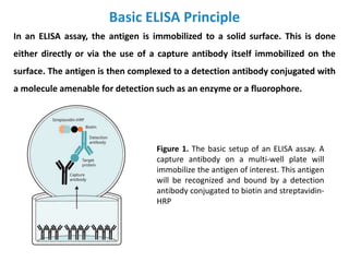
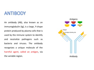
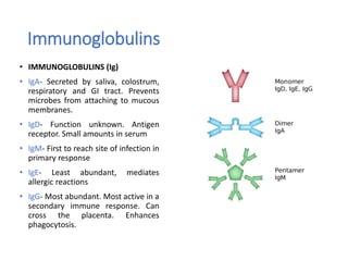
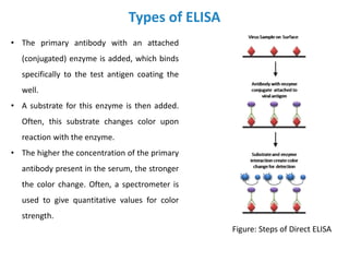
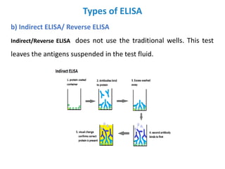
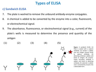
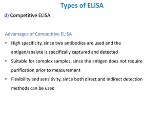
2. Introduction
ELISA (Enzyme-linked immunosorbent assay) is a technique to detect the
presence of antigens in biological samples.
An ELISA, like other types of immunoassays, relies on antibodies to detect a
target antigen using highly specific antibody-antigen interactions.
ELISA is a commonly used analytical biochemistry assay, first described by Eva
Engvall and Peter Perlmann in 1971.
ELISA is a plate-based assay technique designed for detecting and quantifying
peptides, proteins, antibodies and hormones. In an ELISA, an antigen must be
immobilized to a solid surface and then complexed with an antibody that is linked
to a n enzyme.
Detection is accomplished by assessing the conjugated enzyme activity via
incubation with a substrate to produce a measurable product. The most crucial
element of the detection strategy is a highly specific antibody-antigen interaction.
3. Basic ELISA Principle
In an ELISA assay, the antigen is immobilized to a solid surface. This is done
either directly or via the use of a capture antibody itself immobilized on the
surface. The antigen is then complexed to a detection antibody conjugated with
a molecule amenable for detection such as an enzyme or a fluorophore.
Figure 1. The basic setup of an ELISA assay. A
capture antibody on a multi-well plate will
immobilize the antigen of interest. This antigen
will be recognized and bound by a detection
antibody conjugated to biotin and streptavidin-
HRP
4. Basic ELISA Principle
An ELISA assay is typically performed in a multi-well plate (96- or 384-
wells), which provides the solid surface to immobilize the antigen.
Immobilization of the analytes facilitates the separation of the antigen
from the rest of the components in the sample. This characteristic
makes ELISA one of the easiest assays to perform on multiple samples
simultaneously.
5. ANTIBODY
An antibody (AB), also known as an
immunoglobulin (Ig), is a large, Y-shape
protein produced by plasma cells that is
used by the immune system to identify
and neutralize pathogens such as
bacteria and viruses. The antibody
recognizes a unique molecule of the
harmful agent, called an antigen, via
the variable region.
6. Immunoglobulins
• IMMUNOGLOBULINS (Ig)
• IgA- Secreted by saliva, colostrum,
respiratory and GI tract. Prevents
microbes from attaching to mucous
membranes.
• IgD- Function unknown. Antigen
receptor. Small amounts in serum
• IgM- First to reach site of infection in
primary response
• IgE- Least abundant, mediates
allergic reactions
• IgG- Most abundant. Most active in a
secondary immune response. Can
cross the placenta. Enhances
phagocytosis.
7. How Monoclonal antibodies are produced?
Monoclonal antibodies (mAb or
moAb) are monospecific antibodies
that are made by identical immune
cells that are all clones of a unique
parent cell
Monoclonal antibodies have
monovalent affinity, in that they bind
to the same epitope.
polyclonal antibodies are made from
several different immune cells.
8. Types of ELISA
There are four main types of ELISA:
a) direct ELISA,
b) indirect ELISA,
c) sandwich ELISA and
d) competitive ELISA.
Each has unique advantages, disadvantages and suitability.
a) Direct ELISA
The steps of direct ELISA follows the mechanism below:
• A buffered solution of the antigen to be tested for is added to each well (usually 96-well
plates) of a microtiter plate, where it is given time to adhere to the plastic through
charge interactions.
• A solution of nonreacting protein, such as bovine serum albumin or casein, is added to
each well in order to cover any plastic surface in the well which remains uncoated by the
antigen.
9. Types of ELISA
• The primary antibody with an attached
(conjugated) enzyme is added, which binds
specifically to the test antigen coating the
well.
• A substrate for this enzyme is then added.
Often, this substrate changes color upon
reaction with the enzyme.
• The higher the concentration of the primary
antibody present in the serum, the stronger
the color change. Often, a spectrometer is
used to give quantitative values for color
strength.
Figure: Steps of Direct ELISA
10. Types of ELISA
b) Indirect ELISA/ Reverse ELISA
Indirect/Reverse ELISA does not use the traditional wells. This test
leaves the antigens suspended in the test fluid.
11. Types of ELISA
b) Indirect ELISA/ Reverse ELISA
In this type of ELISA test does not use the traditional wells. This test leaves the
antigens suspended in the test fluid.
1. Unlabeled antibody is incubated in the presence of its antigen (sample)
2. A sufficient incubation period is provided to allow the antibodies to bind to the
antigens.
3. The sample is then passed through the Scavenger container. This can be a test
tube or a specifically designed flow through channel. The surface of the
Scavenger container or channel has "Scavenger Antigens" bound to it. These can
be identical or sufficiently similar to the primary antigens that the free antibodies
will bind.
12. Types of ELISA
b) Indirect ELISA/ Reverse ELISA
4. The Scavenger container must have sufficient surface area and sufficient time to
allow the Scavenger Antigens to bind to all the excess Antibodies introduced
into the sample.
5. The sample, that now contains the tagged and bound antibodies, is passed
through a detector. This device can be a flow cytometer or other device that
illuminates the tags and registers the response.
This test allows multiple antigens to be tagged and counted at the same time. This
allows specific strains of bacteria to be identified by two (or more) different color
tags. If both tags are present on a cell, then the cell is that specific strain. If only one
is present, it is not.
This test is done, generally, one test at a time and cannot be done with the
microtiter plate. The equipment needed is usually less complicated and can be used
in the field.
13. Types of ELISA
c) Sandwich ELISA
Sandwich ELISA (or sandwich
immunoassay) is the most commonly used
format. This format requires two
antibodies specific for different epitopes of
the antigen. These two antibodies are
normally referred to as matched antibody
pairs. One of the antibodies is coated on
the surface of the multi-well plate and
used as a capture antibody to facilitate the
immobilization of the antigen. The other
antibody is conjugated and facilitates the
detection of the antigen.
14. Types of ELISA
c) Sandwich ELISA
A "sandwich" ELISA is used to detect sample antigen. The steps are:
1. A surface is prepared with a known quantity of capture antibody.
2. Any nonspecific binding sites on the surface are blocked.
3. The antigen-containing sample is applied to the plate, and captured by
antibody.
4. The plate is washed to remove unbound antigen.
5. A specific antibody is added, and binds to antigen (hence the 'sandwich': the
antigen is stuck between two antibodies). This primary antibody could be in
the serum of a donor, to be tested for reactivity towards the antigen.
6. Enzyme-linked secondary antibodies are applied as detection antibodies,
which bind specifically to the antibody's Fc region (nonspecific).
15. Types of ELISA
c) Sandwich ELISA
7. The plate is washed to remove the unbound antibody-enzyme conjugates.
8. A chemical is added to be converted by the enzyme into a color, fluorescent,
or electrochemical signal.
9. The absorbance, fluorescence, or electrochemical signal (e.g., current) of the
plate's wells is measured to determine the presence and quantity of the
antigen
Figure: A sandwich ELISA. (1)
Plate is coated with a capture
antibody; (2) sample is added,
and any antigen present binds
to capture antibody; (3)
detecting antibody is added, and
binds to antigen; (4) enzyme-
linked secondary antibody is
added, and binds to detecting
antibody; (5) substrate is added,
and is converted by enzyme into
a detectable form.
16. Types of ELISA
c) Sandwich ELISA
Advantages of the sandwich ELISA test
1. The sandwich ELISA test involves the use of two antibodies. And thus, the
antigens are captured and detected more specifically with the help of
sandwich ELISA.
2. This ELISA test is more suitable for complex samples as antigens do not
require purification before the measurement.
3. The sandwich ELISA is 2 to 5 times more sensitive than the direct and
indirect ELISA test. It also gives fast and precise results of the antigens in
an unknown sample.
4. You can use both direct and indirect detection methods in this, and thus,
sandwich ELISA has good sensitivity and flexibility.
17. Types of ELISA
d) Competitive ELISA
This ELISA are somewhat different from the others:
1. Unlabeled antibody is incubated in the presence of its antigen
(sample).
2. These bound antibody/antigen complexes are then added to an
antigen-coated well. The plate is washed, so unbound antibodies
are removed. (The more antigen in the sample, the more Ag-Ab
complexes are formed and so there are less unbound antibodies
available to bind to the antigen in the well, hence
"competition".)
18. Types of ELISA
d) Competitive ELISA
3. The secondary antibody, specific to the primary antibody, is
added. This second antibody is coupled to the enzyme.
4. A substrate is added, and remaining enzymes elicit a
chromogenic or fluorescent signal.
5. The reaction is stopped to prevent eventual saturation of the
signal.
Some competitive ELISA kits include enzyme-linked antigen rather than enzyme-
linked antibody. The labeled antigen competes for primary antibody binding sites
with the sample antigen (unlabeled). The less antigen in the sample, the more
labeled antigen is retained in the well and the stronger the signal.
Commonly, the antigen is not first positioned in the well.
19. Types of ELISA
d) Competitive ELISA
Advantages of Competitive ELISA
• High specificity, since two antibodies are used and the
antigen/analyte is specifically captured and detected
• Suitable for complex samples, since the antigen does not require
purification prior to measurement
• Flexibility and sensitivity, since both direct and indirect detection
methods can be used
21. Commonly Used Enzymatic Markers
The following enzymatic markers commonly used in ELISA assays, which allow
the results of the assay to be measured upon completion.
1. OPD (o-phenylenediamine dihydrochloride) turns amber to detect HRP
(Horseradish Peroxidase), which is often used to as a conjugated protein.
2. TMB (3,3',5,5'-tetramethylbenzidine) turns blue when detecting HRP and
turns yellow after the addition of sulfuric or phosphoric acid.
3. ABTS (2,2'-Azinobis [3-ethylbenzothiazoline-6-sulfonic acid]-diammonium
salt) turns green when detecting HRP.
4. PNPP (p-Nitrophenyl Phosphate, Disodium Salt) turns yellow when
detecting alkaline phosphatase.