
Recommended
More Related Content
What's hot
What's hot (20)
Similar to necrotising fascitiss.pptx
Similar to necrotising fascitiss.pptx (20)
Recently uploaded
Recently uploaded (20)
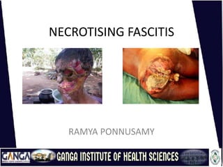
necrotising fascitiss.pptx
- 2. INTRODUCTION
- 3. INTRODUCTION • Necrotizing fasciitis has also been referred to as haemolytic streptococcal gangrene, Meleney ulcer, acute dermal gangrene, hospital gangrene, suppurative fasciitis, and synergistic necrotizing cellulitis. • Fournier gangrene is a form of necrotizing fasciitis that is localized to the scrotum and perineal area.
- 4. DEFINITION • An acute disease in which inflammation of the fasciae of muscles or other organs results in rapid destruction of overlying tissues.
- 5. INCIDENCE Rare disease Incidence: 1000 cases/year, 0.04 cases/1000 person-years Prevalence: one or two cases/career for average practitioner.
- 6. FACTS…. • Necrotising fasciitis is difficult to diagnose in its initial stages, as it mimics cellulitis. • Important early clues are pain, tenderness and systemic illness. • Bullae and ecchymotic skin lesions also point to the condition (and are not normally found with cellulitis). • A high index of suspicion is necessary and suspected cases should be referred immediately. Prompt surgical debridement is essential.
- 9. Classification • Type 1 – poly microbial infection with aerobic and anaerobic bacteria • Type 2 - Group A streptococcus (GAS): • Type 3 - Mono microbial infection: • Type 4 - fungal infection:
- 10. CAUSES • An opening in the skin that allows bacteria to enter the body. This may occur following minor injury (eg small cut, graze, pinprick, injection), or a large wound due to trauma or surgery (eg laparoscopy, sclerotherapy, endoscopic gastrostomy, thoracostomy, caesarean section, hysterectomy). Sometimes no point of entry can be found. • Cervicofacial necrotising fasciitis can follow mandibular fracture or dental infection. • Direct contact with a person who is carrying the bacteria or the bacteria is already present elsewhere on the person.
- 11. CAUSES • Particularly invasive strains of bacteria, eg streptococci that evade the immune system and produce a toxin called cysteine protease , which dissolves tissue. • NF In children may follow varicella zoster infection. • Other causes of necrotising fasciitis in children include omphalitis, necrotising enterocolitis and urachal anomalies.
- 12. Risk factors • Advanced age • Diabetes • Immune suppression • Obesity • Drug abuse • Severe chronic illness • Malignancy
- 13. • Left upper extremity shows necrotizing fasciitis in an individual who used illicit drugs. Cultures grew Streptococcus milleri and anaerobes (Prevotella species). Patient would grease, or lick, the needle before injection
- 14. Sixty-year-old woman who had undergone postvaginal hysterectomy and repair of a rectal prolapse has a massive perineal ulceration with foul- smelling discharge. Cultures revealed Escherichia coli and Bacteroides fragilis. The diagnosis was perineal gangrene.
- 15. Necrotizing fasciitis at a possible site of insulin injection in the left upper part of the thigh in a 50-year-old obese woman with diabetes.
- 16. Pathophysiology Infection of superficial fascia Release of enzymes and proteins Necrosis Horizontal spread of infection(not apparent) Vertical spread of infection9deep structures) Thrombosis Ischemia Tissue necrosis
- 17. SIGNS AND SYMPTOMS • Local pain, swelling and erythema. • Severe, constant pain. • The margins of infection are poorly defined, with tenderness extending beyond the apparent area of involvement (unlike cellulitis).
- 18. SIGNS AND SYMPTOMS After 2-4 DAYS • The area develops tense edema, extending beyond the margin of erythema. • There may be bullae, indicating skin ischemia . These may become hemorrhagic. • Skin becomes discolored, progressing to grey necrosed skin which breaks down. • The subcutaneous tissues have a wooden-hard feel . Fascial planes and muscle groups are not palpable. • There may be crepitus due to subcutaneous gas.
- 19. SIGNS AND SYMPTOMS • Pain sensation may progress from intense tenderness to anesthesia as the nerves are destroyed. • There may be a broad erythematous tract in the skin along the route of the infection. • Lymphangitis is rarely seen . • malaise, tachycardia ± fever and dehydration
- 20. SIGNS AND SYMPTOMS Days 4-5 approximately: • Hypotension and septic shock develop. • Patients become confused and apathetic. • Fournier's gangrene is a rapidly progressive form of infective NF of the perineal, genital or perianal regions, leading to thrombosis of the small subcutaneous vessels and necrosis of the overlying skin.
- 21. INVESTIGATIONS • Blood tests • Bedside finger test • Microbiology • Radiology
- 22. MANAGEMENT • The initial surgery is the most important determinant for survival. The debridement must be extensive, with adequate margins so that no infected tissue remains. • Following initial debridement, the wound must be observed closely. Surgical debridement is repeated daily until the infection is controlled. • When the infection is controlled, daily dressings are required under sedation. • Closure of the wound is by secondary suturing ± skin grafts. Vacuum-assisted wound closing devices may assist healing.
- 23. Antibiotic Regimens • The antibiotic regime will depend on the site of infection, patient allergies and culture results. Examples of recommended regimes include (all drugs given intravenously):Benzylpenicillin plus clindamycin plus gentamicin. • If penicillin-allergic, meropenem plus clindamycin plus gentamicin. Review the need for gentamicin daily. • Piperacillin-tazobactam and clindamycin, or benzylpenicillin and clindamycin.
- 24. Non-surgical treatment • Non-surgical measures include close monitoring and general supportive treatment in an intensive care setting with antimicrobial treatment. • Nutritional support is required from day one, owing to the high protein and fluid loss from the wound (similar to major burns). In severe cases, patients may need twice their basal calorie requirements. Nasogastric feeding may be helpful. • Broad-spectrum antibiotics.
- 25. COMPLICATIONS • NF carries a significant mortality rate, particularly if marine organisms • The deep tissue infection may lead to vascular occlusion, ischemia and tissue necrosis. There may be nerve damage and muscle necrosis. • Large areas of tissue loss may require skin grafting, reconstructive surgery or amputation .
- 26. PROGNOSIS • Even with surgery, the mortality rate is 20- 40%. • Increased mortality is associated with delays in diagnosis, poor surgical technique and diabetes.