
Recommended
More Related Content
What's hot
What's hot (20)
Similar to Chlamydia and mycoplasma
Similar to Chlamydia and mycoplasma (20)
More from Rachna Tewari
More from Rachna Tewari (7)
Recently uploaded
Recently uploaded (20)
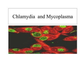
Chlamydia and mycoplasma
- 2. Objectives • To know about Chlamydia and Mycoplasma Classification Morphology Diseases Lab diagnosis
- 3. Classification • Family - Chlamydiacae. • Two genus – Chlamydia and Chlamydophila.. • Has three important human pathogens –C . trachomatis. –Chlamydophila psittaci –Chlamydophila pneumoniae
- 4. Chlamydiacae • Family Chlamydiacae are obligate intracellular bacterial parasites. • They have tropism for squamous epithelial and macrophages of the respiratory tract and GUT. • Depend for energy on host cell .
- 5. General features • Smallest prokayotic cell . • Round to ovoid. • Two lipid bilayers resembling a gram negative envelope. • Non motile. • Gram negative. • Possess both DNA and RNA and ribosome .
- 6. • Differ from other bacteria - No Peptidoglycan content in their cell wall. Lack enzymes of ETC. Require ATP and nutrient sources from host cells.
- 7. Morpholology and growth Chlamydia replicate by unique complex cycle in susceptible host cell - They exist in two morphological forms • Elementary body- extracellular , infectious form, spherical, rigid cellwall • Reticulate body – intracellular, growing , replicative form,oval pliable cell wall,
- 8. Replicative cycle • EB attach to surface – engulfed by cell – Form phagosome – after 6-8 hrs reorganise into RB and replication continues for 18 - 24 hrs. Inclusion body – intracytoplasmic vacuole filled with RB. Can be detected by histologic stains. After 48 hrs – multiplication stops and change to EBs. By 72 hrs – host cell rupture releasing infected EBs.
- 10. Resistance • Being heat labile Inactivated at 56 deg within minutes. – Susceptible to chemicals like ether ,ethanol formalin . – Can be stocked at 4 deg C. – Culture can be preserved frozen at -70 deg.
- 11. Antigenic structure • Two major antigens - Genus specific – lipopolysaccharide.(3-deoxy- manno-octulosonic acid). Important in scarring and fibrosis by inducing cytokines. Antibodies can be detected by IF and CF tests. Species specific- outer membrane proteins . – Elicits protective immunity. – IF Serotype specific - serovars on basis of MOMP.
- 12. Serological typing • Chlamydia- 3 species C.trachomatis, C.suis , C. muridarum C.trachomatis divided into biovars TRIC- trachoma inclusion conjunctivitis LGV - lymphogranuloma venerum BIOVARS are further divided into many serotypes .
- 13. Chlamydophila Has 6 species- • C.pneumonia- no biovars ,no serotypes. • C.psittaci -no biovars ,many serotypes • C.pecorum • C.caviae • C.felis • C.abortus
- 14. DISEASES • C.trachomatis – genitourinary infection and eye infection. • C.psittaci – respiratory infection. • C. pneumoniae – atypical pneumonia.
- 15. Laboratory diagnosis • Specimens- depend on type of lesion (urethra cervix,conjunctiva, lung) –scraping , swabs, blood. • Cytological examination. • Culture and isolation- cell culture/ yolk sac / mice • Detection of chlamydia antigen - Direct Fluoroscent antiody/Elisa • Serodiagnosis - microimmunofluorescence
- 17. Chlamydia trachomatis • Trachoma – keratoconjunctivitis. (A,B,Ba,and C. ) Conjunctival scraping- inclusion bodies known as Helberstaedter- prowazek body • Lymphogranuloma venerum • Inclusion conjunctivitis in adults • Non gonococcal urethritis • Perinatal infections • Neonatal conjunctivitis • Infant pneumonia
- 18. LGV • Sexually transmitted disease characterised by suppurative inguinal adenitis . • C. trachomatis serovars L1,L2 and L3. INCLUSION CONJUNCTIVITIS Sexually active adults it may transfer to eye through genital secretions and lead to paratrachoma and is caused by D to K also known as swimming pool conjunctivitis.
- 19. Non gonococcal urethritis • Caused by serotypes D-K. • Inflammation of genital organs. • Perinatal infection Neonatal conjunctivitis Infant pneumonia
- 20. Laboratory diagnosis • Smears- inclusion body by Giemsa. • Isolation- cell culture HeLa Cells ,McCoy and BHK. • Antigen detection-DirectFluoroscent antibody/ Elisa . • Nucleic acid probe/PCR/LCR. • Serological test –CFT and micro IF.
- 21. Treatment • Sulphonamide and Tetracycline.
- 22. Chlamydophila pneumoniae • Clinical findings- bronchitis and pneumonia,pharyngitis ,coronary heart disease, asthma. • Laboratory diagnosis – same. - demonstration of elimentary bodies. - culture. - antigen , antibodies detection. • Treatment- macrolides.
- 23. Chlamydia psittaci • Source – infected birds . • Clinical presentation – influenza like syndrome ,pneumonia. • Epidemiology – occupational. • Lab diagnosis – culture – levinthal colelillie bodies. • Antigen detection –DIF , EIA • Treatment- tetracycline ,macrolide.
- 24. Mycoplasma • Smallest free living bacteria ,pass bacterial filter. Morphology – gram negative ,pleomorphic. Also known as PPLO (pleuropneumonia like organism ) • Lack cell wall .
- 25. • Enclosed in trilayered membrane having sterol .
- 26. Reproduction • Divide by binary fission. • Grow on cell free media. • Adhere to epithelium of respiratory and urogenital tract.
- 27. Classification • Mycoplasmatacae Mycoplasma do not hydrolyse urea. Ureaplasma hydrolyse urea. • Pathogenic to humans M.hominis M.pneumoniae U.urealyticum
- 28. Cultivation • Facultatively anareobic. • For primary isolation 95 %nitrogen and 5% CO2 • 22-41 deg c. • Media – enriched with 20 percent horse serum and yeast extract. • PPLO broth -yeast extract , serum, glucose ,phenol red. • Solidified by agar , SP-4 media , Mycotrin RS. • Fried egg colony . • Seen by Dienes method
- 30. Biochemical reaction • Mycoplasma – mostly fermentative. • Urea – hydrolysed by ureaplasma. • Not proteolytic.
- 31. Resistance • Heating at 56 deg for 30 min. • Resistant to pencillin and cephalosporin. • Resistant to UV light . • Sensitive to tetracycline, cholorohxidine and cetrimide. • Lyophilization / freezing broth cultures at -70 deg.
- 32. Antigenic structure • Cell membrane constituents - glycolipids act as antigen in vitro in CFT. - protein -P1. Antibodies are found in convalescent serum and respiratory secretion.
- 33. M. pneumoniae • Mild URTI. • LRTI. • Tracheobronchitis and pneumonia (primary atypical pneumonia or walking pneumonia ) • School age children and young adults. • Complications may occur
- 34. M. genitalium • Non gonococcal urethritis and PID . • U. urealyticum causes genital infection. • Transmitted by sexual contact. • M. hominis – lower genital tract. • Cause non gonococcal urethritis .
- 35. Laboratory diagnosis • Specimen – depends on lesion . • Culture –PPLO medium . • Isolation and identification • Colonies- mulberry shaped /fried egg • Species identification Haemadsorption test(G.pig RBC adheres to colonies of M.pneum and not hominis ) • Tetrazolium reduction test- M.pneumoniae appear red. • Serological technique-ELISA and IFA • PCR and DNA probes – high sensitivity
- 36. Antigen detection – IF , EIA ,IMMUNOBLOTTING DNA probes – 16SrRNA sequence in resp. exudate. Serological tests Specific test using mycoplasmal antigens - CFT - ELISA Non specific test Streptococcus MG test- unheated serum and heat killed str. MG suspension Cold Agglutination TEST-antibodies that bind to human erythrocytes at low temp 4 deg
- 37. Treatment • Tetracycline. • Macrolides (azithromycin ). • Fluoroquinolones.
- 38. Further reading • Essentials of medical microbiology. Apurba S Sastry. • Textbook of Microbiology . Ananthannarayan and Paniker.
- 39. Mcqs 1. All of the following are true except a. Elementary body is metablically active b. Reticulate body is the replicating form c. Reticulate body is intracellular form d. Elementary body is infectious form 2 . The most commonly used method for isolation of Chlamydia: a. Culture on artificial media b. Culture on Vero cell line c. Inoculation into guinea pig d. Culture on McCoy cell line
- 40. 3. The most sensitive and specific test for Chlamydia diagnosis: a. Direct immunofluorescence test (DIF) b. Culture on McCoy cell line c. Nucleic acid amplification tests (NAAT) d. Micro-immunofluorescence (MIF) test 4. Which is not a property of Mycoplasma? a. Susceptibility to beta lactams b. Have both DNA and RNA c. Mycoplasmo pneumonioe d. Streptococcus pneumoniae 5. Fried egg colonies are produced by: a. Bacillus cereus b. Haemophilus influenzae c. Neisseria subflavo d. Mycoplosma pneumoniae