1. Hip radiologic imaging
standard projection
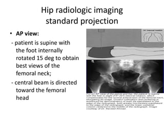
• AP view:
- patient is supine with
the foot internally
rotated 15 deg to obtain
best views of the
femoral neck;
- central beam is directed
toward the femoral
head
2. • Lateral View (cross table
lateral view)
- surgical lateral view:
- this view should be obtained on all
patients suspected of having a hip
fracture or dislocation;
• - patient is supine; the opposite
hip is flexed and abducted;
• - cassette is placed against
the lateral aspect of the affected
hip
• - central beam is directed
horizontally toward the groin with
about 20 degree of cephalic tilt;
3. • Frog leg view:
• - do not order a frog
leg lateral in any patient
suspected of having hip
fracture or dislocation
patient is supine w/ knees
flexed, soles of feet
together, and the thighs
maximally abducted;
- central beam is directed
vertically or with a 10 to 15
deg cephalic tilt to a point
slightly above pubic
symphysis;
4. • Clements-Nakayama view 4
• lateral projection
demonstrating the neck of
the femur without
movement of the either
limb
• the ideal projection for
bilateral hip or femur
trauma
5. • Dunn view
• lataral projection to aid
and diagnose
femoroacetabular
impingement (FAI) due
to its increased
sensitivity for detecting
femoral head-neck
asphericity.
7. Acetabular evaluation
• 45-degree oblique
views of the pelvis
commonly called Judet
views-
– Iliac oblique view
– Obturator oblique view
Editor's Notes
The acetabulum is evaluated radiographically with an anteroposterior pelvic view as well as with the 45-degree oblique views of the pelvis described by Judet and Letournel, commonly called Judet views. In the iliac oblique view, the radiographic beam is roughly perpendicular to the iliac wing. In
the obturator oblique view, the radiographic beam is roughly perpendicular to the obturator foramen.
Inclusion of the opposite hip in the radiographic field on the anteroposterior
and Judet views is essential for evaluation of symmetric contours that may have slight individual variations and to determine the width of the normal articular cartilage in each view.
Six radiographic landmarks were defined by Judet and Letournel and should be appreciated on all plain films.
The iliopubic line, or arcuate line, represents the medial cortex of the anterior column, while the ilioischial line signifies the medial cortex of the posterior column. The radiographic graphic U, more commonly referred to as the teardrop, represents the most inferior and anterior aspect of the acetabular fossa laterally and the anterior aspect of the quadrilateral plate medially. The sourcil represents the acetabular roof and
extends to the lateral aspect of the teardrop superiorly. The anterior and posterior lips represent the most lateral aspect of
the anterior and posterior walls, respectively