Downloaded 814 times


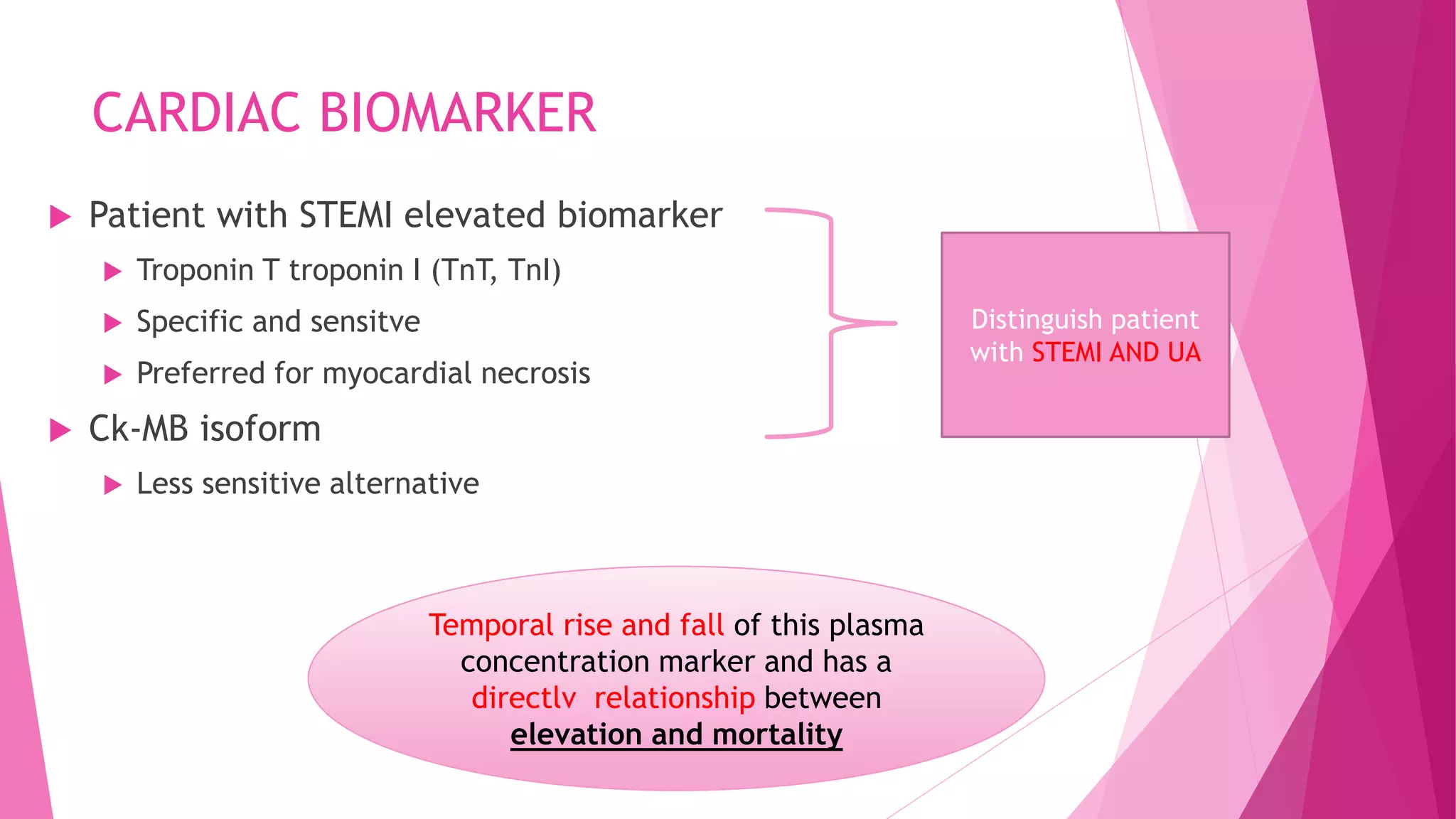
![CKMB TIME
RISE 4-6 HR
PEAK 12 HR
DIASSAP]REA 48-72 HR
TROP T AND TROP I TIME
RISE 4-6 HR
REMAIN ELEVATED 2 WEEKS](https://image.slidesharecdn.com/acutecoronarysyndromeacs-180106041458/75/Acute-coronary-syndrome-acs-20-2048.jpg)


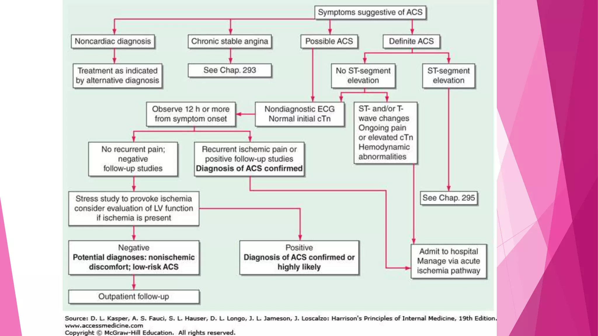
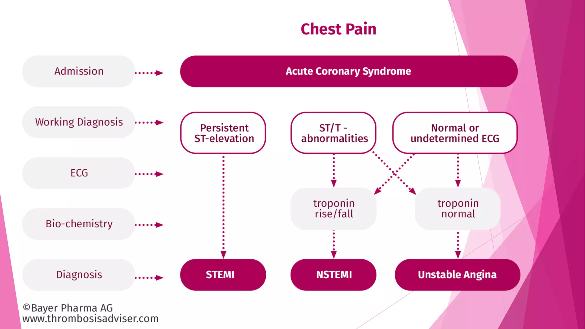

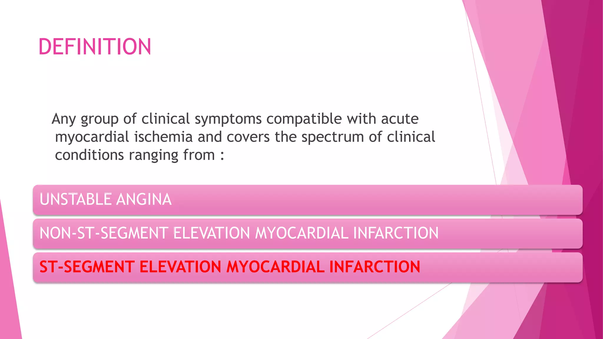
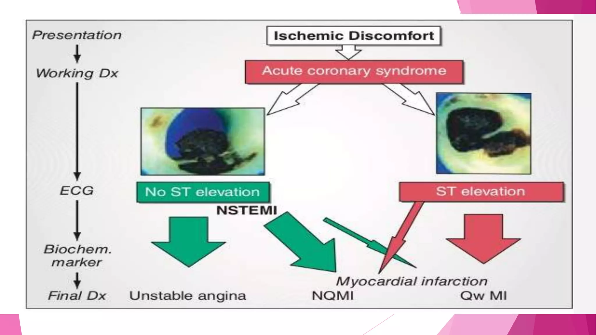

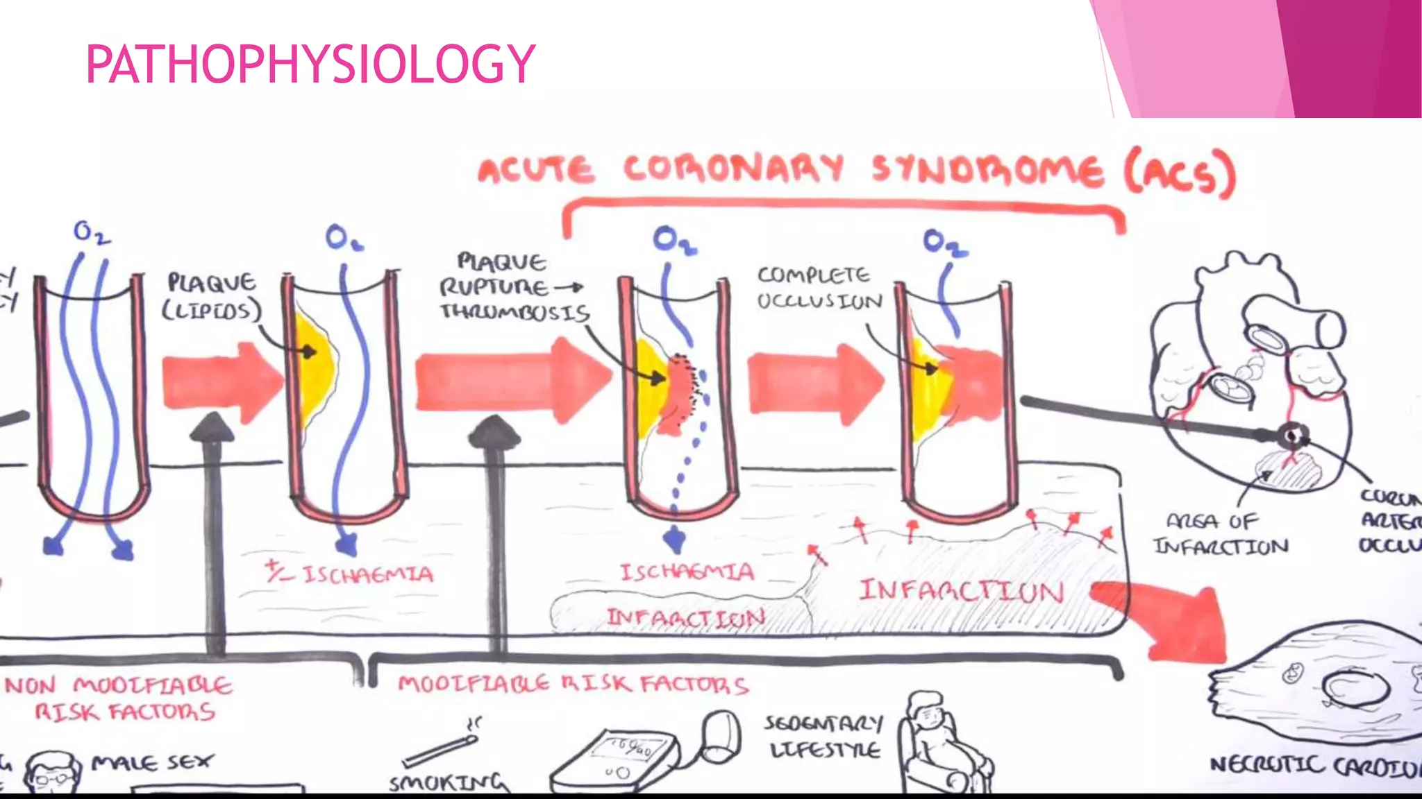


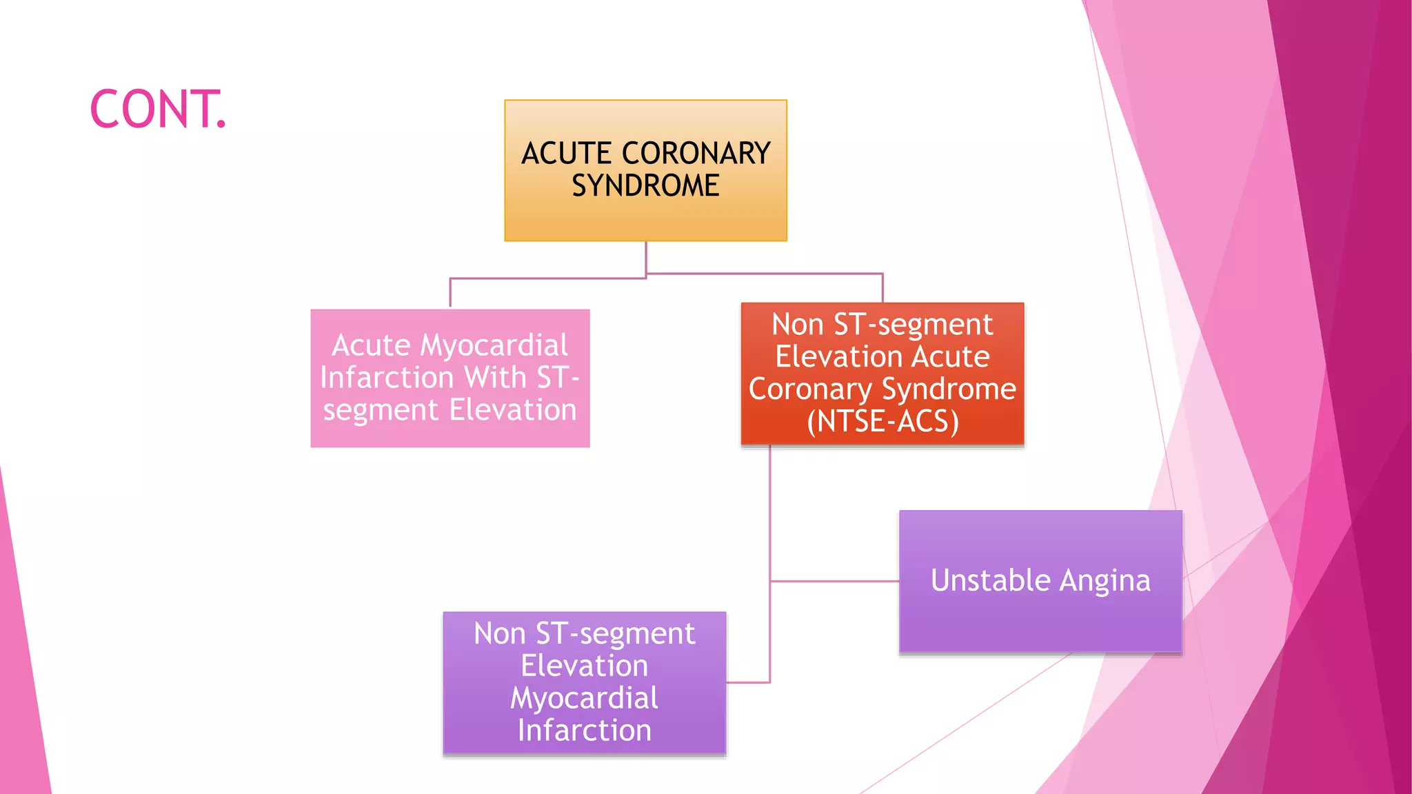
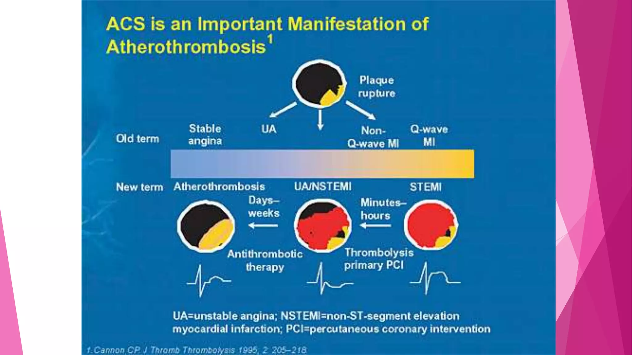
This document discusses acute coronary syndrome (ACS) and non-ST-segment elevation acute coronary syndrome (NSTE-ACS). It defines unstable angina (UA) and non-ST elevation myocardial infarction (NSTEMI) and covers their clinical presentation, diagnostic criteria, laboratory investigation, and management. The key goals of diagnosis and treatment for NSTE-ACS patients are to recognize or exclude myocardial infarction, detect resting ischemia, and identify coronary artery obstruction. Treatment involves anti-ischemic, antithrombotic medications and consideration of coronary revascularization.


