
Recommended
More Related Content
What's hot
What's hot (20)
Viewers also liked
Viewers also liked (8)
Similar to PA Hand Final
Similar to PA Hand Final (20)
Recently uploaded
Recently uploaded (20)
PA Hand Final
- 1. PA Hand By: Nicole DeStefano
- 2. HIPPA Compliance Our image is HIPPA compliant Nowhere on our image does it violate any patient confidentiality
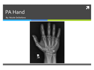
- 3. Marker & Patient ID The correct anatomical side marker is visible in the image. The side marker is placed correctly with the right marker displaying on the viewers left. There are no markers superimposed on any pertinent anatomy.
- 4. Marker & Patient ID There were no additional side markers used but an arrow marker could have been used to indicate sight of pain/injury etc. The image is displayed correctly based on marker placement.
- 5. Radiation Hygiene Acceptable beam restriction requires at least 3 or more sides of collimation. There is no evidence of beam restriction located on our image. Shielding must be provided if the gonads are within 5 cm of the primary beam. There is no evidence of primary shielding on the side closest to the gonads. It is always a good habit to place a lead shield on any patient of child bearing age.
- 6. Completeness of Position/Projection Routine positions/projections for a hand include: PA PA Oblique 45 degree rotation Left fan lateral This is a PA hand which complies with the routine projections/positions. All anatomical parts are correctly visualized on the image.
- 7. Artifact Identification There does not appear that there are any of the following in our image: Physical artifacts Body parts superimposed Hospital paraphernalia Patient clothing/belongings Indwelling artifacts/foreign bodies Our patient does have a fracture of the 5th metacarpal.
- 8. Artifact Identification There does not appear that excess fog is visible and/or degrading overall image quality. There also does not appear that there are any CR/DR artifacts visible in the image.
- 9. Image Sharpness There does not appear that any of the following are in the image: Gross voluntary motion Excessive quantum mottle/image noise Evidence of double exposure Grid lines/cut-off, or artifacts When imaging a hand, it is unnecessary to use a grid.
- 10. Image Sharpness Size distortion does not appear greater than expected because when imaging a hand, the hand lies directly on the IR. The CR should be entering at the third MCP joint. Because the CR is more than 1 cm off- centered to the part, shape distortion is being caused but only to a minimal.
- 11. Accurate Part Positioning The part is adequately aligned to the image media. The part is not accurately centered. The part would have to be moved about an inch to the left. The CR is not centered within 1 cm of the anatomical part. The CR is aligned to the imaging media. The CR does conform to an accepted IR exposure field recognition template.
- 12. Accurate Part Positioning According to Merrill’s Atlas or Radiographic Positioning and Procedures for a PA hand state: Rest the patient’s forearm on the table and place the hand with the palmar surface down on the IR. Center the IR to the MCP joints, and adjust the long axis of the IR with the long axis of the hand and forearm. Spread fingers slightly. Shield gonads. Ask patient to relax hand to avoid motion.
- 13. Evaluation Criteria Evidence of proper collimation No rotation of hand Open MCP and IP joints, indicating hand is placed flat on the IR Slightly separate digits with no soft tissue overlap All anatomy distal to the radius and ulna Soft tissue and bony trabeculation
- 14. Judicious ExposureTechnique Based on criteria, the anatomical part is correctly positioned. The most radiolucent structure visible in the image is the soft tissue of the hand. The most radiopaque structure that is visible on the image is the bony cortex of the hand.
- 15. Judicious ExposureTechnique I think the image displays adequate contrast by displaying short scale contrast. The brightness of our image is adequate as well. Even though we do not know our EI value, I would say our EI value was most likely within normal range.
- 16. Accept or Reject? Based on the evaluation criteria, I would ACCEPT this image. However, for next time: I would make sure I increased my beam restriction especially on the side closest to the gonads. Also appropriately center the CR to correct anatomical part.
- 17. Sources Frank, Eugene D., Bruce W. Long, Barbara J. Smith, Vinita Merrill, and Philip W. Ballinger. Merrill's Atlas of Radiographic Positioning & Procedures. 12th ed. Vol. 1. St. Louis, MO: Mosby/Elsevier, 2007. Print.