2. Is the radiographic image
compliant with the Health
Insurance Portability and
Accountability Act?
The radiographic image appears to
be compliant. It contains no
information that correlates to the
patient or the facility.
It provides data privacy and
security provisions for safeguarding
medical information about that
patient and the x-ray facility.
3. MARKER & PATIENT ID
The correct anatomical side marked is visible.
But superimposed on anatomy. The marker
appears to be annotated.
The radiographer initials are not in the image.
In this case, other additional markers such as
arrows are rarely or not used at all, to point
where the pain is most concentrated. However
positioning markers such as
(AP/PA/Supine/Erect/recumbent) would be used
depending on the facility. Other facilities require
the technologists to annotate date and time, and
place ex: ER, ICU, Pt. Room #, etc.
The image appears to be correctly displayed.
However, since the marker was place in PP;
Image could have been flipped.
4. RT ID
The propper correction for the
placement of makers for this
Radiograph would be:
The placement of the marker on
the IR and on the propper side
in relation to the anamoty of
interest, placed in a anatomical
reading aspect.
An arrow to point out the zone
of interest would not be used,
however other markers such
erect/supine/recumbent could
be used.
Date, time and place could be
also annotated in PP (fallow
facility protocols)
Supine
Portable
ICU
17:35
4/202017
5. RADIATION HYGIENE
A minimum of 3 sides of beam restriction is not
present. Only two appears to be collimated.
The image demonstrates improper restriction of the
collimation field for this projection. However there
is enough information to say there was collimation
present: include as much anatomy of interest as
possible. The red box demonstrates de correct CF.
Pt's gonads would not be shielded for this particular
radiograph; unless is a male pt.
Collimation should be the primary form of shielding
for the patient; Side closer to gonads is not
restricted.
Using a grid for this projection is mandatory in all
hospitals because it would attenuate low energy X-
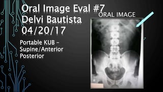
7. This radiographic projection is a routine
projection of the abdomen (lower / KUB). It
complies with the requirements of an AP
projection in particular, and it shows:
1. Evidence of proper collimation / shielding ❌
2. Entire abdominal cavity visualized (without
including diaphragm in its entirety) ❌
3. Symphysis pubis visible ✅
4. Both kidneys visualized ✅
5. No rotation of the pelvis ✅
6. Patient’s arm and clothing out of the
collimated field.✅
7. Bladder silhouette visualized.✅
8. Symmetric lower ribs ❌
Projection/Position
8. Artifact Identification.
There are no artifacts visualized in this radiograph and
superimposition of anatomy of interest with adjacent
body parts is not present.
There is no:
1. Medical paraphernalia,
2. Patient clothing
3. Pt’s Belongings.
Moreover, there is no other types of artifacts or foreign
bodies present.
Fog is present, however, There is no excess fog present
that could have degraded the overall image contrast and
visibility of recorded detail.
There are also no CR/DR artifacts.
A reciprocating /oscillating or high frequency grid was
not used for this radiograph; a stationary grid was
present.
9. IMAGE SHARPNESS
The radiographic image contains
no:
1) quantum mottle ❌
2) Fog is present but not excessively
. ❌
3) Gross voluntary motion ❌
4) double exposure ❌
5) grid lines ✅ (around the
collimated edges), grid artifact❌
grid cut-off✅: a grid is routinely
used for this projection due to
body part being >10cm thick, and
kvp > 70kvp. Stationary grid.
10. IMAGE SHARPNESS
• The image has relatively poor abdominal detail.
1. size distortion is present but minimal. OID
from kidneys and bladder to IR in AP projection
of the abdomen is not minimal, we can expect
some degree of size change.
2. Shape distortion is present; CR was not
perpendicular for this projection. Moreover, the
kidneys have a 45 degrees towards the IR
causing them to appear relatively
foreshortened.
The CR should enter perpendicular to the IR
toward the sagittal plane at the level of the iliac
crest.
T7
11. ACCURATE PART
POSITIONING
The part appears slightly off-centered.
The image appears to be inadequately
obtained due to misrepresenting image
contrast; image looks more on the
short scale contrast level; Hight
brightness of vertebra and pelvis is
visible.
Enough information to say that CR and
part were approximately centered to
the imaging media. There is Shape
distortion, however not grossly
visualized.
12. ACCURATE PART POSITIONING
The part could have been adequately aligned to
the image media: Shape distortion is visible on
the lower ribs and pelvis.
The CR is not centered within 1 cm from the mid-
medial plane at the level of L2-L3.
The CR was not adequately aligned with the
image media: Anatomy of iliac crest look
distorted.
The CR’s alignment seems to not conform to an
accepted IR exposure field recognition
template/field; image looks relatively more on the
short scale contrast. Ex:
T7
13. ACCURATE PART POSITIONING
According to Merrill’s Atlas, Radiographic
Positioning and Procedures for an AP portable
projection, KUB:
Pt would stay laying down and facing up in a supine
position (as flat as possible)
Use grid underneath the patient to show the
abdominal anatomy from the pubic symphysis to the
upper abdominal region in proper contrast.
Place Sponges on the on each side to prevent motion
and have a better positioning of body part .
Keep the grid from tipping side to side by placing it
in the center of the bed and stabilizing it with
blankets or towels / sponges if necessary
Use the patient’s draw sheets to position grid and IR
under the patient and to the pt’s skin to be exposed
directly onto the grid.
Ask the patient to hold breath after expiration / or
hold exposure till pt’s takes an expiration.
15. EXPOSURE TECHNIQUE
Soft tissue and joint spaces would be The
most radiolucent structures (🌗) in this
image.
On the other hand, The most radiopaque
structure(🌕)in this image is the outermost
layer of bony cortex.
🌗
🌕
16. EXPOSURE TECHNIQUE
Enough information is given to prove that the
image was underexposed; however the EI value
is not given. Moreover, In digital radiography a
histogram is compared.
The image looks improperly exposed; outline
of abdominal viscera detail is poorly
demonstrated and kidney shadows are poor
visualized. Vertebral columns look adequately
exposed; improper technique or bilateral grid
cut-off could have caused this
A exposure pre-technique for a portable
abdomen is rarely set before the exposure is
taken when using a portable machine; however
The radiographer could adjusted and fixed
exposure factors according to the CF, anatomy
of interest and pt size; to obtain proper levels
17. EXPOSURE TECHNIQUE
Contrast is determined by window width.
The image seems to portrait inadequate
amount of gray tones.
On the other hand, brightness is control by
window level.
Brightness is inaccurately balanced; image
appears to have short scale contrast. When
imaging the abdomen, the radiograph should
have a long scale contrast for better
demonstration and visualization of soft tissue
in abdominal cavity, most importantly the
urinary system in particular when performing a
KUB.
Image should portrait more shades of gray or
to be darken.
18. REJECT OR ACCEPT !!!!
• According to the evaluation criteria, the anatomy is correctly
positioned however CR appears to be angled caudaly.
Moreover, anatomy of interest is clipped, therefore, the
projection should be repeated.
• Things that would be required to be changed for another
portable KUB:
1. Add Side marker, RT ID before the image is taken, annotate
(recumbent, supine, time, place etc.) if needed
2. Collimation edges should not clip anatomy of interest; DO
NOT crop the tuberculum from iliac crest.
3. Use positioning aid to center body part to grid, collimation
field and to the IR
4. Exposure Technique; according to the anatomy and
exposure field. Moreover, according to the use of grid. Long
scale contrast.
5. Shield if possible; males in specific
6. Use a perpendicular CR, and center to mid-grid.
19. Frank, Eugene D., Bruce W. Long, & Barbara J. Smith. Merrill's Atlas
of Radiographic Positions and Radiographic Procedures: 3-volume
set. 12 ed. St. Louis, MO: Mosby-Elsevier, 2016.
Bontrager, Kenneth L., & John Lampignano. Textbook of
Radiographic Positioning and Related Anatomy. 8 ed. St. Louis,
MO: Mosby-Elsevier, 2016.
Adler, Arlene Mckenna & Richard R. Carlton. Principles Of
Radiographic Imaging: An Art And A Science. 5 ed. Forence, KY:
Thomson Delmar Learning, 2016.
References