2. Instruments. UV−visible spectroscopy was performed using
a Shimadzu UV-3600 UV−vis−NIR spectrophotometer with
Shimadzu UVProbe software. Typical spectra were run from 300
to 900 nm using a single visible light source and a single detector.
The scanning speed was medium with a sample interval of either
0.2 or 1 nm and a slit width of 3 nm. NMR spectra were taken
with a Bruker Avance II 400 MHz spectrometer, and data work-
up was performed using TopSpin 2.1 software.
Preparation of ZnTPyP. Zn(OAc)2 (250 mg, 1.14 mmol) in
MeOH (2 mL) was added to a solution of TPyP (100 mg, 162
μmol) in 50 mL of CHCl3. The solution was heated to 75 °C for
1 h. ZnTPyP was purified by silica gel column chromatography
eluted by CHCl3 with 10% MeOH.
Preparation of PhxPyyP. Pyrrole (1.26 g, 18.7 mmol),
benzaldehyde (1.04 g, 9.8 mmol), and 4-pyridinecarboxaldehyde
(1.14 g, 10.6 mmol) were dissolved in 100 mL of propionic acid.
After the solution was heated to reflux for 1 h, the majority of the
propionic acid was removed by vacuum distillation at 150 °C.
The remaining solution was neutralized using aqueous NaOH,
and the porphyrin was extracted with chloroform. Separation of
the porphyrin solution to isolate Ph3PyP, Ph2Py2P (trans and
cis), and PhPy3P was achieved via column chromatography
(silica gel 230−400 mesh) eluting with CHCl3 and MeOH (2.5−
5%) and 1% acetic acid. The four components were collected in
the second, third, fourth, and fifth red bands, respectively.
Preparation of AxPyyP. Both 4-carbomethoxybenzaldehyde
(1.64 g, 11.1 mmol) and 4-pyridinecarboxaldehyde (3.30 g, 30.8
mmol) were dissolved in 62.5 mL of propionic acid. The solution
was heated to reflux, and pyrrole (2.80 g, 41.8 mmol) was slowly
added. After heating for 1 h and cooling to room temperature, the
mixture was treated with the same work-up procedures as those
in the synthesis of ZnTPyP. The solution was run through
column chromatography (silica gel 230−400 mesh) eluting with
CHCl3 and ethyl acetate (20−100%) and 1% triethylamine to
yield CM3PyP, CM2Py2P (trans and cis), and CMPy3P (where
CM represents the 4-carbomethoxyphenyl substituents). A
Lossen rearrangement was run on each of these individually
isolated porphyrins to prepare the corresponding pyridyl
aminophenyl porphyrins (AxPyyP).26
Each individual CMxPyyP
derivative, and 8 equiv. of NH2OH·HCl was warmed to 100 °C in
polyphosphoric acid, and the solution was slowly heated to 160
°C over 3 h. Upon cooling to room temperature, aqueous NaOH
was slowly added to neutralize the solution, and the porphyrin
was extracted with chloroform. Column chromatography was
then used to remove any residual starting material by eluting with
CHCl3 and MeOH (5%).
Titrations. All porphyrin titrations were performed with
MSA in DMSO. Solutions were purged with nitrogen before the
titrations of the AxPyyP derivatives were conducted. The
titrations began with dilute acid in 2−5 μL increments, and
concentrated acid in higher increments was used for the later
stages. The volume of the initial porphyrin solution was 3.0 mL.
Dilution effects on the concentrations of the porphyrins caused
by the addition of acid were taken into consideration, and all
spectra are reported as extinction coefficients.
■ RESULTS
The spectroscopy observed from the protonation of tetra(4-
pyridylporphyrin) (TPyP) has been reported in the literature.27
It was concluded that the pyridyl groups protonated first, based
on the very small effects on the spectra observed during the initial
aliquots of acid additions, where further additions of acid
generated the characteristic protonated porphyrin spectrum.
While these conclusions are fully consistent with the observed
spectroscopy, because it was particularly important for our study
to know the order of protonation of the various substituents, we
proceeded to confirm the spectroscopic signatures for
protonation of the porphyrin ring as distinguished from
protonation of pyridyl groups on porphyrins.
ZnTPyP. Upon protonation of ZnTPyP (Figure 2), the Soret
band red shifts by 6 nm (426 to 432 nm) and broadens. The two
Q bands show very little change. In this case, the four peripheral
pyridyl groups become protonated, leading to small effects on the
basic porphyrin spectrum.
TMePyP+4
. The spectra observed during acid titration of
TMePyP+4
(Figure 3) show a strong Soret band with a large red
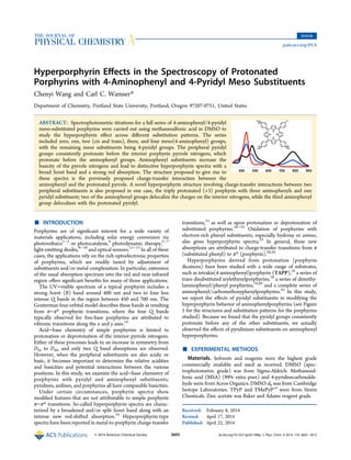
Figure 1. Structures with abbreviations and substitution patterns for the
porphyrins studied. In creating the abbreviations, A = 4-aminophenyl,
CM = carbomethoxy, M = methyl, Ph = phenyl, Py = 4-pyridyl, P =
porphyrin, and T = tetra.
The Journal of Physical Chemistry A Article
dx.doi.org/10.1021/jp501398g | J. Phys. Chem. A 2014, 118, 3605−3615
3606
3. shift (24 nm, from 422 to 446 nm) as well as the collapsing of the
four Q bands into two at 591 and 642 nm, with the Q(0,0) band
substantially stronger. These changes are typical for protonation
of the two internal nitrogens in a free-base porphyrin. It is also
notable that these protonations require acid concentrations
significantly higher than those needed to protonate the
peripheral pyridyl groups above.
Ph2Py2P (Trans and Cis). The spectrophotometric acid
titrations of both the trans and cis isomers of Ph2Py2P (Figures 4
and 5) show spectral changes similar to those observed for TPyP.
The addition of acid first leads to a small red shift (3−5 nm) of
the Soret band as well as the weakening and broadening of the
band. With further addition of acid, the Soret band shifts more
strongly (27−29 nm), and the four Q bands disappear in favor of
the characteristic strong band at about 650 nm. Thus,
unsubstituted phenyl substituents are unremarkable in terms of
any interactions with protonated pyridyl groups or protonated
porphyrins.
APy3P. The protonation of APy3P can be tracked in three
distinct stages as the pyridyl groups, porphyrin, and amino all
protonate separately (Figure 6). Initial aliquots of acid generated
a small red shift of the Soret band from 418 to 423 nm,
characteristic of protonation of the three pyridyl groups only; Q
bands were essentially unaffected during this initial stage of the
titration. Additional acid generated a weak, broad absorption
between 700 and 800 nm indicative of a hyperporphyrin; the
Soret band slightly broadened without shifting. In the final stage,
acid protonated the amino group, effectively destroying the mild
hyperporphyrin effect. The final spectrum shows a red-shifted
Soret and a strong 658 nm band characteristic of a fully
protonated porphyrin.
A2Py2P (Trans and Cis). Only two distinct stages can be
discerned in the acid titrations of both the trans and cis isomers of
A2Py2P (Figures 7 and 8). Initially, the Soret bands (421 nm in
trans and 419 nm in cis) increase and red shift to 422 nm, while
Figure 2. Spectrophotometric titration of ZnTPyP (3.37 μM) with
MSA (0−12 μmol) in DMSO.
Figure 3. Spectrophotometric titration of TMePyP+4
(7.5 μM) with
MSA (0−15 mmol) in DMSO.
Figure 4. Spectrophotometric titration of trans-Ph2Py2P (3.24 μM)
with MSA in DMSO. PH2
+2
represents protonation of the two
peripheral pyridyl groups (0−66 μmol MSA), while PH4
+4
represents
protonation of the internal porphyrin nitrogens (0.066−3.95 mmol
MSA).
Figure 5. Spectrophotometric titration of cis-Ph2Py2P (3.24 μM) with
MSA in DMSO. PH2
+2
represents protonation of the two peripheral
pyridyl groups (0−70 μmol MSA), while PH4
+4
represents protonation
of the internal porphyrin nitrogens (0.07−2.3 mmol MSA).
The Journal of Physical Chemistry A Article
dx.doi.org/10.1021/jp501398g | J. Phys. Chem. A 2014, 118, 3605−3615
3607
4. all of the Q bands also undergo red shifts by 6−20 nm. At
wavelengths above 700 nm, broad hyperporphyrin absorbances
appear in both spectra, in which the cis isomer shows
considerably stronger absorption than the trans isomer. These
features indicate that the pyrrole groups have been protonated
and are interacting with the aminophenyl groups to generate
hyperporphyrin spectra. The small changes associated with
protonation of only the pyridyl groups, that is, a +2 diprotonated
state, cannot be distinguished during the titration. This implies
that the pyrrole groups of the porphyrin have become more basic
with the presence of the two aminophenyl substituents and the
pyrroles protonate together with the pyridyl groups. In the
second stage of the titrations, new Soret bands (448 nm in trans
and 450 nm in cis) grow in, the Q bands collapse into one major
absorption (673 nm in trans and 656 nm in cis), and the
hyperporphyrin peaks disappear, characteristic of a fully
protonated porphyrin with all groups noninteracting.
A3PyP. The spectrophotometric acid titration of A3PyP
(Figure 9) shows three distinct stages. A simple +1 protonated
state, indicating protonation of the pyridyl group only, was not
observable. The first protonation stage is characterized by clear
Figure 6. Spectrophotometric titration of APy3P (3.24 μM) with MSA
in DMSO. PH3
+3
represents protonation of the three peripheral pyridyl
groups (0−385 μmol MSA), PH5
+5
represents protonation of the
internal porphyrin nitrogens (0.385−2.77 mmol MSA), and PH6
+6
represents protonation of the peripheral aminophenyl group (2.77−9.7
mmol MSA).
Figure 7. Spectrophotometric titration of trans-A2Py2P (9.28 μM) with
MSA in DMSO. PH4
+4
represents protonation of the two peripheral
pyridyl groups as well as the two internal porphyrin nitrogens (0−230
μmol MSA), and PH6
+6
includes protonation of the two peripheral
amino groups as well (0.23−5.1 mmol MSA).
Figure 8. Spectrophotometric titration of cis-A2Py2P (7.73 μM) with
MSA in DMSO. PH4
+4
represents protonation of the two peripheral
pyridyl groups as well as the two internal porphyrin nitrogens (0−155
μmol MSA), and PH6
+6
includes protonation of the two peripheral
amino groups as well (0.155−6.2 mmol MSA).
The Journal of Physical Chemistry A Article
dx.doi.org/10.1021/jp501398g | J. Phys. Chem. A 2014, 118, 3605−3615
3608
5. isosbestic points marking the development of a split Soret band
(402 and 478 nm; intensity ratio 1.84), loss of the four Q bands,
and growth of a huge red absorbance peaking at 781 nm, all
features indicating very strong hyperporphyrin interactions. This
all takes place at a significantly lower acid concentration than any
of the previous titrations; thus, the molecule has been made
significantly more basic by the presence of the multiple amino
groups. We assign this spectrum to a +3 state, assuming that the
pyridyl substituent is also protonated, a topic discussed later
while considering the contributing resonance forms. During the
second stage of the acid titration, there are no clear isosbestic
points as multiple effects seem to be taking place. The Soret band
at 402 nm grows into a higher absorption at increasing
wavelength, while the Soret band at 478 nm decreases and
moves to longer wavelength. The red hyperporphyrin
absorbance decreases and shifts to shorter wavelengths. We
consider this to be the protonation of one of the three amino
groups, possibly at different positions, to form +4 isomers, as well
as protonation of two amino groups to form a +5 state. The
hyperporphyrin effect is weakened but remains remarkably
strong throughout these changes. As the acid titration completes,
distinct isosbestic points can be observed, leaving only one Soret
band at 477 nm and one Q band at 658 nm, indicative of the fully
protonated porphyrin. We attribute the final stage to protonation
of the last remaining amino group.
TAPP. The acid titration of TAPP is shown in Figure 10,
which is consistent with titrations reported in the litera-
ture.16,19,20,25
TAPP is significantly more basic than all other
porphyrins in this study. Initially, the Soret band splits into a
weak absorption at 392 nm and a strong one at 467 nm, as well as
a strong red absorption peaking at 813 nm. Protonation to the +4
state still leaves a remarkably strong hyperporphyrin spectrum.
Complete protonation leads to a Soret band at 443 nm and a Q
band at 660 nm.
Comparison of Hyperporphyrin Spectra. Figure 11
highlights the spectra of the different hyperporphyrins obtained
from the variously substituted aminophenyl/pyridyl porphyrins.
With increasing numbers of aminophenyl groups, clear trends are
Figure 9. Spectrophotometric titration of A3PyP (7.57 μM) with MSA
in DMSO. PH3
+3
represents protonation of the one peripheral pyridyl
group as well as the two internal porphyrin nitrogens (0−7.7 μmol
MSA), PH5
+5
represents protonation of two of the three peripheral
amino groups (0.077−1.57 mmol MSA), and PH6
+6
represents
protonation of all of the three peripheral amino groups (1.57−7.7
mmol MSA).
Figure 10. Spectrophotometric titration of TAPP (5.93 μM) with MSA
in DMSO. PH2
+2
represents protonation of the two internal porphyrin
nitrogens (0−690 nmol MSA), PH4
+4
represents protonation of two
peripheral amino groups (0.69−126 μmol MSA), and PH6
+6
represents
protonation of the last two peripheral amino groups (0.126−7.1 mmol
MSA).
The Journal of Physical Chemistry A Article
dx.doi.org/10.1021/jp501398g | J. Phys. Chem. A 2014, 118, 3605−3615
3609
6. observed in terms of the wavelength and intensity of the red
hyperporphyrin band as well as the wavelength and intensity of
the split Soret band. Table 1 summarizes these spectral features,
which are discussed further in terms of resonance delocalization
in the next section.
Relative Basicities. Because most of the various protonated
forms are distinguishable spectroscopically, it is possible to
determine the relative order of basicity of the different nitrogen
bases present. Generally, the peripheral pyridyl groups protonate
first, followed by the interior porphyrin pyrrole groups, followed
by the peripheral amino groups. When there are two or more
aminophenyl substituents, the interior porphyrin pyrrole groups
become more basic and protonate along with the pyridyl groups
(we could not distinguish separate protonation stages for A3PyP
or cis- or trans-A2Py2P). The aminophenyl groups consistently
protonate last; in the molecules under study, they are involved in
electron donation to the hyperporphyrin and bear partial positive
charge.
Quantitative measures of basicities were determined by
monitoring the acid titrations at three or more distinct
wavelengths to distinguish the relative concentrations of different
species at different points in the titrations, using the method of
Splan18
and Rudine.25
An apparent pKa value was obtained from
the interpolated point at which the acid concentration was
sufficient to generate equal concentrations of both the acid and
conjugate base forms. These are considered apparent pKa values
because of the nonaqueous solvent (DMSO) and the
unidentified state of ionization of MSA. Nevertheless, they are
useful indicators of the relative basicity of the different
derivatives, which are tabulated in Table 2. Because of the
unique spectrum obtained for A3PyPH3
+3
, we also investigated
other triamino-substituted porphyrins, and these are included in
the table as well.
■ DISCUSSION
The most common examples of hyperporphyrin effects occur
with strong electron-donating groups available to promote
charge-transfer interactions with positively charged sites on the
interior of a protonated porphyrin. We have previously reported
such hyperporphyrin effects of 4-aminophenyl groups, which are
strong electron donors by resonance effects, studying a complete
series of derivatives with either 4-aminophenyl or 4-carbome-
thoxyphenyl substituents.25
In this study, we have examined a
similar series with either 4-aminophenyl or 4-pyridyl as the
substituents. Although the pyridyl groups are similarly electron-
withdrawing, comparable to carbomethoxyphenyl, the additional
complexity is that the pyridyl groups are readily protonated,
creating an even stronger electron-withdrawing effect. We
propose that under certain circumstances, the protonated pyridyl
groups can also take part in charge-transfer interactions with the
aminophenyl groups via resonance effects through the porphyrin
ring.
In the series that we studied, a typical porphyrin has multiple
possible sites for protonation, the interior of the porphyrin, the
pyridyl substituents, and the aminophenyl substituents. The
reported acid titration of TPyP led to the conclusion that the
peripheral pyridyl groups protonate before the porphyrin
interior,27
which is consistent with our observations of the
spectroscopy of protonated ZnTPyP (which cannot protonate
on the interior) (Figure 2) and TMPyP+4
(where the methylated
pyridinium groups simulate protonated pyridyl groups) (Figure
3).
Acid titration of APy3P showed subtle but distinctive spectra
indicating all three protonations, first the three pyridyl groups,
then the porphyrin interior, and then the amino group (Figure
6). Protonation of the pyridyl groups first was consistently
observed until two or more aminophenyl substituents were
present; these groups apparently enhance the basicity of the
interior porphyrin pyrrole groups; therefore, for the cis and trans
isomers of A2Py2P as well as for A3PyP, it was not possible to
detect separately the protonation of the pyridyl groups and the
protonation of the porphyrin pyrroles.
Examining the trends in pKa values in Table 2, pyridyl groups
are relatively consistent in their basicity, although multiple
electron-donating amino groups distinctly improve basicity.
Basicity of the porphyrin pyrrole groups, however, is strongly
affected by amino groups. As one to four amino groups are added,
relative pKa values increase, 0.37, 2.43 or 2.38, 3.5, 4.57. Part of
this trend is likely due to the effects of the electron-withdrawing
pyridinium substituents that would decrease basicity; as more
amino groups are added, fewer pyridinium groups are necessarily
present. This effect can be normalized by comparison with the
unsubstituted phenyl derivatives; even with two pyridinium
substituents, the pKa of the porphyrin of trans- or cis-Ph2Py2P
(0.72 or 1.0) is significantly lower (less basic) than that of
A2Py2P (2.43 or 2.38). Thus, the amino groups provide the
dominant effect in increasing basicity.
Aminophenyl groups always protonate last. Because the
aminophenyl groups provide effective stabilization of the
protonated porphyrin via hyperporphyrin resonance, they
already bear a partial positive charge, and protonation to an
ammonium ion would cause a loss of the resonance stabilization.
The pKa values roughly correlate with the overall charge on the
molecule, that is, forming a +6 state requires more acid than
forming a +5 or +4 state.
The importance of the amino groups for the hyperporphyrin
spectral effect is also emphasized by comparisons with the acid
titrations of the Ph2Py2P derivatives (Figures 4 and 5). These
porphyrins protonate essentially identically to TPyP, that is, first
the pyridyl groups protonate, and then the porphyrin pyrroles
protonate. No hyperporphyrin spectral effects are detectable at
any stage, indicating that simple phenyl groups are not suitable to
generate hyperporphyrin effects.
Significant hyperporphyrin effects are seen with two or more
aminophenyl groups (Figures 7−11). Although the initial
protonations on the pyridyl groups are relatively localized, the
two positive charges generated by protonation of the interior
pyrrole groups can be delocalized. A variety of different
resonance forms can be generated to illustrate the sites for
delocalization of these positive charges. For this, we apply (and
extend) the Gouterman framework for porphyrin spectroscopy,
specifically for anomalous porphyrin spectroscopy such as
Figure 11. Hyperporphyrin spectra of the various porphyrins studied.
The Journal of Physical Chemistry A Article
dx.doi.org/10.1021/jp501398g | J. Phys. Chem. A 2014, 118, 3605−3615
3610
7. hyperporphyrins.14,20
In this framework, A-type spectra are
normal porphyrin spectra, in which the four meso substituents do
not interact significantly with the porphyrin ring; there are both
neutral and diprotonated A-type spectra. When meso sub-
stituents are capable of strong interaction with the porphyrin π
system, however, the chromophore can be extended significantly
beyond the porphyrin core, and anomalous spectra result. In a B-
type spectrum, one such substituent interacts, while in a C-type
spectrum, two such substituents interact.
Figure 12 illustrates both A and B types of resonance forms for
trans-A2Py2PH4
+4
. Note that we are correlating the terminology
of the anomalous spectral types in the Gouterman formalism
with the resonance forms that we propose give rise to the
anomalous spectra. The parenthetical numbers in Figures 12−14
indicate the number of unique sites to which the charge may be
delocalized using that type of resonance; the total number of
resonance forms will be much larger. In the case of trans-
A2Py2PH4
+4
, there are two unique aminophenyl groups that
could delocalize the charge as in the B-type form. Note that fully
protonated porphyrins will necessarily revert back to the A-type
spectrum because the strongly interacting amino groups become
noninteracting ammonium ions once protonated.
Note that a single amino substituent, as in APy3P, will give rise
to a set of resonance forms similar to those of trans (although
only one B possibility), and APy3PH5
+5
displays a similarly weak
hyperporphyrin effect.
Table 1. Peak Wavelengths and Extinction Coefficients (λmax (nm), ε (× 103
M−1
cm−1
)) of the Porphyrins and Protonated
Porphyrins, Along with the Initial Porphyrin Concentration (3 mL volume) and the Amount of Acid (MSA in DMSO) Used to
Create the Maximum Concentration of That Particular Species and Give Rise to the Reported Spectruma
P λmax (ε × 103
) PH2
+2
λmax (ε × 103
) PH3
+3
λmax (ε × 103
) PH4
+4b
λmax (ε × 103
) PH6
+6
λmax (ε × 103
)
0 mmol 66 μmol 4.0 mmol
trans-Ph2Py2P (3.24 μM) 419 (183) 422 (163) 449 (141)
515 (10) 517 (9) 603 (6)
550 (5) 552 (5) 666 (14)
590 (4) 590 (4)
644 (3) 645 (4)
0 mmol 70 μmol 2.3 mmol
cis-Ph2Py2P (3.24 μM) 417 (261) 422 (172) 451 (162)
514 (10) 518 (9) 603 (6)
547 (4) 552 (4) 654 (20)
588 (4) 589 (4)
642 (3) 645 (2)
0 mmol 385 μmol +5b
2.8 mmol 9.7 mmol
APy3P (4.75 μM) 418 (254) 423 (259) 423 (223) 451 (184)
515 (11) 516 (13) 517 (15) 658 (16)
554 (3) 551 (3) 552 (9)
589 (4) 589 (3) 585 (5)
650 (1) 650 (1) 650 (2)
726 (6)
0 mmol 230 μmol 5.1 mmol
trans-A2Py2P (9.28 μM) 421 (105) 422 (179) 448 (117)
519 (9) 516 (16) 673 (16)
567 (9) 547 (9)
657 (5) 589 (5)
646 (4)
726 (4)
0 mmol 155 μmol 6.2 mmol
cis-A2Py2P (7.73 μM) 419 (138) 422 (185) 450 (158)
518 (11) 512 (27) 656 (21)
571 (10) 551 (12)
657 (5) 589 (10)
644 (7)
767 (16)
0 mmol 7.7 μmol +5b
1.6 mmol 7.7 mmol
A3PyP (7.57 μM) 434 (194) 403 (64) 438 (138) 447 (283)
525 (15) 478 (118) 496 (65) 658 (37)
575 (21) 781 (84) 740 (49)
664 (12)
0 mmol 690 nmol 126 μmol 7.1 mmol
TAPP (5.93 μM) 438 (174) 390 (38) 417 (54) 443 (271)
530 (11) 467 (130) 474 (93) 658 (35)
580 (20) 813 (89) 786 (67)
669 (11)
a
The far-red hyperporphyrin bands are highlighted in boldface. b
The +5 states for APy3P and A3PyP appear in the +4 column for ease of display.
The Journal of Physical Chemistry A Article
dx.doi.org/10.1021/jp501398g | J. Phys. Chem. A 2014, 118, 3605−3615
3611
8. Careful manipulation of the available resonance forms will
show that delocalization of both positive charges cannot be
accomplished when the donor groups are trans. For donor
groups at cis meso positions, however, both charges may be
delocalized simultaneously, generating the C-type resonance
form (Figure 13). Thus, cis-A2Py2PH4
+4
has an additional type of
resonance form, unavailable to the trans isomer, and the
observation is a much stronger hyperporphyrin effect (compare
Figures 7 and 8).
With three aminophenyl groups available, as in A3PyP, a novel
situation arises (Figure 14). There are three unique aminophenyl
sites for type B resonance and two different cis orientations that
could lead to C-type resonance. However, the type C form leaves
still one more available aminophenyl group. We propose that yet
another type of resonance form is possible, which we call type D.
In this case, the third aminophenyl group provides electron
donation to the protonated pyridinium group, leading to a novel
type of resonance form in which all four meso groups are
interacting through the porphyrin core. The hyperporphyrin
effect in A3PyPH3
+3
is remarkably strong, comparable to that
observed for TAPPH2
+2
, and we propose that this additional type
D resonance delocalization contributes to the strengthened
hyperporphyrin effect. As with TAPPH2
+2
, all four substituents
can be involved in resonance with the porphyrin core.
Overall, the spectral trends observed can be correlated by the
wavelength position and intensity of the red hyperporphyrin
band as well as the wavelength position and intensity of the split
Soret band. Increasing numbers of aminophenyl groups lead to a
more intense and red-shifted hyperporphyrin band, as well as a
more intense split Soret band. We tabulate these as ratios of
Table 2. Apparent pKa Values (half-equivalence points) for the Various Protonated Forms of the Porphyrinsa
porphyrin (P) PH2
+2
PH3
+3
PH4
+4
PH5
+5
PH6
+6
trans-Ph2Py2P 2.53 (Py) 0.72 (P)
cis-Ph2Py2P 2.68 (Py) 1.0 (P)
APy3P 1.82 (Py) 0.37 (P) −0.32 (A)
trans-A2Py2P 2.43 (Py,P) 0.21 (A)
cis-A2Py2P 2.38 (Py,P) 0.16 (A)
A3PyP 3.5 (Py,P) 0.84 (A) −0.03 (A)
TAPP 4.57 (P) 1.86 (A) 0.37 (A)
A3CMPP 3.8b
(P) 0.5b
(A)
A3MPP 4.0 (P) 0.78 (A)
ZnTPyP 3.72 (Py)
a
Parenthetical indications denote whether the protonation is assigned to pyridyl (Py), porphyrin pyrrole (P), or aminophenyl (A) groups. b
Data
from ref 25.
Figure 12. Hyperporphyrin resonance forms for trans-A2Py2PH4
+4
. A and B types are discussed in the text.
Figure 13. Hyperporphyrin resonance forms for cis-A2Py2PH4
+4
. A, B, and C types are discussed in the text.
The Journal of Physical Chemistry A Article
dx.doi.org/10.1021/jp501398g | J. Phys. Chem. A 2014, 118, 3605−3615
3612
9. extinction coefficients in Table 3 and also indicate the number of
unique positions for each type of resonance form as an indicator
of the extent of resonance delocalization possible.
In order to determine whether the protonation behavior of
A3PyP was unusual, we also examined other cases of tris(p-
aminophenyl)porphyrins. We had earlier published results
including a carbomethoxy derivative (A3CMP).25
However,
like a pyridinium group, the carbomethoxy group is electron-
withdrawing and could engage in a similar push−pull resonance
effect. Therefore, we synthesized a methyl derivative for this
study (A3MP), where the methyl group is electron-donating and
cannot partake in resonance effects. Results from both are
included in Table 3 below. A3PyP showed the strongest
hyperporphyrin band of the three.
Where there are more than two amino groups available for
resonance, it becomes possible to protonate one or more while
still leaving two available for hyperporphyrin effects. Thus, in the
case of TAPP, it is possible to observe a secondary hyper-
porphyrin spectrum in which two amino groups remain
unprotonated, that is, TAPPH4
+4
. In the case of A3PyP, we
could not clearly identify such a spectrum, but it seemed clear
that there were other hyperporphyrin states present between the
+3 and +5 states (see Figure 9). Data for these are included in the
comparisons with the other hyperporphyrins below.
Comparing the hyperporphyrin effects of the three triamino
derivatives, we conclude that the pyridinium group has a
significant effect on the hyperporphyrin band extinction
coefficient (84 000 versus 56 000 or 53 000 M−1
cm−1
) as well
as the wavelength of the high-energy Soret peak (403 versus 420
nm). Thus, we propose that forms of type D are uniquely
significant in the structure of this particular hyperporphyrin.
Structures of type D have been observed before and are generally
categorized as oxoporphyrinogens.28
Porphyrinogens are satu-
rated at the meso positions, while oxoporphyrinogens show
exocyclic π bonds at the meso positions. Oxoporphyrinogens are
often tautomeric forms, for example, of p-hydroxyphenylpor-
phyrins.28
We have been particularly interested in pyridyl derivatives as
part of push−pull photosensitizers, considering that structures of
type D might be able to utilize the reduced pyridine ring as a
hydride donor. As such, electron-rich pyridinium could serve as a
two-electron reducing agent, somewhat analogous to NADH.
Hydride loss would lead to restoration of the pyridine aromatic
Figure 14. Hyperporphyrin resonance forms for A3PyPH3
+3
. A, B, C, and D types are discussed in the text.
Table 3. Relative Positions and Intensities of the Split Soret
Band and the Far-Red Band in the Hyperporphyrin Spectra of
the Various Derivatives, Correlated with the Number of
Unique Resonance Forms of Each Type
hyperporphyrin
split Soret
(ratio)a
far-red band
(ε)b
type B type C type D
APy3PH5
+5
423,
517 (0.067)
726 (6) 1 0 0
trans-
A2Py2PH4
+4
422,
516 (0.089)
726 (4) 2 0 0
cis-A2Py2PH4
+4
422,
512 (0.145)
767 (16) 2 1 0
TAPPH2
+2
390, 467 (3.42) 813 (89) 4 4 0
TAPPH4
+4
417, 474 (1.72) 786 (67) 2 1 0
A3PyPH3
+3
403, 478 (1.84) 781 (84) 3 2 1
A3PyPH5
+5
438, 496 (0.47) 740 (49) 2 1 0
A3MPH2
+2
420, 474 (1.35) 785 (56) 3 2 0
A3CMPH2
+2,c
420, 480 (1.21) 784 (53) 3 2 0
a
Soret peaks (nm) and the ratio of extinction coefficients (longer
wavelength/shorter wavelength). b
Far-red hyperporphyrin band (nm)
and extinction coefficient (× 103
M−1
cm−1
). c
Data from ref 25.
The Journal of Physical Chemistry A Article
dx.doi.org/10.1021/jp501398g | J. Phys. Chem. A 2014, 118, 3605−3615
3613
10. ring and oxidation of the porphyrin, as indicated in Figure 15.
Experiments are underway to determine if this proposed
chemistry can be made to occur in either stoichiometric or
catalytic fashion, using either electrochemical or photochemical
stimulation. We cite the example of CO2 reduction because
pyridine has been reported as an effective catalyst for
photoreduction of CO2.29,30
■ CONCLUSIONS
Acid titrations of aminophenylporphyrins with 4-pyridyl meso
substituents show significant hyperporphyrin effects, indicated
by split Soret bands and a strong, broad absorption in the red
region. In these cases, the spectroscopic effects are attributed to
delocalization of interior positive charges to exterior amino-
phenyl groups, as observed with other aminophenylporphyrins.
In addition, we propose that a novel hyperporphyrin structure,
which we call type D, can also be formed in which charge is
delocalized between a meso 4-pyridinium ion and a meso 4-
aminophenyl group, resulting in a situation in which all four meso
substituents interact via the porphyrin π system.
■ AUTHOR INFORMATION
Corresponding Author
*E-mail: wamserc@pdx.edu.
Notes
The authors declare no competing financial interest.
■ ACKNOWLEDGMENTS
Support from the Oregon Nanoscience and Microtechnologies
Institute (ONAMI) and the National Science Foundation
(Grant CHE-0911186) is gratefully acknowledged.
■ REFERENCES
(1) Imahori, H.; Kurotobi, K.; Walter, M. G.; Rudine, A. B.; Wamser, C.
C. Porphyrin- and Phthalocyanine-Based Solar Cells. Handb. Porphyrin
Sci. 2012, 18, 58−123.
(2) Diau, E. W.-G.; Li, L.-L. Porphyrin-Sensitized Solar Cells. Handb.
Porphyrin Sci. 2012, 28, 279−318.
(3) Hasobe, T. Porphyrin-Based Supramolecular Nanoarchitectures
for Solar Energy Conversion. J. Phys. Chem. Lett. 2013, 4, 1771−1780.
(4) Shelnutt, J. A.; Tian, Y.; Martin, K. E.; Medforth, C. J. Binary Ionic
Porphyrin Nanomaterials for Energy from Sunlight. Handb. Porphyrin
Sci. 2012, 28, 228−278.
(5) Denis, T. G. S.; Huang, Y.-Y.; Hamblin, M. R. Cyclic Tetrapyrroles
in Photodynamic Therapy: The Chemistry of Porphyrins and Related
Compounds in Medicine. Handb. Porphyrin Sci. 2012, 27, 256−303.
(6) Giuntini, F.; Boyle, R.; Sibrian-Vazquez, M.; Vicente, M. G. H.
Porphyrin Conjugates for Cancer Therapy. Handb. Porphyrin Sci. 2012,
27, 304−416.
(7) Alonso, C.; Boyle, R. W. Bioconjugates of Porphyrins and Related
Molecules for Photodynamic Therapy. Handb. Porphyrin Sci. 2010, 4,
121−190.
(8) Fenwick, O.; Sprafke, J. K.; Binas, J.; Kondratuk, D. V.; Di Stasio, F.;
Anderson, H. L.; Cacialli, F. Linear and Cyclic Porphyrin Hexamers as
Near-Infrared Emitters in Organic Light-Emitting Diodes. Nano Lett.
2011, 11, 2451−2456.
(9) Graham, K. R.; Yang, Y.; Sommer, J. R.; Shelton, A. H.; Schanze, K.
S.; Xue, J.; Reynolds, J. R. Extended Conjugation Platinum(II)
Porphyrins for use in Near-Infrared Emitting Organic Light Emitting
Diodes. Chem. Mater. 2011, 23, 5305−5312.
(10) Ryan, A.; Tuffy, B.; Horn, S.; Blau, W. J.; Senge, M. O. Carbazole-
Linked Porphyrin Dimers for Organic Light Emitting Diodes: Synthesis
and Initial Photophysical Studies. Tetrahedron 2011, 67, 8248−8254.
(11) Lei, J.; Ju, H. Porphyrin-Based Nanocomposites for Biosensing.
Handb. Porphyrin Sci. 2012, 18, 170−212.
(12) Paolesse, R.; Monti, D.; Nardis, S.; Natale, C. D. Porphyrin-Based
Chemical Sensors. Handb. Porphyrin Sci. 2010, 12, 54.
(13) Kemling, J. W.; Qavi, A. J.; Bailey, R. C.; Suslick, K. S.
Nanostructured Substrates for Optical Sensing. J. Phys. Chem. Lett. 2011,
2, 2934−2944.
(14) Gouterman, M. Optical Spectra and Electronic Structure of
Porphyrins and Related Rings. In The Porphyrins; Dolphin, D., Ed.;
Academic Press: New York, 1978; Vol. III, pp 1−165.
(15) Sayer, P.; Gouterman, M.; Connell, C. R. Metalloid Porphyrins
and Phthalocyanines. Acc. Chem. Res. 1982, 15, 73−79.
(16) Weinkauf, J. R.; Cooper, S. W.; Schweiger, A.; Wamser, C. C.
Substituent and Solvent Effects on the Hyperporphyrin Spectra of
Diprotonated Tetraphenylporphyrins. J. Phys. Chem. A 2003, 107,
3486−3496.
(17) Wasbotten, I.; Conradie, J.; Ghosh, A. Electronic Absorption and
Resonance Raman Signatures of Hyperporphyrins and Nonplanar
Porphyrins. J. Phys. Chem. B 2003, 107, 3613−3623.
(18) Goldberg, P. K.; Pundsack, T. J.; Splan, K. E. Photophysical
Investigation of Neutral and Diprotonated Free-Base Bis(arylethynyl)-
porphyrins. J. Phys. Chem. A 2011, 115, 10452−10460.
(19) Walter, R. I.; Ojadi, E. C. A.; Linschitz, H. A Proton NMR Study of
the Reactions with Acid of meso-Tetraphenylporphyrins with Various
Numbers of 4-Dimethylamino Groups. J. Phys. Chem. 1993, 97, 13308−
13312.
(20) Ojadi, E. C. A.; Linschitz, H.; Gouterman, M.; Walter, R. I.;
Lindsey, J. S.; Wagner, R. W.; Droupadi, P. R.; Wang, W. Sequential
Protonation of meso-[p-(Dimethylamino)phenyl]porphyrins: Charge-
Transfer Excited States Producing Hyperporphyrins. J. Phys. Chem.
1993, 97, 13192−13197.
(21) De Luca, G.; Romeo, A.; Scolaro, L. M. Aggregation Properties of
Hyperporphyrins with Hydroxyphenyl Substituents. J. Phys. Chem. B
2006, 110, 14135−14141.
Figure 15. Proposed use of a type D hyperporphyrin structure as a hydride donor.
The Journal of Physical Chemistry A Article
dx.doi.org/10.1021/jp501398g | J. Phys. Chem. A 2014, 118, 3605−3615
3614
11. (22) Guo, H.; Jiang, J.; Shi, Y.; Wang, Y.; Wang, Y.; Dong, S. Sequential
Deprotonation of meso-(p-Hydroxyphenyl)porphyrins in DMF: From
Hyperporphyrins to Sodium Porphyrin Complexes. J. Phys. Chem. B
2006, 110, 587−594.
(23) Walter, M. G.; Wamser, C. C. Synthesis and Characterization of
Electropolymerized Nanostructured Aminophenylporphyrin Films. J.
Phys. Chem. C 2010, 114, 7563−7574.
(24) Vitasovic, M.; Gouterman, M.; Linschitz, H. Calculations on the
Origin of Hyperporphyrin Spectra in Sequentially Protonated meso-
(Dimethylaminophenyl)porphyrins. J. Porphyrins Phthalocyanines 2001,
5, 191−197.
(25) Rudine, A. B.; DelFatti, B. D.; Wamser, C. C. Spectroscopy of
Protonated Tetraphenylporphyrins with Amino/Carbomethoxy Sub-
stituents: Hyperporphyrin Effects and Evidence for a Monoprotonated
Porphyrin. J. Org. Chem. 2013, 78, 6040−6049.
(26) Walter, M. G.; Wamser, C. C.; Ruwitch, J.; Zhao, Y.; Braden, D.;
Stevens, M.; Denman, A.; Pi, R.; Rudine, A.; Pessiki, P. J. Syntheses and
Optoelectronic Properties of Amino/Carboxyphenylporphyrins for
Potential Use in Dye-Sensitized TiO2 Solar Cells. J. Porphyrins
Phthalocyanines 2007, 11, 601−612.
(27) De Luca, G.; Romeo, A.; Scolaro, L. M. Role of Counteranions in
Acid-Induced Aggregation of Isomeric Tetrapyridylporphyrins in
Organic Solvents. J. Phys. Chem. B 2005, 109, 7149−7158.
(28) Hill, J. P.; Ariga, K.; D’Souza, F. Structures and Properties of
Hemiquinone-Substituted Oxoporphyrinogens. J. Porphyrins Phthalo-
cyanines 2009, 13, 60−69.
(29) Morris, A. J.; McGibbon, R. T.; Bocarsly, A. B. Electrocatalytic
Carbon Dioxide Activation: The Rate-Determining Step of Pyridinium-
Catalyzed CO2 Reduction. ChemSusChem 2011, 4, 191−196.
(30) Barton Cole, E.; Lakkaraju, P. S.; Rampulla, D. M.; Morris, A. J.;
Abelev, E.; Bocarsly, A. B. Using a One-Electron Shuttle for the
Multielectron Reduction of CO2 to Methanol: Kinetic, Mechanistic, and
Structural Insights. J. Am. Chem. Soc. 2010, 132, 11539−11551.
The Journal of Physical Chemistry A Article
dx.doi.org/10.1021/jp501398g | J. Phys. Chem. A 2014, 118, 3605−3615
3615