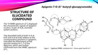
Research Journal
Phyllanthus Emlica. L
(Emblica officinalis)
Article Information
A new cytotoxic acylated apigenin glucoside from
Phyllanthus emblica L.
S. K. EL-DESOUKY, SHI YOUNG RYU and YOUNG-KYOON KIM
College of Forest Science, Kookmin University, 861-1 Sungbuk-gu, Jungnung
dong, Seoul 136-702, Korea
Korea Research Institute of Chemical Technology, Taejeon 305-606, Korea
(Received 15 January 2007; in final form 24 July 2007)
Natural Product Research, Vol.22, No.1,
10 January 2008, 91–95
INTRODUCTION OF PLANT
Synonym: Phyllanthus emblica Linn.
Family: Euphorbiaceae
Habitat: Native to tropical Southeast, Asia; distributed
throughout India; also planted in public parks.
English: Emblic, Indian gooseberry.
Ayurvedic: Aaamalaki, Aaamalaka, Dhaatri,
Kaayasthaa, Amoghaa, Amritaphala, Amla, Aaamalaa,
Dhaatriphala, Vayasyaa, Vrshya, Shiva, Hattha.
Unani: Aamalaa, Amlaj.
Urdu:Aamla, Amla, Aonla
AGRO ECOLOGY (CULTIVATION)
 Indian gooseberry grows in tropical and subtropical regions from near
sea-level to 1,500 m altitude.
 It grows equally well in arid and wet or humid conditions.
 It is light demanding plant, common in grassy areas, brush and village
groves.
 It is photosensitive, only producing flowers at a day-length between
12-13.5 h.
 The species is not fastidious of soil type and grows on a wide range of
soil types ranging from sandy loam to clay, light or heavy, and slightly
acidic to slightly alkaline.
 It is fire-resistant and is one of the first trees to recover after a fire.
 The tree is rather slow-growing, bearing fruit after 5–8 years.
BIOGRAPHY (BOTANY)
Height:
A small to medium-sized, low, deciduous tree, 5–25 m tall; trunk often crooked and gnarled, upto 35 cm
in diameter.
Bark:
Bark thin, smooth, grey, peeling in patches, with numerous knobs, main branches angular and densely
pubescent.
Leaves:
Leaves on the main branches are deltoid-squami form, resembling stipules.
Flowers:
Flower are greenish-yellow, fascicled in axils of leaves or fallen leaves, unisexual.
Fruit:
Fruit a depressed globose drupe 2–4 cm in diameter, pale green changing to
yellow. The fruit skin is thin, translucent and adherent to the very crisp, juicy, concolorous flesh.
Seed:
A slightly hexagonal stone is tightly embedded in the center of the flesh; stone has three slightly
dehiscent compartments, each usually containing two trigonous, 4–5 mm × 2–3 mm seeds.
Leaves
Flower
Fruit
Bark
MEDICINAL USES OF PHYLLANTHUS
EMBLICA
Every part of the plant is useful in antidotal treatment of snakebite and scorpion-sting.
The fruits of the plant are known to be a rich source of vitamin C and are frequently
used in making pickles, preserves and jellies. The unripe fruit is cooling, diuretic and
laxative.
The leaves are used in aphrodisiac, antipyretic, useful in biliousness, asthma,
bronchitis, leucorrhoea and vomiting. The leaves are also used in cases of chronic
dysentery and are also considered as a bitter tonic.
Antithermic lotions have also been prepared from leaves of the plant.
The roots, the bark and the ripe fruit are astringent.
Juice of the fresh bark with honey and turmeric is given in gonorrhoea.
The flowers are refrigerant and aperient.
The extract of the plant also showed antioxidant and antiatheroscleotic activities.
Juice with turmeric powder and honey is prescribed in diabetes insipidus.
ISOLATION AND
STRUCTURE ELUCIDATION
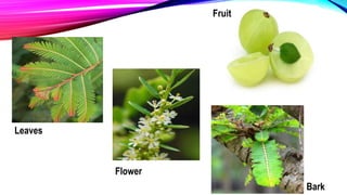
Several reports about this plant have shown that the fruits are rich in,
Vitamin C (Aescorbic Acid) [1], Mucic Acid and Tannins [2,3], Flavanone Glycosides [4], Phenolic
glycosides [5], Sesquiterpenoids [6], Norsesquiterpenoids [7], Phenolic Acids [8] and Flavonol
Glycosides [9].
In this report the isolation and structure elucidation of a new acylated flavone glucoside from the leaves of
P. emblica,
Isolated compounds From leaves Phyllanthus Emlica:
1) apigenin-7-O-(6”-butyryl--glucopyranoside) (1)
2) gallic acid (2),
3) methyl gallate (3),
4) 1,2,3,4,6-penta-O-galloylglucose (4),
5) luteolin-40-O-neohesperidoside (5). (rare flavone glycoside)
The structure elucidation of these compounds was established on the basis of 1D and 2D NMR experiments.
Among the isolated compounds, (1) exhibited a potent cytotoxicity against cultured tumor cell lines in vitro
using the SRB (sulforhodamine-B) method for cytotoxicity evaluation.
STRUCTURE OF
ELUCIDATED
COMPOUND
The 1H NMR spectrum of 1 displayed
the characteristic signals of apigenin
nucleus. A pair of A2B2 aromatic
system protons.
The downfield shift of both H-6 at
6.45 and H-8 at 6.85 relative to the
corresponding positions in apigenin,
indicated the attachment of the
glucose residue to C-7 of the
aglycone, which was further
confirmed from the HMBC spectrum
of 1.
Apigenin-7-O-(6”-butyryl-glucopyranoside)
EXTRACTION METHOD
The air dried leaves of P. emblica (400 g) were extracted with 70% methanol (3 x 3L) at 80C. The
combined extracts were evaporated under reduced pressure at 45C, which yielded 70g of the
residue. The methanolic extract was chromatographed on polyamide CC, first with an increasing
polar gradient of water methanol, obtaining four fractions (I - IV).
Fraction I:
Fraction I (10% MeOH) was chromatographed on silica gel CC using CH2Cl2–MeOH (4 : 1, v/v) to
afford 2 (23 mg) and 3 (40 mg).
Fraction II:
Fraction II was subjected to Sephadex LH-20 CC and eluted with methanol to give a yellow powder
of 1 (13 mg).
Fraction III:
Fraction III was chromatographed using EtOAc and EtOAc–MeOH increasing gradient polarity on
silica gel CC to afford 4 (120 mg) and 5 (65 mg).
SPECTROSCOPIC METHODS
UV- Spectroscopy:
 Compound 1 was obtained as a yellow amorphous powder, which
appeared violet under UV light of 366 nm.
 The UV spectrum with usual shift reagents indicated that 1 is an acylated
apigenin glycoside.
 The glycosylation site of the sugar moiety to the apigenin was found to
be at C-7, since free OH groups were detected at C-5 by a bathochromic
shift in band II of 25 nm with AlCl3 and at C-4’ by a bathochromic shift of
band I of 45 nm without a decrease in the intensity with NaOCH3.
NMR Data Table of Compound
 The 1H NMR spectrum of 1 displayed the characteristic signals of apigenin nucleus.
 A pair of A2B2 aromatic system protons at 7.93 (d, J=8.4 Hz, 2H) and 6.93 (d, J=8.4 Hz, 2H) for
H-2’,6’ and H-3’,5’ respectively.
 Two meta coupling signals at 6.45 (d, J=2.1 Hz, 1H) for H-6 and at 6.85 (d, J=2.1 Hz, 1H) for
H-8 and a singlet centered at 6.91 for H-3.
 The downfield shift of both H-6 at 6.45 and H-8 at 6.85 relative to the corresponding
positions in apigenin indicated the attachment of the glucose residue to C-7 of the aglycone,
which was further confirmed from the HMBC spectrum of 1 by the presence of a three bond
long-range correlation between the anomeric glucose proton H-1” at 5.28 and C-7 of
apigenin at 162.34.
 The 1H NMR spectrum of 1 also revealed the anomeric proton of glucose moiety at 5.28 (d,
J=7.5 Hz, 1H), showing the -configuration of glucose, with the remaining glucose protons
appearing in the range of 3.33– 4.16.
 The signals of the butyryl moiety as indicated by the presence of two triplets centered at 0.82
(t, J=7.2 Hz, 3H) and 1.53 (t, J=7.2 Hz, 2H) and one multiplet centered at 1.30 (m, 2H) (Table
1).
 All the protons were assigned unambiguously from the HSQC experiment in which all the
protons were correlated with those of the corresponding carbons.
1H NMR spectrum
13C NMR SPECTRUM
WITH
(DEPT) The presence of the butyryl residue was further confirmed by 13C NMR
spectral data and DEPT measurements of 1 (table 1).
 This revealed the presence of 25 carbon signals, out of which
 15 carbon signals represented the aglycone moiety (7 methine and 8
quaternary carbons),
 6 carbon signals for the glucose unit (1 oxymethylene and 5 methine
carbons) and
 4 carbon signals for the butyryl moiety (1 quaternary carbonyl, 1 methyl and
2 methylene carbons).
 The attachment of the butyryl residue to C-6” of glucose was deduced from
the downfield shift of C-6” of glucose ( 64.21) in the 13C NMR spectrum of
1 and was further confirmed by the three bond HMBC correlation between
the carbonyl carbon of the butyryl residue at 168.47 and the C-6” protons
of glucose at 4.16 and 4.08.
SUMMARY OF
METHODS
The ESI-MS (Electrospray Ionization Mass Spectrometry) spectrum (negative mode- ion) of 1
depicted the [M-1]+peak at 501 m/z, confirming the molecular formula C25H26O11 of compound 1.
Mass Spectrum
From these results, compound 1 was identified as:
apigenin-7-O-(600-butyryl--glucopyranoside
The rest of the compounds were identified by comparing their spectral data (1H NMR,13C NMR
and ESI-MS) to those published, gallic acid, methyl gallate, 1,2,3,4,6-penta galloylglucose
and luteolin-4’- neohespridoside.
Summary
References:
Electrospray and MALDI Mass Spectrometry Fundamentals, Instrumentation, Practicalities, and
Biological Applications Second Edition Edited by Richard B. Cole (WILEY)
Indian Medicinal Plants An Illustrated Dictionary Author C.P. Khare (Springer)
INTRODUCTION TO SPECTROSCOPY 5th Ed Donald L. Pavia
International Journal Of Ayurvedic And Herbal Medicine 2:5 (2012) 828:834
Research journal

Research journal

  • 2.
    Research Journal Phyllanthus Emlica.L (Emblica officinalis)
  • 3.
    Article Information A newcytotoxic acylated apigenin glucoside from Phyllanthus emblica L. S. K. EL-DESOUKY, SHI YOUNG RYU and YOUNG-KYOON KIM College of Forest Science, Kookmin University, 861-1 Sungbuk-gu, Jungnung dong, Seoul 136-702, Korea Korea Research Institute of Chemical Technology, Taejeon 305-606, Korea (Received 15 January 2007; in final form 24 July 2007) Natural Product Research, Vol.22, No.1, 10 January 2008, 91–95
  • 4.
    INTRODUCTION OF PLANT Synonym:Phyllanthus emblica Linn. Family: Euphorbiaceae Habitat: Native to tropical Southeast, Asia; distributed throughout India; also planted in public parks. English: Emblic, Indian gooseberry. Ayurvedic: Aaamalaki, Aaamalaka, Dhaatri, Kaayasthaa, Amoghaa, Amritaphala, Amla, Aaamalaa, Dhaatriphala, Vayasyaa, Vrshya, Shiva, Hattha. Unani: Aamalaa, Amlaj. Urdu:Aamla, Amla, Aonla
  • 5.
    AGRO ECOLOGY (CULTIVATION) Indian gooseberry grows in tropical and subtropical regions from near sea-level to 1,500 m altitude.  It grows equally well in arid and wet or humid conditions.  It is light demanding plant, common in grassy areas, brush and village groves.  It is photosensitive, only producing flowers at a day-length between 12-13.5 h.  The species is not fastidious of soil type and grows on a wide range of soil types ranging from sandy loam to clay, light or heavy, and slightly acidic to slightly alkaline.  It is fire-resistant and is one of the first trees to recover after a fire.  The tree is rather slow-growing, bearing fruit after 5–8 years.
  • 6.
    BIOGRAPHY (BOTANY) Height: A smallto medium-sized, low, deciduous tree, 5–25 m tall; trunk often crooked and gnarled, upto 35 cm in diameter. Bark: Bark thin, smooth, grey, peeling in patches, with numerous knobs, main branches angular and densely pubescent. Leaves: Leaves on the main branches are deltoid-squami form, resembling stipules. Flowers: Flower are greenish-yellow, fascicled in axils of leaves or fallen leaves, unisexual. Fruit: Fruit a depressed globose drupe 2–4 cm in diameter, pale green changing to yellow. The fruit skin is thin, translucent and adherent to the very crisp, juicy, concolorous flesh. Seed: A slightly hexagonal stone is tightly embedded in the center of the flesh; stone has three slightly dehiscent compartments, each usually containing two trigonous, 4–5 mm × 2–3 mm seeds.
  • 7.
  • 8.
    MEDICINAL USES OFPHYLLANTHUS EMBLICA Every part of the plant is useful in antidotal treatment of snakebite and scorpion-sting. The fruits of the plant are known to be a rich source of vitamin C and are frequently used in making pickles, preserves and jellies. The unripe fruit is cooling, diuretic and laxative. The leaves are used in aphrodisiac, antipyretic, useful in biliousness, asthma, bronchitis, leucorrhoea and vomiting. The leaves are also used in cases of chronic dysentery and are also considered as a bitter tonic. Antithermic lotions have also been prepared from leaves of the plant. The roots, the bark and the ripe fruit are astringent. Juice of the fresh bark with honey and turmeric is given in gonorrhoea. The flowers are refrigerant and aperient. The extract of the plant also showed antioxidant and antiatheroscleotic activities. Juice with turmeric powder and honey is prescribed in diabetes insipidus.
  • 9.
    ISOLATION AND STRUCTURE ELUCIDATION Severalreports about this plant have shown that the fruits are rich in, Vitamin C (Aescorbic Acid) [1], Mucic Acid and Tannins [2,3], Flavanone Glycosides [4], Phenolic glycosides [5], Sesquiterpenoids [6], Norsesquiterpenoids [7], Phenolic Acids [8] and Flavonol Glycosides [9]. In this report the isolation and structure elucidation of a new acylated flavone glucoside from the leaves of P. emblica, Isolated compounds From leaves Phyllanthus Emlica: 1) apigenin-7-O-(6”-butyryl--glucopyranoside) (1) 2) gallic acid (2), 3) methyl gallate (3), 4) 1,2,3,4,6-penta-O-galloylglucose (4), 5) luteolin-40-O-neohesperidoside (5). (rare flavone glycoside) The structure elucidation of these compounds was established on the basis of 1D and 2D NMR experiments. Among the isolated compounds, (1) exhibited a potent cytotoxicity against cultured tumor cell lines in vitro using the SRB (sulforhodamine-B) method for cytotoxicity evaluation.
  • 10.
    STRUCTURE OF ELUCIDATED COMPOUND The 1HNMR spectrum of 1 displayed the characteristic signals of apigenin nucleus. A pair of A2B2 aromatic system protons. The downfield shift of both H-6 at 6.45 and H-8 at 6.85 relative to the corresponding positions in apigenin, indicated the attachment of the glucose residue to C-7 of the aglycone, which was further confirmed from the HMBC spectrum of 1. Apigenin-7-O-(6”-butyryl-glucopyranoside)
  • 11.
    EXTRACTION METHOD The airdried leaves of P. emblica (400 g) were extracted with 70% methanol (3 x 3L) at 80C. The combined extracts were evaporated under reduced pressure at 45C, which yielded 70g of the residue. The methanolic extract was chromatographed on polyamide CC, first with an increasing polar gradient of water methanol, obtaining four fractions (I - IV). Fraction I: Fraction I (10% MeOH) was chromatographed on silica gel CC using CH2Cl2–MeOH (4 : 1, v/v) to afford 2 (23 mg) and 3 (40 mg). Fraction II: Fraction II was subjected to Sephadex LH-20 CC and eluted with methanol to give a yellow powder of 1 (13 mg). Fraction III: Fraction III was chromatographed using EtOAc and EtOAc–MeOH increasing gradient polarity on silica gel CC to afford 4 (120 mg) and 5 (65 mg).
  • 12.
    SPECTROSCOPIC METHODS UV- Spectroscopy: Compound 1 was obtained as a yellow amorphous powder, which appeared violet under UV light of 366 nm.  The UV spectrum with usual shift reagents indicated that 1 is an acylated apigenin glycoside.  The glycosylation site of the sugar moiety to the apigenin was found to be at C-7, since free OH groups were detected at C-5 by a bathochromic shift in band II of 25 nm with AlCl3 and at C-4’ by a bathochromic shift of band I of 45 nm without a decrease in the intensity with NaOCH3.
  • 13.
    NMR Data Tableof Compound
  • 14.
     The 1HNMR spectrum of 1 displayed the characteristic signals of apigenin nucleus.  A pair of A2B2 aromatic system protons at 7.93 (d, J=8.4 Hz, 2H) and 6.93 (d, J=8.4 Hz, 2H) for H-2’,6’ and H-3’,5’ respectively.  Two meta coupling signals at 6.45 (d, J=2.1 Hz, 1H) for H-6 and at 6.85 (d, J=2.1 Hz, 1H) for H-8 and a singlet centered at 6.91 for H-3.  The downfield shift of both H-6 at 6.45 and H-8 at 6.85 relative to the corresponding positions in apigenin indicated the attachment of the glucose residue to C-7 of the aglycone, which was further confirmed from the HMBC spectrum of 1 by the presence of a three bond long-range correlation between the anomeric glucose proton H-1” at 5.28 and C-7 of apigenin at 162.34.  The 1H NMR spectrum of 1 also revealed the anomeric proton of glucose moiety at 5.28 (d, J=7.5 Hz, 1H), showing the -configuration of glucose, with the remaining glucose protons appearing in the range of 3.33– 4.16.  The signals of the butyryl moiety as indicated by the presence of two triplets centered at 0.82 (t, J=7.2 Hz, 3H) and 1.53 (t, J=7.2 Hz, 2H) and one multiplet centered at 1.30 (m, 2H) (Table 1).  All the protons were assigned unambiguously from the HSQC experiment in which all the protons were correlated with those of the corresponding carbons. 1H NMR spectrum
  • 15.
    13C NMR SPECTRUM WITH (DEPT)The presence of the butyryl residue was further confirmed by 13C NMR spectral data and DEPT measurements of 1 (table 1).  This revealed the presence of 25 carbon signals, out of which  15 carbon signals represented the aglycone moiety (7 methine and 8 quaternary carbons),  6 carbon signals for the glucose unit (1 oxymethylene and 5 methine carbons) and  4 carbon signals for the butyryl moiety (1 quaternary carbonyl, 1 methyl and 2 methylene carbons).  The attachment of the butyryl residue to C-6” of glucose was deduced from the downfield shift of C-6” of glucose ( 64.21) in the 13C NMR spectrum of 1 and was further confirmed by the three bond HMBC correlation between the carbonyl carbon of the butyryl residue at 168.47 and the C-6” protons of glucose at 4.16 and 4.08.
  • 16.
    SUMMARY OF METHODS The ESI-MS(Electrospray Ionization Mass Spectrometry) spectrum (negative mode- ion) of 1 depicted the [M-1]+peak at 501 m/z, confirming the molecular formula C25H26O11 of compound 1. Mass Spectrum From these results, compound 1 was identified as: apigenin-7-O-(600-butyryl--glucopyranoside The rest of the compounds were identified by comparing their spectral data (1H NMR,13C NMR and ESI-MS) to those published, gallic acid, methyl gallate, 1,2,3,4,6-penta galloylglucose and luteolin-4’- neohespridoside. Summary References: Electrospray and MALDI Mass Spectrometry Fundamentals, Instrumentation, Practicalities, and Biological Applications Second Edition Edited by Richard B. Cole (WILEY) Indian Medicinal Plants An Illustrated Dictionary Author C.P. Khare (Springer) INTRODUCTION TO SPECTROSCOPY 5th Ed Donald L. Pavia International Journal Of Ayurvedic And Herbal Medicine 2:5 (2012) 828:834