Insect Cuticle or The Insect Integument.pptx
•
1 like•769 views

Insect Cuticle or The Insect Integument

Recommended
Recommended
More Related Content
What's hot
What's hot (20)
Similar to Insect Cuticle or The Insect Integument.pptx
Similar to Insect Cuticle or The Insect Integument.pptx (20)
More from Dr. Mandeep Rathee, KVK Kaithal, CCSHAU Hisar
More from Dr. Mandeep Rathee, KVK Kaithal, CCSHAU Hisar (10)
Recently uploaded
Recently uploaded (20)
Insect Cuticle or The Insect Integument.pptx
- 2. INTEGUMENTARY SYSTEM •Introduction •Function of the integument •Evolutionary significance to arthropods •Structure of the cuticle •Chemical composition •Sclerotization •Physical properties of cuticle •Specializations in cuticle •Coloration and melanization
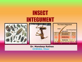
- 3. The Integument The outer covering or cuticle of an insect, plus the epidermal cells that secrete the cuticle is called as integument. Many of the characteristics that make insects unique are attributable to the integument which is mainly composed of: • Cuticle • Epidermis • Basement membrane
- 4. Transmission Electron Micrograph of Cuticle – Adult female of Heliothis zea Epicuticle Exocuticle Endocutie Epidermal cell Basement membrane
- 5. Functions of The Integument • Protection of internal organs and tissues. • Protective barrier against entry of pathogens, pesticides, parasites, and predators. • Preventive barrier against water loss. • Provides for the insect the sensory “windows to the outside world” • Also lines the tracheae, tracheoles, salivary glands and portions of reproductive tract. At the molt, all of this is shed. • Protective barrier for foregut and hindgut. • Integument functions in locomotion, feeding, excretion, protection from desiccation, breathing.
- 6. Immature insects are able to encapsulate certain parasites and get rid of them at the molts. Here, a house fly larva is shown with two encapsulated nematodes that are then moved to the area beneath the cuticle. At the molt, these parasites are expelled with the old exocuticle. Parasitic expulsion
- 7. Evolutionary significance of arthropod integument POSITIVE EFFECTS • Prevention of water loss because of wax layer and cuticular composition. • Smaller the organism the greater the amount of surface area per unit volume-greater tendency to lose water. • Preventative barrier for pathogens, parasites and predators. NEGATIVE EFFECTS • As size increases, problems with cuticle being too heavy and also gaseous exchange (oxygen uptake) adversely affected • Evolutionary experiment with Meganeuron shows that cuticle is responsible for smaller size of insects.
- 9. 1. The Cuticle • In all insects there is a thin layer of cuticle with special properties and is comprised of following layers : 1. Epicuticle 2. Exocuticle 3. Endocuticle NOTE It can be as thin as 1 µm in the hindgut and over gills (e.g. Ephemeropteran larvae) and as thick as 200 + µm (elytra of large Coleoptera). There is always an epicuticle layer and the epidermal cell layer, but the exocuticle and endocuticle may be greatly reduced or absent in particular parts of the cuticle of the same or different insects. Often there are additional cuticle layers within one or more of the three principal layers as evidenced by electron density in cross sections viewed in the transmission electron microscope.
- 10. A. The Epicuticle Complex and the outermost layer and probably the most important layer. Responsible for water proofing and general impermeability of cuticle. Produced by epidermis and dermal glands. Insects needing a renewable layer Soil dwelling insects due to erosion of the cuticle by abrasion
- 11. Significance of each layer of epicuticle 1. Cement layer : • Outermost layer. Closely associated with wax layer and may serve to protect it. • Not found in all insects. 2. Wax layer: • Hydrocarbons constitute 90% of this layer. Important to insects for water loss, thus waterproofing of cuticle. • In some(e.g., Fulgoridae and scales), the insects produce a large bloom of wax on outside. • Bees have special glands, wax glands, on ventral abdominal segments 4- 7 that produce wax, which is then formed into flakes used by the bees to make their cells.
- 12. 3. Outer epicuticle- • Cuticulin is a very thin layer of protein about0.0075 μm thick, which is the typical thickness of an animal cell membrane. It is impregnated with lipids, some of which may be covalently bound to proteins as lipoproteins. • First layer formed following the molt and is the layer that protects the new procuticle from digestion by molting enzymes (i.e., chitinases and proteinases). Also called cuticulin layer. 4. Inner epicuticle- • Function not that clear but it is a much thicker layer(1µm) than the outer epicuticle. 5. Wax canal filament- • A filament of wax that is produced by the hypodermal cells and extends to the inner part of the epicuticle.
- 13. • wax channels about 0.006 μm–0.013 μm in diameter) that are 10–20 times smaller than pore canals. 6. Pore canals- ● Tiny pores that run from the hypodermal cells to the inner part of the inner epicuticular layer. Inside the canals are wax filaments that extend up to the epicuticular layer. Probably serve as a transport passage for wax from hypodermal cells up to wax layer. ● Pore canals are passageways from 0.1 μm to 0.15 μm diameter extending from the epidermal cells through the endo and exocuticle. The pore canals can be very numerous; for example, -1.2 × 10⁶/mm2 in some cockroaches, or -as few as 15,000/mm2 in a flesh fly larva.
- 15. Electron micrograph of a transverse section of the cuticle from a larva of Galleria showing the fibrous structure of the lamellae in the endocuticle and pore canals each containing an axial filament. pcf, pore canal filament ; pc, pore canal. (Osmium tetroxide, Araldite). X 42,000.
- 16. Electron micrograph of a thick transverse section to show the main features. Ep, epicuticlc; End, endocuticlc; Epith, epithelium. (Osmium tctroxide, Araldite). X 3,200.
- 17. B. The Exocuticle •The exocuticle contains chitin and protein. • It lies just beneath the epicuticle. •It is highly sclerotized and is therefore hard and rigid. •Layers within the exocuticle may refract light in such a way to produce structural colors in some insects. Many of the iridescent greens and blues of insects are structural colors due to refracted light rather than to pigments. •The thickness of the exocuticle is variable and species specific. Adult insects generally have a thicker and more sclerotized exocuticle than larval insects.
- 18. • Region in which cross-linking of occurs to give cuticle hardness. • Region not broken down by proteases at molt and is what usually remains in form of exuviae. • Melanin and other pigments found in this area. Sclerotization mainly occurs here. • Also arthropodin and sclerotin. Cross-section of Oncopeltus showing exocuticle --------»
- 19. C. The Endocuticle •Is continually being synthesized (in a dark/light way-24 hrs) and often is laid down in layers, thus can often be used to age-grade some insects. •Contains most of the chitin, which is broken-down at the molt by chitinase. Little cross-linking of proteins, thus most is broken-down at molting and reabsorbed. •In soft-bodied insects and regions of flexibility (in the intersegmental membranes), this layer is well developed and not the exocuticle.
- 20. 2. The Epidermis • A single layer of cells, underlying the cuticle and is responsible for its production. • It the only living portion of the integument that is ectodermal in origin. • These cells can be modified to form dermal glands, sensory receptors and oenocytes. • Epidermal cells are involved in wound repair, and can move from an area of undamaged cells into an area of destroyed cells.
- 21. • The sex pheromone in most female Lepidoptera is secreted by a small patch of tall, columnar epidermal cells located beneath the cuticle of the ventral inter-segmental membrane in the 8–9th segment of the abdomen. • The Golgi complex is prominent in epidermal cells, and probably serves several functions including the following: (i) processing of secretory substances necessary to synthesize cuticle, (ii) production of material for the plasma membrane of the cell, (iii) processing and packaging of lysozymes needed for autophagy.
- 22. • Oenocytes are large, prominent cells scattered among the epidermal cells, and also clustered at spiracles, near origin of larger tracheae, and scattered among fat body cells. • These cells are usually large, polyploid, and always have extensive tubular smooth endoplasmic reticulum and well developed plasma membrane reticular system. • Their function is not very clear, but their morphology suggests lipid secretion and lipid metabolism.
- 23. Basement membrane or Basal lamina • Muco-polysaccharide layer that is secreted by the hemocytes . • It is penetrated by nerves and tracheae going to hypodermis, and is a selective barrier between haemolymph and epidermal cells. • Hormones and other nutrients can pass through this selectively permeable layer to reach the hypodermal cells. • Its important in the recognition of ‘self’; thus, the insect’s blood cells do not recognize it as ‘foreign.’ • Molecules in this layer are charged and probably act like a molecular sieve. • Extremely important in self recognition and cuticular integrity.
- 24. In this micrograph we added cationized ferritin molecules and one can see that they are attracted to the anionized sites in the basal lamina. This was in Phormia regina follicle cells (fc). He=hemolymph
- 25. 3. Chemical composition of the cuticle Lipids • Waxes- Wax blooms produced by several groups of insects • Cuticular hydrocarbons- used by systematists. Carbohydrates • Chitin-polysaccharide - with units of N-acetyglucosamine residues. Chitin gives the cuticle its strength, not its hardness. Proteins • Arthropodin -Untanned protein that during sclerotization is crosslinked to produce sclerotin and give the cuticle its hardness.
- 27. • Sclerotin- Tanned protein that gives cuticle hardness. This is brought about by a process called sclerotization and involves eclosion hormone and bursicon, another rhormone. • Chitinase- Enzyme involved in digestion of chitin at the molt. It is released and produced by the epidermal cells. • Proteinases- Enzymes other than chitinase that aid in the digestion of the endocuticle at the molts.
- 28. Resilin- Colorless rubber-like protein. Can be stretched and stores energy due to tension. Places where it is found: Wing hinges; Food pump of reduviid bugs; Hind legs of jumpers; Aids in inspiration in beetles, which lack inspiration muscles. resilin pad.
- 29. Transverse section through the thoracic wall and wing base of a grasshopper showing the position of the wing hinge and the resilin pad.
- 30. Sclerotization of cuticle • The process whereby untanned or unlinked proteins become cross- linked to form strong linkages that give the cuticle its hardness. This process occurs mainly in the exocuticle area. • The greater the degree of sclerotization, the harder the cuticle. The content of chitin does not control hardness of the cuticle, but sclerotization does. • Because of the sclerotization, little or none of the exocuticle is digested by molting fluid, and it is shed, along with the epicuticle, at molting.
- 31. 5. Physical properties of the cuticle • Hardness • Strength • Flexibility • Plasticization • Ability to heal or repair wounds • Hydrophobicity
- 32. • Hardness- Is due to the amount of sclerotization or tanning of the proteins that takes place. This involves cross-linking of the proteins. A process often called tanning or sclerotization. Mainly occurs in the exocuticle. • Strength- Is due to the presence of chitin in the endocuticle. • Flexibility It is due to less proteins being sclerotized and is usually due to more endocuticle being present than exocuticle. Also certain proteins, such as resilin provide flexibility.
- 33. Scanning electron micrograph of the inner surface of a mandible. X-ray microanalysis of the same mandible showing the presence of Zinc in the mandibular cusps, which gives greater strength to the mandible.
- 34. Relationship between sclerotization and cuticular stiffness in the abdominal tergite of the honeybee around time of eclosion. Sclerotization is expressed as the inverse of the % of protein that is extractable. At the same time, the stiffness of the cuticle also increases.
- 35. Flexibility of the cuticle. Extrinsic articulations where thes clerotized parts meet outside the membrane. FLEXIBLE REGION
- 36. At the time of laying eggs, the desert locust’s abdominal cuticle becomes plasticized. This permits the ovipositor to extend further into the soil so that the eggs can be laid as deeply as possible. Plasticization
- 37. Openings or specializations in the cuticle 1. Openings in the cuticle a. Pore canals b. ‘Sweat pores’ of Sonoran desert cicada 2. Specialized areas of cuticle a. Anal organ of dipterous larvae b. Sense organs c. Pheromone or deterrent chemical site release
- 38. Pore canals-Tiny pores that extend from the top of the hypodermal cells to the inner most part of the epicuticle. Believed to be involved in the transport of lipids from the hypodermal cells to the epicuticle. Pore canals
- 39. Sonoran desert cicada. Pores 7X size of pore canals located on dorsal mesonotum. These are connected to special dermal glands via cuticular ducts are involved in water transport to the surface. Cooling of 2-5oC below ambient of42-45oC.
- 40. Anal organ of dipterous larvae Organ is involved in osmoregulation in the dipterous larva. Structure tells you something about its function- thinner cuticle, larger epidermal cells, and separaation from rest of cuticle.
- 41. Formation of sensilla Trichogen cell- Creates the shaft of hair Tormogen cell- Creates the socket of the hair or sensillum Thecogen cell- Creates the sheath that surrounds the neurons and isolates them and provides the neuron with ions and nutrients.
- 42. 6. Coloration and melanization • Insect colors are due to either: a. Pigments in the exocuticle such as melanin and they are usually lacking in endocuticle b. Physical structure of the cuticle to form defraction gradients that defract light in various ways. This results in the iridescence of the blue morpho butterflies and gold of beetles, gold on monarch chrysalis, and color of tiger beetles, etc.
- 43. REFRENCES: • Chapman, R.F., 1998. The Insects: Structure and Function, 4th ed., Cambridge University Press, Cambridge, UK. • Chitra K. C., 2006. A Text Book of Insect Physiology, Xpress Graphics, Delhi, India. • Wigglesworth, V. B., 1965. The Principles of Insect Physiology, English Language Book Society and Methven and Co. Ltd. • Images and pdf on insect cuticle from www.google.com.