1. HISTOLOGY of THE
INTEGUMENTARY SYSTEM
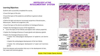
Learning Objectives
Students who successfully completed this course;
• Count the layers of the skin.
• Count the layers of the epidermis and defines its general cellular
properties.
• Define the light and electron microscopic properties of Keratinocytes ,
Melanocytes , Merkel and Langerhans cells.
• Count the layers of the dermis and defines its histological features.
• Explain the histological structure of the hypodermis.
Count the layers of hair follicles and explains their cellular properties.
• Explain the histological features of sweat glands and sebaceous glands.
• Explain the histological features of nails.
• Explain the embryological development of epidermis and dermis
layer.
• Explain the embryological development of the hair follicle.
• Explain the embryological development of sweat and mammary
glands.
Count the diseases and anomalies that occur in the development of skin,
hair, sweat and mammary glands.
Assist. Prof. Ender Deniz Asmaz
2. OVERVIEW OF THE INTEGUMENTARY SYSTEM
o The integumentary system consist of the
skin (integument) and skin derivatives
o the external covering of the body
o is the largest organ
3. The skin consists of two main layers:
1.The epidermis : is composed of a keratinized stratified
squamous epithelium that grows continuously but maintains
its normal thickness by the process of desquamation.
-Epidermis is derived from
ectoderm.
2.The dermis: is composed of a dense connective tissue that
imparts mechanical support, strength, and thickness to the
skin.
- Dermis is derived from
mesoderm.
4. hypodermis lies deep to the dermis.
hypodermis contains adipose tissue arranged into lobules
separated by connective tissue septa.
The epidermal derivatives of the skin (epithelial skin
appendages) include the following structures
I.Hair follicles and hair
II.Sweat glands
III.Sebaceous glands
IV.Nail
5. Major functions of the skin include the
following:
1.Barrier: protects against
physical, chemical, and biologic
agents in the external
environment
2.It provides immunologic
information obtained during
antigen processing to the
appropriate effector cells in the
lymphatic tissue.
3.Homeostasis: by regulating
body temperature and water loss.
6. 4.It conveys sensory information about the
external environment to the nervous
system.
5.It performs endocrine functions by
secreting hormones, cytokines, and growth
factors
6.It functions in excretion through the
exocrine secretion of sweat, and sebaceous
glands.
7. Skin is categorized as thick or thin, a reflection of thickness
and location
The thickness of the skin varies over the
surface of the body.
The terms thick skin and thin skin, as
used in histologic description,
It is only related to the thickness of the
epidermal layer.
9. LAYERS OF THE SKIN
Epidermis
The epidermis is composed of kr. stratified
squamous epithelium in which four distinct
layers can be identified.
In the case of thick skin, a fifth layer is
observed
10. Beginning with the deepest layer,
these are as follow:
1.the stratum basale also called the
stratum germinativum because of the
presence of mitotically active cells, the stem cells of the
epidermis;
2.the stratum spinosum, also called the
spinous layer or prickle cell layer because of the
characteristic light micro- scopic appearance of short
processes extending from cell to cell;
3.the stratum granulosum, which
contains numerous intensely staining
11. 4. the stratum lucidum,
which is limited to thick
skin and considered a
subdivision of the stratum
corneum
5. the stratum corneum,
which is composed of
keratinized cells.
12. 1. The stratum basale
-The stratum basale is represented
by a single layer of cells that rests
on the basal lamina.
-provides cell for epidermal
renewal.
-It contains the stem cells from which
new cells, the keratinocytes, arise
by mitotic division.
13. -the stratum basale is also called
the
stratum germinativum.
-because of the
presence of mitotically
active cells, the stem
cells of the epidermis
14. The cells are small and
cuboidal to low columnar.
They have less cytoplasm
The basal cells also
contain various amounts of
melanin
15. Basal cells exhibit extensive cell junctions;
they are connected to each other and to
keratinocytes by desmosomes
and to the underlying basal lamina by
hemidesmosomes.
16. As new keratinocytes arise in this
layer by mitotic division, they move
into the next layer, thus beginning
their process of upward migration.
This process terminates when the
cell becomes a mature keratinized
cell, which is eventually sloughed off
at the skin surface.
17. 2. The stratum spinosum
The stratum
spinosum is at least
several cells thick.
Keratinocytes in this
layer are larger than
those of the stratum
basale.
They exhibit numerous
cytoplasmic
processes or spines,
which gives this layer
its name
18.
19. As the cells mature and move to the
surface, they increase in size and
become flattened in a plane parallel to
the surface.
in the most superficial spinous cells,
the nuclei also become elongate
instead of ovoid.
20. 3. The stratum granulosum
Keratinocytes in this layer contain numerous
keratohyalin granules, hence the name of the
layer.
This layer varies from one to three cells thick.
These granules contain cystine-rich and histidine-
rich proteins.
Keratohyalin granules are irregular in shape and
variable in size.
Because of their intense basophilic staining, they
are readily seen in routine histologic sections.
21. 4. Stratum corneum
The stratum corneum consists of
flattened, anucleate squamous
cells largely filled with keratin
filaments.
They lose their nucleus and
cytoplasmic organelles and
become filled almost entirely
with keratin filaments.
The thickness of this layer is more in
22. Stratum lucidum
The stratum lucidum, considered a subdivision
of the stratum corneum only well seen in thick
skin.
In the light microscope, it has a refractile
appearance and may stain poorly.
keratinization is well advanced in these cells
The nucleus and cytoplasmic organelles
become disrupted and disappear as the cell
gradually fills with keratin.
23. Dermis
-The junction between the dermis
and epidermis is seen in the light
microscope as an uneven
boundary.
-Sections of skin reveal numerous
fingerlike connective tissue
protrusions, dermal papillae,
that project into the undersurface
of the epidermis.
24. The dermis is composed of two layers: The
papillary layer and the reticular layer.
1. The papillary layer,
the more superficial layer, consists of
loose connective tissue beneath the
epidermis
type I and type III collagen molecules.
the elastic fibers form an irregular network.
25. layer is relatively thin and
includes the dermal papillae and
dermal ridges.
the blood vessels and sensory
nerve endings apparent in the
dermal papillae.
26. 2.The reticular layer lies deep
to the papillary layer.
•Although its thickness varies in
different parts of the body,
•it is always considerably thicker and
less cellular than the papillary layer.
•It is characterized by thick, irregular
bundles of mostly type I collagen
and elastic fibers.
27. Hypodermis
Layers of adipose tissue, smooth
muscle, and, in some sites, striated
muscle may be found just beneath the
reticular layer.
This layer serves as a major energy
storage site and also provides
insulation.
It is particularly thick in individuals who
28. CELLS OF THE EPIDERMIS
The cells of the epidermis consist of four
different cell types:
1.Keratinocytes are highly specialized
epithelial cells
2.Melanocytes are the pigment-producing cells
of the epidermis.
3.Langerhans’ cells are involved in signaling in
the immune system.
4.Merkel’s cells are associated with sensory
nerve endings.
30. keratinocytes has two activities:
1.They participate in the formation of
the epidermal water barrier.
2. They produce keratins major
structural proteins of the epidermis.
31. The keratinocytes in the
basal layer contain free
ribosomes, intermediate
(keratin) filaments, a
small Golgi apparatus,
mitochondria, and rER.
Keratins form
intermediate filaments;
they constitute almost
85% of fully differentiated
keratinocytes.
32. In the upper part of the stratum spinosum,
the free ribosomes within the keratinocytes begin
to synthesize keratohyalin granules that become
the distinctive feature of the cells in the stratum
granulosum.
Lamellar bodies contribute to the
formation of the intercellular epidermal
water barrier.
An epidermal water barrier is essential
for mammalian “dry” epithelia and is
responsible for maintaining body
homeostasis.
33. 2.Melanocytes
Neural crest–derived
melanocytes are scattered among
the basal cells of the stratum
basale.
The epidermal melanocyte is a dendritic cell
They are called dendritic cells
because the rounded cell body
extends long processes between the
34. In routine H&E preparations, melanocytes
are seen in the stratum basale with
elongated nuclei surrounded by a clear
cytoplasm.
The epidermal melanocytes produce
and secrete the pigment melanin into
keratinocytes.
The most important function of
melanin is to protect the organism against
the damaging effects of nonionizing
35. 3.Langerhans’ Cells
Langerhans’ cells are dendritic-appearing,
antigen-presenting cells in the epidermis.
They originate from common lymphoid
progenitor (CLP) cells in bone marrow,
migrate via the bloodstream, and enter the
epidermis where they differentiate into
immunocompetent cells.
36. Langerhans’ cells process antigens
entering through the skin.
Therefore, they constitute part of the
mononuclear phagocytotic system.
Once antigen is phagocytized,
processed, and displayed on the
surface of the Langerhans’ cell,
the cell migrates from the epidermis
to a regional lymph node where it
interacts with T lymphocytes
37. Langerhans’ cells cannot be
distinguished with in routine H&E–
stained paraffin sections.
immunostaining with antibody
against CD1a molecules,
Langerhans’ cells can be readily seen
in the stratum spinosum.
38. 4.Merkel’s Cells
Merkel’s cells are dendritic cells
located in the stratum basale.
The origin of Merkel’s cells is
unknown; they have antigenic
markers of both epidermal and
neural type.
They are most abundant in skin
where sensory perception is acute
such as the fingertips.
39. Merkel’s cells are bound to keratinocytes by
desmosomes and contain intermediate
(keratin) filaments in their cytoplasm.
The nucleus is lobed, and the cytoplasm is
denser
Merkel’s cells are closely associated with the
expanded terminal bulb of afferent
myelinated nerve fibers
40. STRUCTURES OF SKIN
Nerve Supply
Sensory nerve
endings with connective
tissue sheaths are
called encapsulated
endings.
Many encapsulated endings are located in the skin
I.Pacinian corpuscles
II.Meissner’s corpuscles
III.Ruffini’s corpuscles,
IV.Krause’s end bulb,
41. I.Pacinian corpuscles
Pacinian corpuscles are large
ovoid structures found in the deeper
dermis and hypodermis (especially
in the fingertips),
Pacinian corpuscles respond to
pressure and vibration
They are composed of a
myelinated nerve ending
surrounded by a capsule structure
in connective tissue, and in association
with joints, periosteum, and internal
organs
42. II. Meissner’s corpuscles
Meissner’s corpuscles are present in the
dermal papillae
Within these receptors, one or two
unmyelinated nerve endings follow spiral
paths in the corpuscle.
Meissner’s corpuscles are touch receptors
that are particularly responsive to low
frequency stimuli in the papillary layer of
hairless skin (e.g., the lips and of the
fingers and toes).
43. III. Ruffini’s corpuscles
They have an elongated fusiform
shape.
consists of a single
myelinated fiber that enters
the capsule, where it loses its
myelin sheath and branches
to form a dense axonal
endings
44. IV. Krause’s end bulb
a specialized sensory nerve ending
enclosed in a capsule in the skin.
tongue and dermis of external
genitalia
Round corpuscle
Its nerve fiber is myelinated
penetrates the corpuscle give
numerous nonmyelinated branches
that end by bulbous terminations.
45. Epidermal Skin Appendages
Skin appendages
- derived from down growths of epidermal epithelium
during development.
-They include the following:
1.Hair follicles and their product, hair
2.Sebaceous glands and their product,
sebum
3.Eccrine sweat glands and their product, sweat
4.Apocrine sweat glands their mixed product sweat
46. 1.Hair Follicles and Hair
Hair follicles and hairs are
invagination of the epidermis
they are absent only palmar
surfaces of the hands, plantar
surfaces of the feet, the lips,
and the region around the
urogenital orifices.
The hair follicle is responsible
for the production and growth of
a hair
47. The hair follicle is divided into three
segments:
1.The infundibulum extends from the
surface opening of the follicle to the level
of the opening of its sebaceous gland.
The infundibulum is a part of the
pilosebaceous canal, which is used for
the discharge of the oily sebum.
48. 2.The isthmus extends from the
infundibulum to the arrector pili
muscle.
3.The inferior segment
expands to form the bulb.
The base of the bulb is
invaginated by vascularized
loose connective tissue called, a
49. Histology of the hair follicle and
hair
Hairs are composed of keratinized cells
that develop from hair follicles
The internal root sheath is a multilayered
cellular covering that surrounds the deep
part of the hair.
The internal root sheath, consisting of
soft keratin.
the outermost part of the hair follicle, is
designated the external root sheath.
50. A thick basal lamina,
called the glassy
membrane, separates the
hair follicle from the
dermis.
Surrounding the follicle is
a dense irregular
connective tissue sheath
containing follicular
bulge.
The arrector pili
muscle is attached to
51. Hairs are elongated also consist of three layers:
1.The medulla forms the central part of the shaft
and contains large vacuolated cells.
2.The cortex is located peripherally to the medulla
and contains cuboidal cells.
-These cells undergo differentiation into keratin-
filled cells.
3.The cuticle of the hair shaft contains squamous
cells that form the outermost layer of the hair.
52. 2. Sebaceous Glands
Sebaceous glands secrete sebum that
coats the hair and skin surface.
Sebaceous glands develop as outgrowths
of the external root sheath of the hair follicle
The oily substance produced in the gland,
sebum, is the product of holocrine secretion.
The entire cell produces and becomes filled
with the fatty product while it undergoes
programmed cell death (apoptosis) as the
product fills the cell.
53. both the secretory
product and cell debris
are discharged from the
gland by pilosebaceous
canal with the short duct
of the sebaceous gland.
54. 3. Eccrine sweat glands
Sweat glands are classified on the bases of
their structure and the nature of their secretion.
Two types of sweat glands are recognized:
1.Eccrine sweat glands are distributed over the
entire body surface except for the lips and part of
the external genitalia.
2.Apocrine sweat glands are limited to the
areola, and nipple of the mammary gland; skin
around the anus; and the external genitalia.
55.
56. Eccrine sweat glands
Eccrine sweat glands play a major role in
temperature regulation
Eccrine sweat glands are independent
structures
Each eccrine gland is arranged as a blind-
ended, simple, coiled tubular structure.
It consists of two segments:
A secretory segment located deep in the
dermis
less coiled duct segment that leads to the
57. The secretory segment of the eccrine sweat gland
contains three cell types.
1.Clear cells
2.Dark cells, both of which are secretory
epithelial cells
3.Myoepithelial cells are contractile epithelial
cells.
All of the cells rest on the basal lamina;
their arrangement is that of a
pseudostratified epithelium.
58. I. Clear cells are characterized by
abundant glycogen.
The glycogen stain intensely with the
periodic acid–Schiff (PAS) method.
In routine H&E preparations, the
cytoplasm of clear cells stains poorly.
Membranous organelles include
numerous mitochondria, profiles of
sER, and a relatively small Golgi
apparatus.
59. the lateral and apical surfaces
and the basal surface of the cell
possesses infoldings,
The morphology of these cells
indicates that they produce the
watery component of sweat.
60. 2. Dark cells are characterized by
abundant rER and secretory
granules.
The Golgi apparatus is relatively
large, a feature consistent with
the glycoprotein secretion of
these cells.
The apical cytoplasm contains
mature secretory granules
61. 3. Myoepithelial cells are limited to the basal
aspect of the secretory segment.
They lie between the secretory cells
The cytoplasm contains numerous contractile
filaments (actin) that stain deeply with eosin, thus
making them readily identifiable in routine H&E
specimens.
Contraction of these cells is responsible for rapid
expression of sweat from the gland.
62. The duct segment of
eccrine glands is lined by
stratified cuboidal
epithelium and lacks
myoepithelial cells.
63. 4.Apocrine sweat glands
Apocrine glands are large lumen tubular
glands associated with hair follicles.
Apocrine sweat glands develop from the same
down growths of epidermis that give rise to hair
follicles.
Apocrine glands are coiled tubular glands.
They are sometimes branched.
The secretory portion of the gland is located
deep in the dermis or, in the upper region of the
64. The secretory portion of apocrine glands has a wider lumen than
that of eccrine glands
Apocrine glands store their secretory product in the lumen.
The secretory portion of the gland is composed of simple epithelium.
Only one cell type is present, and the cytoplasm of the cell is
eosinophilic.
65. The apical cytoplasm contains numerous small
granules that are the secretory component
within the cell and are discharged by
exocytosis.
The duct portion of apocrine
glands is lined by stratified
cuboidal epithelium and lacks
myoepithelial cells.
The duct of the apocrine gland is similar to
that of the eccrine duct; it has a
narrow lumen. However, it continues
from the secretory portion of the
gland in a relatively straight path to
empty into the follicle canal.
66. Because of its straight course,
the probability of viewing both
the duct and the secretory
portion of an apocrine gland in
the same histologic section is
reduced.
Also in contrast to the eccrine
duct, resorption does not take
place in the apocrine duct. The
secretory product is not altered
in its passage through the duct.
67. Myoepithelial cells are also present in
the secretory portion of the gland and are
situated between the secretory cells
and the adjacent basal lamina.
contraction of the processes of myoid
cells facilitates expulsion of the secretory
product from the gland.
Apocrine glands produce a secretion
that contains protein, carbohydrate, lipid
68. The nails are hard keratin plates on
the dorsal surfaces of the terminal
phalanges of the fingers and toes.
The nail plate covers the nail bed,
the surface of the skin that consists of
the stratum basale and stratum
spinosum only.
The proximal edge of the plate is the
root or matrix of the nail,where the
whitish crescent-shaped lunula is
located.
Nail
69. The proximal edge of the nail
plate is covered by the
eponychium, a projecting
fold of the stratum corneum
of the skin, the cuticle.
Under the distal and free
edge of the nail plate, the
stratum corneum of the
epidermis forms a thick
structure, the hyponychium.
The hyponychium protects
the matrix bed of the nail
from bacterial and fungal
70. Development of Integumentary System
SKIN
The skin is the largest organ in the
body and has a dual origin:
(1)Epidermis: A superficial layer,
develops from the surface ectoderm.
(2)Dermis: A deep layer, develops from
the underlying mesenchyme.
71. Epidermis
Initially, the embryo is covered by
a single layer of ectodermal cells.
In the beginning of the second
month, this epithelium divides, and
a layer of flattened cells, the
periderm is laid down on the
surface
With further proliferation of cells
in the basal layer, a third,
intermediate zone is formed
72. Finally, at the end of the fourth month, four layers can be
distinguished in the epidermis
1.The basal layer, or germinative
layer, is responsible for production of
new cells.
2.A thick spinous layer consists of
large polyhedral cells
3.The granular layer contains small
keratohyalin granules in its cells.
4.The horny layer, made up of closely
packed dead cells containing keratin.
73. Dermis
•Dermis is derived from
mesenchyme that has 3 sources:
1.Lateral plate mesoderm:
supplying cells for dermis in the
limbs and body wall
2.Paraxial mesoderm supplying
cells for dermis in the back
3.Neural crest cells supplying cells
for dermis in the face and neck
74. During third and
fourth months
dermis forms many
irregular papillary
structures the
dermal papillae,
upword into the
75. HAIR
Hairs begin development
as solid epidermal
proliferations from the
germinative layer that
penetrates the underlying
dermis
At their terminal ends, hair buds
invaginate.
76. The invaginations, the hair
papillae, are rapidly filled
with mesoderm in which
vessels and nerve endings
develop
Soon, cells in the center of
the hair buds become
keratinized, forming the hair
shaft,
while peripheral cells
become cuboidal, giving rise
to the epithelial hair sheath
77. The dermal root
sheath is formed by the
surrounding
mesenchyme.
The epithelial wall of
the hair follicle usually
shows a small bud
penetrating the
surrounding mesoderm
Cells from these buds
form the sebaceous
79. Eccrine sweat glands form in
the skin as buds from
germinative layer of the
epidermis.
These buds grow into the
dermis and their end coils to
form the secretory parts of the
gland.
Smooth muscle cells
associated with the glands also
80. Apocrine sweat glands develop
anywhere there is body hair, including the
face, axilla and pubic region.
They begin to develop during puberty
and arise from the same epidermal buds
that produce hair follicle.
These sweat glands open onto hair
follicles.
The glands produce lipids,
81. Which of the following is the layer in the epidermis where keratinocytes expel lamellar granules from the
cytoplasm?
A)stratum basale B.stratum spinosum C. stratum granulosum
D. Stratum lucidium E. Stratum corneum
Which of the following is seen predominantly in thick skin on histological examination of the epidermis?
A. Stratum lucidium B. Stratum spinosum C. Stratum granulosum
D. Stratum basale E.Stratum corneum
82. In which layer of the skin can we see the Meissner corpuscle?
A. Stratum corneum B. Stratum lucidium C. Stratum granulosum
D. Dermal papillae E.Stratum spinosum
Which of the following epidermis cells serves as antigen presenting cells in the skin?
A. Langerhans’ cells B. Melanocytes C. Merkel’s cells
D. Stratum basale cells E. Stratum lucidium cells
83. Which of the following is not found in the reticular and papillary layers of the dermis?
Langerhans cells are mostly found in which of the following epidermis layers?
A)stratum germinavituvum B.stratum spinosum C. stratum granulosum
D. Stratum lucidium E. Stratum corneum
A)Blood vessels B. Keratohyalin C. Nerves D. Glands E. Arrector pili muscle
84. Which of the following is not found in the structure of the epidermis?
In which of the following structures are myoepithelial cells found in the skin?
A. Stratum lucidium B. Stratum spinosum C. Stratum corneum
D. Stratum Reticularis E.Stratum corneum basale
A. Eccrine sweat gland B. Hair Folicle C. Stratum granulosum
D. Stratum basale E. Sebaceous gland
85. Which of the following cells stores melanin in the skin?
I. Epidermis
II. Hair Folicle
III. Sebaceous gland
IV. Eccrine sweat gland
Which of the above structures in the skin contain melanocytes?
A. I,II B. I, III C. II, III D. II, IV E. I,III,IV
A. sweat gland epithelium B. Keratinocyte C. Langerhans’ cells
D. Merkel’s cells E. Henle cell