Successfully reported this slideshow.
Your SlideShare is downloading. ×

Openfracture

Ad
Ad
Ad
Ad
Ad
Ad
Ad
Ad
Ad
Ad
Ad
Upcoming SlideShare
Managment of Open fractures
Loading in …3
×

Check these out next

1 of 37 Ad

More Related Content

Advertisement

Similar to Openfracture (20)

Advertisement

Recently uploaded (20)

Openfracture

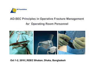
  1. 1. AO-SEC Principles in Operative Fracture Management<br />for Operating Room Personnel<br />Oct 1-2, 2010 | RDEC Bhaban, Dhaka, Bangladesh <br />
  2. 2. Open Fractures<br />Prof. Muhammad Shahiduzzaman<br />Head, Department of Orthopaedics & Traumatology<br />Dhaka Medical College<br />
  3. 3. Objectives<br /><ul><li>Open fracture classification
  4. 4. Patient evaluation
  5. 5. Surgical management
  6. 6. In the emergency department
  7. 7. First visit to the OT
  8. 8. Definitive management
  9. 9. Soft tissue
  10. 10. Fracture</li></li></ul><li>Definition<br />“An open fracture is one that communicates with the outside environment.”<br />
  11. 11. Classification<br />
  12. 12. Gustilo type I<br /><ul><li>Wound less than 1cm
  13. 13. Minimal soft tissue injury
  14. 14. Minimal contamination
  15. 15. Fracture usually simple transverse, short oblique fracture
  16. 16. Low energy injury</li></li></ul><li>Gustilo type II<br /><ul><li>Wound greater than 1 cm
  17. 17. Moderate soft tissue injury
  18. 18. Slight or moderate crush
  19. 19. No extensive soft tissue damage, flaps or avulsions
  20. 20. Simple transverse short oblique fracture with moderate comminution</li></li></ul><li>Gustilo type IIIA<br /><ul><li>Adequate soft tissue coverage of the bone
  21. 21. Includes segmental and severely comminuted fractures</li></li></ul><li>Gustilo type IIIB<br /><ul><li>Extensive periosteal stripping and bone exposure
  22. 22. Massive contamination
  23. 23. Severe comminution with high energy injury
  24. 24. Requires free or local flap for bone coverage</li></li></ul><li>Gustilo type IIIC<br /><ul><li>Any open fracture that is associated with an arterial injury that must be repaired regardless of the degree of soft tissue injury</li></li></ul><li>Management<br /><ul><li>Prevent Infection
  25. 25. Soft tissue and bone healing without complications
  26. 26. Restoration of function
  27. 27. Infection
  28. 28. Delayed and Non union
  29. 29. Loss of extremity</li></li></ul><li>Emergency assessment<br /><ul><li>Assessment of patient
  30. 30. ATLS guidelines
  31. 31. Manage life threatening injuries first.
  32. 32. Examination of the injury
  33. 33. Wound (photos)
  34. 34. Neurological status
  35. 35. Vascular status
  36. 36. Compartments :Open fractures can still develop compartment syndrome</li></li></ul><li>Emergency assessment…<br />
  37. 37. Emergency Management<br /><ul><li>Bleeding – control with direct pressure
  38. 38. Remove gross debride, gentle small volume irrigation, sterile dressing (normal saline)
  39. 39. Reduce bone or joint
  40. 40. Splint limb
  41. 41. Intravenous antibiotics</li></li></ul><li>Principle of treatment<br /><ul><li>Treat any open fracture as an emergency.
  42. 42. Evaluate the patient to diagnose other life threatening injury.
  43. 43. Institute appropriate and adequate antibiotic.
  44. 44. Adequate wound excision.
  45. 45. Stabilize the fracture.
  46. 46. Perform delayed closure of the wound within 3-7 days.
  47. 47. Decide on early amputation.
  48. 48. Rehabilitate the involved extremity. </li></li></ul><li>This is a surgical emergency<br /><ul><li>Treatment of open fracture is second only to life threatening and arterial injury.
  49. 49. It is imperative to immediately treat open fracture in order to reduce or prevent wound sepsis.
  50. 50. All open fractures of more than 8 hours should be considered infected.</li></li></ul><li>Antibiotic therapy<br /><ul><li>Prevention of wound sepsis is the primary objectives in the treatment of open fracture
  51. 51. Tetanus prophylaxis is indicated.
  52. 52. Both gram-negative and aerobic gram positive are major pathogens in open #
  53. 53. Penicillin is added for Agricluturalinjuries.</li></li></ul><li>Common pathogen<br />
  54. 54. Wound excision<br />Wound excision to be under taken under strict aseptic condition, must be systematic and complete.<br />Lavage is done with normal saline or distilled water. For final irrigation mixture of bacitracin and polymyxin solution is preferable.<br />Wound incision must be large enough to facilitate exposure and inspection.<br />The following structures are debrided : <br />skin, fascia and tendons,muscles, and bones.<br />
  55. 55. Debridement<br /><ul><li>Prior to irrigation
  56. 56. Requires extension of the wound
  57. 57. Longitudinal
  58. 58. Systematic fashion
  59. 59. Skin
  60. 60. Subcutaneous fat
  61. 61. Fascia
  62. 62. Muscle
  63. 63. Bone</li></li></ul><li>Debridement-skin<br /><ul><li>Prior to irrigation
  64. 64. Requires extension of the wound
  65. 65. Longitudinal
  66. 66. Systematic fashion
  67. 67. Skin
  68. 68. Subcutaneous fat
  69. 69. Fascia
  70. 70. Muscle
  71. 71. Bone</li></li></ul><li>Debridement-muscle<br /><ul><li>Open the fascial compartments to see the extent of injury
  72. 72. Remove muscle that is dead or necrotic
  73. 73. Based on colour and turgor of muscle (bleeding not as good)
  74. 74. If in doubt can leave and relook in 24 to 48 hours</li></li></ul><li>Debridement-bones<br /><ul><li>Remove bone that has no soft tissue attachment
  75. 75. Keep large articular fragments
  76. 76. consider fixing at the initial debridement</li></li></ul><li>Irrigation<br /><ul><li>After the debridement
  77. 77. Options:
  78. 78. Low pressure versus high pressure (pulse lavage)
  79. 79. Saline alone versus additives (antiseptics, antibiotics, or soap/detergents)</li></li></ul><li>Wound management<br /><ul><li>“If there is the slightest doubt in the surgeon’s mind as to whether there has been adequate debridement of the wound after an open fracture, the wound should not be closed regardless of the type of open fracture. For the surgeon who manages only an occasional open fracture, the safe rule is not to close the wound”
  80. 80. Gustilo and Anderson JBJS 1974</li></li></ul><li>VAC system<br /><ul><li>Provides closed suction system
  81. 81. Reduces oedema and bacterial counts
  82. 82. Enhances granulation tissue
  83. 83. Carefully does not prevent primary wound closure
  84. 84. Not a substitute for early definitive coverage
  85. 85. Plastics & Reconstructive Surgery 2008</li></li></ul><li>Antibiotic bead pouch<br /><ul><li>Antibiotic cement beads
  86. 86. Fill dead space
  87. 87. High local antibiotic concentration
  88. 88. Seal wound from further contamination
  89. 89. Infection rates decreased from 12.0% to 3.7% in 1085 fractures (Ostermann JBJS 1995)</li></li></ul><li>Repeat Debridement<br /><ul><li>Can include opening a wound that was primarily closed (eg subcutaneous border of the tibia)
  90. 90. Remember that this does interfere with fracture healing to some degree
  91. 91. High grade injury
  92. 92. Severe contamination
  93. 93. Questionable tissue viability
  94. 94. Repeat 24-48 hours until wound viable</li></li></ul><li>Wound Coverage and closure<br /><ul><li>For type I and type II open fracture delayed primary closure or skin graft can be accomplished in 3 to 5 days.
  95. 95. For type III B and III C open fracture with significant soft tissue loss and exposed bone often require two or three debridement before flap coverage.</li></ul>Early soft tissue converge is key to minimize woundsepsis.<br />
  96. 96. Soft tissue Coverage<br /><ul><li>Primary closure
  97. 97. Delayed primary closure
  98. 98. Skin graft
  99. 99. Local flaps
  100. 100. Fasciocutaneous flaps
  101. 101. Muscle pedicle
  102. 102. Free flaps
  103. 103. Standard (eg lat dorsi)
  104. 104. Fasciocutaneous (eg lateral thigh flap)</li></li></ul><li>Stabilization of open fracture<br /><ul><li>Plaster immobilization
  105. 105. Skeletal traction
  106. 106. Internal fixation with implant
  107. 107. External fixation</li></li></ul><li>Stabilization of open fracture…<br /><ul><li>Achieving fracture stability is just as important as wound excision.
  108. 108. Stable fracture fixation preserves the integrity of the remaining soft tissues, muscles and neurovascular structures.
  109. 109. Facilitates care of the wound and contributes to the well-being of the whole patient.
  110. 110. Allows joint motion and muscle exercise program.</li></li></ul><li>Fixation<br /><ul><li>Important to protect the soft tissues from additional injury by fracture fragments
  111. 111. Aids in wound care
  112. 112. Allows early mobilisation and rehabilitation of the patient</li></li></ul><li>Skeletal traction<br /><ul><li>Skeletal traction is indicated for type I and type II open fracture of femoral shaft.
  113. 113. Wound is allowed to heal followed by internal fixation in 10 to 14 days.</li></li></ul><li>External Fixation<br /><ul><li>Safe and reliable method of achieving bony stability in open fracture.
  114. 114. Major disadvantage is pin tract infection, but can be considerably reduced with proper pin insertion and care.</li></li></ul><li>Amputation<br />Can a functional,viable extremity be achieved ? <br />Can the the time and expense of saving the extremity be justified ?<br />Absolute indication of amputation :<br /> Type III C injury with posterior tibial nerve disruption.<br /> Type III C injury with soft tissue loss, massive contamination with severely comminuted segmented fracture or massive bone loss.<br />
  115. 115. Rehabilitation<br /><ul><li>Immediate objectives of rehabilitation are to prevent muscle atrophy, prevent joint stiffness and improve circulation in the extremity.
  116. 116. The ultimate objective, of course, is to restore the extremity to the greatest degree of function of which it is capable.
  117. 117. A well-organized rehabilitation program initiated early will help return the patient to a functional status.</li>

×