SlideShare a Scribd company logo
1 of 11
Download to read offline
Cell Stem Cell
Protocol Review
Guidelines and Techniques for the Generation
of Induced Pluripotent Stem Cells
Nimet Maherali1,2 and Konrad Hochedlinger1,*
1Massachusetts General Hospital Center for Regenerative Medicine and Cancer Center, Harvard Stem Cell Institute,
Department of Stem Cell and Regenerative Biology, 185 Cambridge Street, Boston, MA 02114, USA
2Department of Molecular and Cellular Biology, Harvard University, 7 Divinity Avenue, Cambridge, MA 02138, USA
*Correspondence: khochedlinger@helix.mgh.harvard.edu
DOI 10.1016/j.stem.2008.11.008
Direct reprogramming of somatic cells to induced pluripotent stem cells (iPSCs) provides an invaluable
resource for regenerative medicine, enabling the generation of patient-specific cells of any lineage without
the use of embryonic material. A variety of methods exist for iPSC derivation, all reliant upon manipulation
of a select group of transcription factors. We compare the currently reported protocols, identify essential
steps common to these methods, and suggest minimal criteria for defining fully reprogrammed cells. In ad-
dition, specific procedures aimed to optimize reproducible iPSC derivation are presented, with an emphasis
on standardization of certain parameters for accurate comparison between independent experiments.
The generation of pluripotent cells from differentiated adult cells
has vast therapeutic implications, particularly in the context of
in vitro disease modeling, pharmaceutical screening, and cellular
replacement therapies. In addition, the ability to revert somatic
cells to an embryonic state provides a unique tool to dissect
the molecular events that permit the conversion of one cell
type to another.
Previously devised strategies to induce pluripotency, such as
somatic cell nuclear transfer (cloning) or fusion of somatic cells
with embryonic stem cells (ESCs) (Hochedlinger and Jaenisch,
2006) are fraught with technical, ethical, and logistical barriers
that impede the use of the resulting pluripotent cells in both
research and therapy. Thus, the direct generation of pluripotent
cells without the use of embryonic material has been deemed
a more suitable approach that lends itself well to mechanistic
analysis and has fewer ethical implications.
The direct reprogramming of somatic cells to pluripotency
was accomplished in 2006, when Takahashi and Yamanaka
converted adult mouse fibroblasts to iPSCs through ectopic ex-
pression of a select group of transcription factors. Subsequent
reports optimized this technique, demonstrating that iPSCs
were indeed highly similar to ESCs when tested across a rigorous
set of assays (Maherali et al., 2007; Okita et al., 2007; Wernig
et al., 2007). In 2007, direct reprogramming was achieved in
human cells (Takahashi et al., 2007b; Yu et al., 2007), providing
an invaluable contribution to the field of regenerative medicine.
While the establishment of iPSC lines is conceptually and
technically simple, direct reprogramming is a slow and inefficient
process consisting of largely unknown events. Several variables
must be considered in order to reproducibly obtain iPSCs, which
include (1) the choice of factors used to reprogram cells; (2) the
methods used to deliver these factors; (3) the choice of target cell
type; (4) the parameters of factor expression, such as timing and
levels; (5) the culture conditions used to derive iPSCs; and the
methods of (6) identifying and (7) characterizing reprogrammed
cells. This review addresses each of these steps in detail and
is summarized as an overview in Figure 1. A discussion on the
efficiency of reprogramming has also been included to promote
standardization in the calculation and reporting of efficiencies.
Choice of Reprogramming Factors
Direct reprogramming was initially performed in mouse fibro-
blasts through retroviral transduction of 24 candidate genes
that were all implicated in the establishment and maintenance
of the pluripotent state. This pool of 24 genes was ultimately nar-
rowed down to four transcription factors, Oct4 (Pou5f1), Sox2,
c-Myc, and Klf4, that were sufficient to mediate reprogramming
(Takahashi and Yamanaka, 2006). This core set of factors has
been shown to work across a multitude of mouse cell types (Aoi
et al., 2008; Eminli et al., 2008; Hanna et al., 2008; Kim et al.,
2008; Stadtfeld et al., 2008a, 2008c; Wernig et al., 2008a), as
well as rhesus monkey (Liu et al., 2008) and human cells (Park
et al., 2008a; Takahashi et al., 2007b; Lowry et al., 2008).
Variations on the four-factor cocktail have been used to suc-
cessfully reprogram cells. In mouse fibroblasts, Sox1 and Sox3
can replace Sox2, albeit with a decrease in reprogramming effi-
ciency; Klf2 can replace Klf4, and L-Myc and N-Myc can replace
c-Myc (Blelloch et al., 2007; Nakagawa et al., 2008). It has also
been reported that a partially different set of factors, OCT4,
SOX2, NANOG, and LIN28, is sufficient to reprogram human
fibroblasts (Yu et al., 2007).
The endogenous expression of certain reprogramming factors
in different cell types has permitted their exclusion from the
factor cocktail. For example, fibroblasts express c-Myc and
Klf4, and it has been demonstrated that exogenous c-Myc is
not necessary for the reprogramming of mouse and human fibro-
blasts, although the efficiency is much lower and reprogramming
requires more time (Nakagawa et al., 2008; Wernig et al., 2008b).
Neural progenitor cells, which express Sox2 and c-Myc at levels
higher than in ESCs, have been reprogrammed using only Oct4/
Klf4 or Oct4/c-Myc, though at lower efficiency than with four
factors (Kim et al., 2008).
While the original suite of four factors remains the standard
for direct reprogramming, a handful of small molecules and
Cell Stem Cell 3, December 4, 2008 ª2008 Elsevier Inc. 595
additional factors have been reported to enhance the reprog-
ramming process and/or functionally replace the role of some
of the transcription factors (Table 1). The identification of such
mediators is beginning to yield insight into the mechanisms by
which reprogramming occurs, and many similar studies are likely
to follow. The use of small molecules and soluble factors is par-
ticularly appealing given their ease of use and lack of permanent
genome modification that constrains the use of retro- and lenti-
viruses (further described in the following section); however, it is
currently unknown whether small molecules alone can recapitu-
late the series of transcriptional and epigenetic changes brought
about by the four transcription factors. An important caveat to
the increasing use of epigenetic modifiers is that their broad and
nonspecific effects may elicit an overall dysregulation of gene
expression. For instance, 5-azacytidine is mutagenic (Jackson-
Grusby et al., 1997), and mice with global alterations in DNA
methylation levels develop tumors at a high frequency (Gaudet
et al., 2003). In general, the use of alternative and adjunct factors
is growing in popularity, and newer methods should be subject to
rigorous testing to ensure quality of the resulting iPSC lines.
Methods of Factor Delivery
The production of iPSCs has so far been achieved through nu-
cleic-acid-based delivery of the reprogramming factors. Initial
generations of mouse and human iPSCs employed retroviral vec-
tors (Takahashi et al., 2007b; Takahashi and Yamanaka, 2006)
and constitutive lentiviruses (Blelloch et al., 2007; Yu et al.,
2007), while later generations were produced using inducible
Figure 1. Overview of the iPSC Derivation
Process
lentiviruses (Brambrink et al., 2008;
Hockemeyer et al., 2008; Maherali et al.,
2008; Stadtfeld et al., 2008b). These viral
systems, however, have been criticized
for their permanent integration into the
genome, and endeavors to make iPSCs
more therapeutically applicable have
led to the pursuit of nonintegrating
approaches (Table 2). Two such ap-
proaches, adenoviral delivery and tran-
sient transfection, have been success-
fully used in the reprogramming of
mouse cells (Okita et al., 2008; Stadtfeld
et al., 2008c), lending promise to the
eventual use of transient delivery
methods in human iPSC derivation.
The first attempts at direct reprogram-
ming employed Moloney-based retroviral
vectors that are known to undergo silenc-
ing in the ESC state (Jahner et al., 1982;
Wolf and Goff, 2007); this self-silencing
property provided an advantage for initial
attempts as the temporal requirement of
factor expression was undefined. How-
ever, several drawbacks in addition to
genome integration preclude the use of these retroviruses: (1)
their infectivity is limited to dividing cells (Miller et al., 1990),
thus restricting the range of cell types that can be reprogrammed;
(2) silencing occurs gradually during the course of iPSC induction,
resulting in a lowered efficiency of conversion compared to
nonsilencing viral methods (Stadtfeld et al., 2008b); and (3) iPSCs
made with retroviruses often maintain viral gene expression
(Dimos et al., 2008; Park et al., 2008c), thus limiting their utility.
While lentiviruses permit the transduction of nondividing cell
types with high expression levels (Naldini et al., 1996), they are
poorly silenced in the pluripotent state (Lois et al., 2002), making
the constitutive versions less suitable for reprogramming at-
tempts. Although iPSCs made with constitutive lentiviruses
have been reported (Blelloch et al., 2007; Yu et al., 2007), it is un-
clear how differentiation proceeds during continued transgene
expression (Brambrink et al., 2008).
Drug-inducible lentiviruses have provided a more attractive
approach, as they permit temporal control over factor expres-
sion. Although these viruses also integrate in the host genome,
they are particularly useful for conducting mechanistic analyses
(Brambrink et al., 2008; Stadtfeld et al., 2008b). For instance, the
use of such viruses has led to the establishment of ‘‘secondary
systems,’’ whereby iPSC-derived differentiated cells harbor the
proviral integrations in the same pattern that enabled primary
iPSC induction. Upon reinduction, the viral transgenes are
homogeneously reactivated, leading to a >100-fold increase in
secondary iPSC production (Hockemeyer et al., 2008; Maherali
et al., 2008; Wernig et al., 2008a). Such systems provide
596 Cell Stem Cell 3, December 4, 2008 ª2008 Elsevier Inc.
Cell Stem Cell
Protocol Review
a powerful tool to aid in chemical and genetic screening for fac-
tors that enhance reprogramming, as well as the optimization of
iPSC derivation conditions. Newer techniques that build on this
approach may include the targeting of reprogramming factor
DNA to known regions of the genome (Hochedlinger et al.,
2005), thus preventing positional effects caused by random
integration; methods to link all four reprogramming factors on
one transcript may facilitate such targeting efforts (Okita et al.,
2008).
The use of integrating viruses for iPSC induction has repre-
sented a major roadblock in the pursuit of clinically relevant ap-
plications, as genomic insertion has been shown to alter gene
function (Kustikova et al., 2005), and viral transgene reactivation
in iPSC-derived chimeric mice has been implicated in tumori-
genesis (Nakagawa et al., 2008). Analysis of integration sites in
iPSCs yielded no common targets or pathways, indicating that
genomic integration is not necessary for reprogramming (Aoi
et al., 2008; Varas et al., 2008). The derivation of mouse iPSCs
Table 1. Small Molecules and Other Factors Used to Enhance the Reprogramming Process
Molecule/Factor Target/Mode of Action Role in Reprogramming References
Valproic Acid histone deacetylase inhibitor enhances reprogramming efficiency
with four factors (O/S/M/K) in mouse
fibroblasts
Huangfu et al., 2008a, 2008b
restores reprogramming efficiency
in mouse fibroblasts without c-Myc
(O/S/K only)
permits reprogramming of human
fibroblasts treated with OCT4 and
SOX2, though at extremely low
efficiency
5-azacytidine; shRNA
against Dnmt1
DNA demethylating agent 4-fold enhancement of
reprogramming efficiency with four
factors (O/S/M/K) in mouse
fibroblasts
Mikkelsen et al., 2008
no effect if applied too early; toxic
to differentiated cells
BIX01294 histone methyltransferase inhibitor restores reprogramming efficiency
in mouse neural progenitor cells
with Oct4/Klf4 to four-factor level
(O/S/M/K)
Shi et al., 2008b
permits reprogramming of mouse
neural progenitor cells in the
absence of Oct4, though at
extremely low efficiency and
requires the presence of the
other three factors (S/M/K)
BayK8644 L-type calcium channel agonist cooperates with BIX01294 to
enable reprogramming of mouse
embryonic fibroblasts with
Oct4/Klf4
Shi et al., 2008a
Wnt3a cell signaling molecule;
transcriptional activation of
multiple downstream targets,
including c-Myc
1.2-fold enhancement of
reprogramming efficiency with
four factors (O/S/M/K) in mouse
fibroblasts
Marson et al., 2008
20-fold enhancement with three
factors (O/S/K); partially restores
efficiency in the absence of c-Myc
siRNA against p53
and Utf1 cDNA
tumor suppressor; ESC-specific
cofactor, respectively
modest increase in four-factor (O/S/
M/K) reprogramming efficiency of
human fibroblasts with individual
factors (4 + p53siRNA or 4 + UTF1)
Zhao et al., 2008
100-fold enhancement with the
combined factors (O/S/M/K +
p53siRNA + UTF1); further slight
enhancement by the exclusion of
c-MYC (O/S/K + p53siRNA/UTF1)
O, Oct4; S, Sox2; M, c-Myc; K, Klf4; Dnmt1, DNA methyltransferase 1.
Cell Stem Cell 3, December 4, 2008 ª2008 Elsevier Inc. 597
Cell Stem Cell
Protocol Review
using transient delivery approaches has confirmed this notion
(Okita et al., 2008; Stadtfeld et al., 2008c) and has provided
a solid foundation on which such techniques might be optimized
for human cells.
Choice of Cell Type
For the first reprogramming attempts in both mouse and human,
fibroblasts were used as the starting cell population. Adult fibro-
blasts have been previously shown to be amenable to reprog-
ramming by nuclear transfer in mouse (Wakayama et al., 1998)
and cell fusion in both mouse and human (Cowan et al., 2005;
Tada et al., 2001). Further, the derivation of fibroblasts is techni-
cally simple (Nagy et al., 2003; Park et al., 2008b), and disease-
specific human fibroblasts are readily available through cell
repositories such as Coriell. Fibroblasts are also compatible
with ESC culture conditions and are used as feeder layers for
ESC growth, making them a feasible starting candidate for direct
reprogramming efforts.
Table 2. Methods of Factor Delivery
Method Advantages Disadvantages References
Moloney-based retrovirus silenced in pluripotent cells genomic integration; risk of insertional
mutagenesis
Takahashi and Yamanaka, 2006
self-silencing eliminates need
for timed factor withdrawal
limited to dividing cells
expression often maintained in iPSCs;
increased tumor incidence in chimeric
mice due to transgene reactivation
HIV-based lentivirus Constitutive
transduction of both dividing
and nondividing cells
genomic integration; risk of insertional
mutagenesis
Brambrink et al., 2008;
Blelloch et al., 2007;
Yu et al., 2007
lack of silencing in pluripotent state
Inducible
temporal control over factor
expression
genomic integration; risk of
insertional mutagenesis
Stadtfeld et al., 2008b;
Brambrink et al., 2008
possibility of leaky expression
Integrase-Deficient
low frequency of genomic
integration
lower expression levels than
integrated form
Nightingale et al., 2006
integration provides selective
advantage and necessitates clone
screening
not yet reported for iPSC production
Transient transfection no viral components multiple rounds of transfection are
required
Okita et al., 2008
low frequency of genomic
integration
lower levels of expression than
when integrated
technically simple procedure delayed kinetics of reprogramming
integration provides selective
advantage and necessitates clone
screening
Adenovirus low frequency of genomic
integration
repeated infection required for
certain cell types
Stadtfeld et al., 2008c
delayed kinetics of reprogramming
some generation of tetraploid cells
Small molecules transient controllable activity issue of toxicity versus efficacy Huangfu et al., 2008a, 2008b;
Mikkelsen et al., 2008
technically easy to work with undefined/nonspecific effects
not yet reported for iPSC production
Protein transduction direct delivery of transcription
factors avoids complications of
nucleic-acid-based delivery
short half-life; multiple applications
required
Gump and Dowdy, 2007;
Bosnali and Edenhofer, 2008
some proteins difficult to purify
not yet reported for iPSC production
598 Cell Stem Cell 3, December 4, 2008 ª2008 Elsevier Inc.
Cell Stem Cell
Protocol Review
Since the success of fibroblast reprogramming, a multitude of
mouse cell types, including stomach cells (Aoi et al., 2008), liver
cells (Aoi et al., 2008; Stadtfeld et al., 2008c), pancreatic b cells
(Stadtfeld et al., 2008a), lymphocytes (Hanna et al., 2008), and
neural progenitor cells (Eminli et al., 2008; Kim et al., 2008), as
well as human keratinocytes (Aasen et al., 2008; Maherali et al.,
2008), have been reprogrammed. Many of these experiments
employed genetic labeling or other techniques to confirm the
identity of the donor cell, ruling out the possibility of contaminat-
ing resident fibroblasts as the cell of origin.
What has emerged from these studies is that there is a strong
influence of cell type on reprogrammability, including the effi-
ciency and kinetics of the process as well as the ease at which
reprogramming factors can be delivered. For example, mouse
stomach and liver cells showed reactivation of the ESC-specific
Fbx15 gene during reprogramming much faster than fibroblasts
and contained fewer viral integrations (Aoi et al., 2008), and
human keratinocytes reprogrammed faster and more efficiently
than human fibroblasts (Aasen et al., 2008; Maherali et al.,
2008). The effective delivery of factors has also played a role in
the reprogramming of a given cell type. For instance, the reprog-
ramming of mouse fibroblasts with adenoviral vectors required
100- to 200-fold higher titers than that of liver cells (Stadtfeld
et al., 2008c).
Several factors must therefore be considered in determining
the optimal cell type for a given application: (1) the ease at which
reprogramming factors can be introduced, which varies both by
cell type and delivery approach; (2) the availability and ease of
derivation of the given cell type; and (3) the age and source of
the cell. Older cells or those that have undergone several pas-
sages in culture may harbor genetic lesions that undermine the
therapeutic potential of the resulting iPSCs; similarly, cells
obtained from organs that are more likely to have acquired
DNA damage, such as skin cells that may have accumulated
UV-induced mutations, would be less suitable for clinical appli-
cations. Thus, while fibroblasts are likely to remain the choice
cell type in basic research efforts to mechanistically dissect
the reprogramming process, iPSCs derived for therapeutic
purposes will require the donor cells to be easily attainable,
less likely to contain genetic aberrations, and easy to reprogram
with transient approaches.
Parameters of Factor Expression
The shift toward nonintegrating delivery approaches has neces-
sitated a better understanding of how the factors coordinate their
efforts to orchestrate reprogramming. To improve the process of
iPSC derivation, it is important to define the temporal require-
ment of factor expression, as well as the optimal factor levels
and stoichiometry. Additionally, the quantification of factor deliv-
ery is important for exploiting such knowledge and can ensure
reproducibility as well as allow proper comparison between
independent experiments.
The length of time required for cells to become independent of
factor expression has been addressed using doxycycline-induc-
ible systems. The kinetics of factor requirements has been quan-
tified in mouse fibroblasts, which require at least 8–12 days of
factor exposure (Brambrink et al., 2008; Stadtfeld et al., 2008b),
and in human keratinocytes, which require 10 days (Maherali
et al., 2008). Although prolonged exogenous factor expression
beyond the minimal amount of time results in enhanced colony
recovery (Brambrink et al., 2008; Stadtfeld et al., 2008b; Wernig
et al., 2008a), persistent expression in the pluripotent state may
be detrimental (Mikkelsen et al., 2008). Thus, a general guideline
to follow is that exogenous factor expression should be discon-
tinued as soon as genuine iPSCs become apparent (further
discussed in the section ‘‘Methods to Identify Reprogrammed
Cells’’). While the kinetics of reprogramming is highly influenced
by the starting cell type, in all instances reprogramming requires
several days to proceed. The wide temporal ranges reported for
each cell type suggest that aspects in addition to cell identity can
influence the kinetics, which can most likely be attributed to
differential factor delivery (levels and stoichiometry), but may
also reflect cell-intrinsic differences such as cell cycle stage,
differentiation status, and passage number.
The precise expression levels and stoichiometry required for
reprogramming have been difficult to examine. Variance in factor
delivery, coupled with the low efficiency of conversion, has made
it impossible to retrospectively analyze the individual contribu-
tion of each factor that causes a single differentiated cell to ac-
quire a pluripotent state. However, achieving optimal expression
levels is indeed important for the reprogramming process; for
example, the conversion of neural progenitor cells into iPSCs
proceeds more efficiently when Sox2 is omitted from the four-
factor cocktail, indicating that transgene-driven Sox2 expression
in addition to high endogenous Sox2 levels is detrimental (Eminli
et al., 2008; Silva et al., 2008). The establishment of secondary
systems in which each clone has a distinct but reliable pattern
of factor reactivation offers a more reliable tool to dissect the
precise contribution of individual factors (Hockemeyer et al.,
2008; Maherali et al., 2008; Wernig et al., 2008a).
Though the optimal expression levels and stoichiometry of
factors still remain poorly defined, ensuring that cells receive all
factors is an important step in achieving reprogramming, and
quantification of individual factor expression within the chosen
delivery system is critical for reproducibly obtaining iPSCs. No
reports of iPSC generation have thoroughly quantified factor
delivery; in most instances this has been addressed indirectly
through the use of separately delivered reporter constructs,
such as GFP-encoding vectors. However, the use of such surro-
gate markers provides an inaccurate readout. For example,
reporter proteins are quite stable with long half-lives and are
not subject to the same cellular handling experienced by tran-
scription factors; this difference is particularly relevant to tran-
sient delivery methods where multiple applications of factors
are required (Okita et al., 2008; Stadtfeld et al., 2008c), and reap-
plication depends on the length of time each factor is expressed.
For viral-based methods, titers are influenced by the gene of in-
terest, as the gene product is expressed at high levels during
packaging and can potentially alter the function of the packaging
cells (Tiscornia et al., 2006). Thus, even with constant transfection
parameters, viral titer is highly variable. The best method for
quantification is a direct analysis of expression in the cell type
of interest; this assessment can be accomplished by using a re-
porter-linked construct, such as IRES-GFP, or through immunos-
taining, which permits analysis at a single-cell level. For a more
accurate measure of factor delivery, one can also assess coinfec-
tivity to determine the percentage of cells receiving all factors.
While testing the expression of each factor in every batch
Cell Stem Cell 3, December 4, 2008 ª2008 Elsevier Inc. 599
Cell Stem Cell
Protocol Review
produced requires a considerable amount of work, there is a high
payoff in reproducibility, and controlling for factor input facilitates
the transition between different delivery methods.
As viral-based gene delivery methods remain popular for iPSC
derivation, the ability to produce high-titer virus is of great value.
Detailed protocols specifically written for iPSC derivation provide
a good starting resource (Park et al., 2008b; Takahashi et al.,
2007a); though limited to retroviruses, these basic protocols
are highly adaptive and can be tailored to optimize factor delivery
in different contexts. More extensive reviews covering a broad
scope of viral methods serve as excellent references (Ramezani
and Hawley, 2002; Rosenzweig, 2007; Tiscornia et al., 2006).
An alternative is to have viruses produced commercially, which
is more suitable for applications that solely require iPSCs for
downstream analysis and do not require flexibility of manipulation
during the derivation process.
Culture and Derivation Conditions
Both mouse and human iPSC derivation proceed under the
same culture conditions used for ESC maintenance (Akutsu
et al., 2006; Cowan et al., 2004; Lerou et al., 2008; Nagy et al.,
2003), and it is important to ensure that the selected conditions
support ESC growth. While alternative conditions for reprogram-
ming have not yet been reported, there will indeed be a push
toward creating defined and xeno-free cultures in efforts to es-
tablish iPSCs that will be more suitable for clinical applications.
As ESC conditions are sufficient to obtain iPSCs from most cell
types, it has been speculated that conditions used to facilitate
ESC derivation may also enhance iPSC derivation. For instance,
the use of knockout serum replacement instead of fetal bovine
serum greatly facilitates mouse ESC (mESC) derivation (Cheng
et al., 2004), and it has also been reported to improve the reprog-
ramming of mouse fibroblasts (Blelloch et al., 2007). The use of
knockout serum replacement provides an alternative culture
condition for the reprogramming of various cell types for which
standard serum is unsuitable. An important point to note, how-
ever, is that the use of undefined media components such as
serum introduces batch-to-batch variability and may not elicit
reproducible effects. Thus, it is important to screen individual
batches for ESC supportive capacity.
ESCs rely on fibroblast-derived factors to support their growth,
particularly human ESCs (hESCs). mESCs can be derived and
cultured on gelatin in the absence of feeders and additional
growth factors (Ying et al., 2008), and similarly, mouse iPSCs
can be derived under feeder-free conditions (Stadtfeld et al.,
2008b; Wernig et al., 2008a). While defined culture conditions
for hESCs have also been established (Amit and Itskovitz-Eldor,
2006), the derivation of human iPSCs without feeder cells has
not yet been reported, though it remains a clinically relevant
goal to avoid the use of animal products.
A key aspect for creating favorable derivation conditions is to
achieve an optimal cell density. Cells seeded at very low densi-
ties may senesce and be less amenable to reprogramming, while
cells seeded at high density can quickly become overconfluent,
hindering the growth of new colonies and posing the risk of the
cell layer lifting, particularly after the prolonged culture times
required for reprogramming. This risk has been illustrated in sec-
ondary systems where reprogramming occurs at high efficiency;
at high cell densities, the frequency of colony formation drops
despite identical expression of the reprogramming factors
(Maherali et al., 2008; Wernig et al., 2008a), indicating a nonlinear
relationship between cell density and reprogramming efficiency.
While the optimal cell density must be experimentally deter-
mined, a general guideline to follow is to seed the infected target
cells at 10% confluence and to use a feeder density less than
2.5 to 5 3 104
cells/cm2
.
Determining appropriate culture conditions for the reprogram-
ming of nonfibroblast cell types presents a specialized case that
must be tailored to satisfy the needs of both the donor cell and
the arising iPSC. Accordingly, the reprogramming factors are
typically introduced into the donor cells under their native condi-
tions and then switched to ESC conditions during the course of
reprogramming, the timing of which must be experimentally
determined. For instance, the reprogramming of mouse neural
progenitor cells requires a switch from serum-free conditions to
serum-containing ESC conditions; if switched too early, no
iPSCs are obtained (Wernig et al., 2008a). In some instances it
is possible to employ cultures that support the growth of both
the donor cell and iPSC; for example, in the reprogramming of
lymphocytes, a combination of B lineage growth factors and
LIF was used, making the culture environment suitable for
both hematopoietic cells and iPSCs, respectively (Hanna et al.,
2008).
Human iPSC (hiPSC) derivation also represents a unique case,
as the cells are more sensitive than their mouse counterparts to
the conditions under which they are grown. For example,
hiPSCs/hESCs display some sensitivity to doxycycline exposure
(Maherali et al., 2008), which must be accounted for when using
such inducible systems. hiPSCs/hESCs also exhibit poor sur-
vival when grown as single cells; accordingly, the addition of
small molecules that enhance single-cell survival in established
hiPSC/hESC cultures, such as the Rho-associated kinase
(ROCK) inhibitor (Watanabe et al., 2007) have been suggested
to facilitate hiPSC derivation (Park et al., 2008b), although their
use is not required for successful reprogramming.
Methods to Identify Reprogrammed Cells
In the first attempts at reprogramming, it was anticipated that the
introduction of multiple factors would yield multiple cell fates,
thus necessitating the use of a selection system whereby only
cells that reactivated ESC-specific genes could survive (Takaha-
shi and Yamanaka, 2006). Such systems, however, entailed per-
manent genetic modification through the introduction of reporter
alleles, rendering the system unfeasible for reprogramming in
a clinical setting. Several advances have since been made,
leading to effective and therapeutically compatible methods to
identify and obtain reprogrammed cells.
The first generation of mouse iPSCs was obtained via selec-
tion for the ESC-specific, but nonessential, gene Fbx15 (Takaha-
shi and Yamanaka, 2006). While the resulting cells demonstrated
pluripotency in the context of teratoma formation, they were not
fully reprogrammed, as they could not generate chimeric mice,
and their gene expression profiles and DNA methylation status
were distinct from ESCs. It was later found that selection for
the essential ESC-specific genes, Nanog and Oct4, permitted
the generation of iPSCs that were much more similar to ESCs
(Maherali et al., 2007; Okita et al., 2007; Wernig et al., 2007).
With this finding also came the result that delayed onset of
600 Cell Stem Cell 3, December 4, 2008 ª2008 Elsevier Inc.
Cell Stem Cell
Protocol Review
selection was key to generating fully reprogrammed cells, ulti-
mately leading to the discovery that selection methods were un-
necessary and actually counterproductive (Blelloch et al., 2007;
Maherali et al., 2007; Meissner et al., 2007).
The identification of iPSC colonies based solely upon morpho-
logical criteria requires a considerable degree of ESC expertise.
In general, mouse ESC colonies can be distinguished by their re-
fractive, or ‘‘shiny,’’ appearance and tight, well-defined borders,
while human ESC colonies display a cobblestone appearance
with prominent nucleoli and pronounced individual cell borders.
The stepwise morphological changes that occur during reprog-
ramming have been depicted in both systems (Figure 2). Of
importance to note is that morphologically similar but non-
iPSC colonies also arise during the course of human fibroblast
reprogramming. These colonies are often mistaken for iPSCs,
particularly by novices in the field, but are distinguishable from
iPSCs becuase they are loose and granular in appearance,
contain phase-bright cells, and appear earlier in the process
(after 1–2 weeks) (Lowry et al., 2008; Takahashi et al., 2007b).
Additional methods to identify iPSCs have been described;
these techniques become useful when one is dealing with cell
types that provide a high background of non-iPSC colonies (for
example, those formed during human fibroblast reprogramming),
or when ESC expertise is lacking. Two such methods have used
ESC-specific surface antigen expression and loss of transgene
dependence as strategies to identify reprogrammed cells. For
example, isolation of the Thy-1-SSEA-1+ population during the
course of mouse fibroblast reprogramming greatly enriches for
cells poised to become iPSCs (Stadtfeld et al., 2008b), and live
staining of cultures for the hESC-specific surface antigen Tra-
1-81 has aided in the identification of genuine hiPSC colonies de-
rivedfromhumanfibroblasts(Lowryetal.,2008).Thelossoftrans-
gene dependence, which correlates with full reprogramming, can
be assessed through the use of reporters that are silenced in the
pluripotent state (Stadtfeld et al., 2008b; Zhao et al., 2008), or
through factorwithdrawal, whichrequiresaninducibleortransient
delivery system. Upon withdrawal, cells that rely on continued
factor expression are eliminated, thus permitting selective expan-
sion of fully reprogrammed cells (Maherali et al., 2008).
Expansion and Characterization of Cells
The steps involved in taking a new colony to a fully established
iPSC line are identical to those for ESC derivation, which have
been described in detail elsewhere (Akutsu et al., 2006; Lerou
et al., 2008; Nagy et al., 2003). Of particular importance to note,
however, are the methods used to passage mouse and human
iPSCs, as well as those used to ensure purity of the resulting
iPSC lines.
Mouse iPSCs/ESCs can withstand single-cell dissociation,
and newly derived colonies can be immediately subjected to
enzymatic passaging, thus facilitating their quick expansion
into lines. Human iPSCs/ESCs, however, survive poorly as single
cells, and initial passaging of new colonies must be done me-
chanically; several passages (approximately five to ten) are
required before the cells can be adapted to enzymatic dissocia-
tion (Lerou et al., 2008). As hiPSCs/ESCs are highly prone to
differentiation, especially within the first few passages, it is im-
portant to continually remove differentiated structures to prevent
0d 6d3d
9d
BA
DC
17d16d14d
8d 12d10d
18d 19d 20d
i
ii
Figure 2. Progressive Formation of iPSC
Colonies
(A) Time course of reprogramming in primary
infected mouse fibroblasts. The arrow at 3d indi-
cates a nascent colony. At day 9, two adjacent
colonies are shown; only one has reactivated the
endogenous Oct4-GFP reporter allele, represent-
ing a true iPSC colony. Bottom right image de-
picts iPSC colonies from an established line
(Stadtfeld et al., 2008b).
(B) Two representative colonies derived from pri-
mary infected mouse fibroblasts. (Bi) iPSC colony,
characterized by a tight well-defined border and
Oct4-GFP expression; (Bii) differentiated colony,
consisting of loosely packed cells and lacking
expression of Oct4-GFP (Stadtfeld et al., 2008b).
(C) Colony tracking during the reprogramming of
secondary human fibroblasts with a doxycy-
cline-inducible system. After doxycycline with-
drawal at 14 days, colony regression occurs,
and at day 17, a hESC-like colony is seen to
emerge (indicated by arrow). This colony contin-
ued to develop (denoted by arrows) and, by day
20, showed signs of differentiation typical for
hESCs (Maherali et al., 2008).
(D) Primary human iPSC colony, depicting the flat
colony structure with cobblestone morphology
(top panel). Lower panel illustrates the character-
istic morphology with pronounced individual cell
borders.
Cell Stem Cell 3, December 4, 2008 ª2008 Elsevier Inc. 601
Cell Stem Cell
Protocol Review
them from being carried forward in the expansion. While use of
a ROCK inhibitor can greatly facilitate hiPSC/hESC line
expansion (Watanabe et al., 2007), it is not known to safeguard
against differentiation and may result in carryover of differenti-
ated cells; thus, its use is left to the discretion of the experi-
menter. Both mouse and human iPSC cultures can harbor initial
contamination with improperly reprogrammed or differentiated
cells, and subcloning may be necessary to ensure the quality
of newly derived lines (Maherali et al., 2007; Wernig et al.,
2007). This has been of particular importance in iPSC clones
that maintain transgene expression, for instance, in partially
reprogrammed cells (Mikkelsen et al., 2008).
Several criteria have been set forth to ascertain whether a fully
reprogrammed state has been achieved, which include an array
of unique features associated with pluripotency, encompassing
morphological, molecular, and functional attributes (Figure 3).
Morphologically, iPSCs must appear identical to ESCs and dem-
onstrate unlimited self-renewal.
Weeks 1-2 (106 cells available)
- Analysis of pluripotency gene expression
- RT-PCR,immunostaining
- DNA methylation analysis
- Bisulfite sequencing
- Begin in vitro differentiation
- Perform blastocyst injections (also
tetraploid complementation)
Weeks 14-16
- Determine germline transmission
Weeks 6-8
- Analyze in vivo differentiation:
Teratomas
Chimeras
(Tetraploid complementation)
Weeks 2-3
- Analyze in vitro differentiation
- Inject teratomas (~106 cells)
- Other molecular assays:
- Transcriptional profiling
- Karyotype
- ESC-like histone modifications (ChIP)
- X chromosome reactivation (FISH)
Weeks 10-12
- Mate to test germline transmission
~2 weeks 3-4 weeks
Weeks 16-20
- Analyze teratomas
Weeks 2-3 (20 colonies available)
- Analysis of pluripotency gene expression
- RT-PCR,immunostaining
- DNA methylation analysis
- Bisulfite sequencing
- Begin in vitro differentiation
Weeks 6-8
- Inject teratomas (~107 cells)
- Other molecular assays:
- Transcriptional profiling
- Karyotype
- ESC-like histone modifications (ChIP)
- X chromosome reactivation (FISH)
Weeks 4-6
- Analyze in vitro differentiation
Picking of primary iPSC colonies (time = 0)
3 wks
6 wks
12 wks
18 wks
Morphological analysis
Continued
- Analysis of self-renewal
Continued
- Analysis of self-renewal
Figure 3. Timeline for the Characterization
of Mouse and Human iPSC Lines
Suggested time points for key assays are indi-
cated, providing a rough outline for a general
characterization of iPSCs. Red text indicates
morphological assays, green indicates molecular
assays, and blue indicates functional character-
ization. ChIP, chromatin immunoprecipitation;
FISH, fluorescent in situ hybridization.
On a molecular level, iPSCs must dis-
play gene expression profiles that are
indistinguishable from ESCs, which ex-
tends to the display of other associated
features, including (1) protein-level ex-
pression of key pluripotency factors (e.g.,
Oct4, Nanog) and ESC-specific surface
antigens (e.g., SSEA-1 in mouse; SSEA-
3/-4,Tra-1-60/-81inhuman);(2)functional
telomeraseexpression;and(3)expression
of genes involved in retroviral silencing,
such as de novo methyltransferases and
Trim28 (Lei et al., 1996; Wolf and Goff,
2007). Accordingly, genuine iPSCs must
be independent of transgene expression
and thus lack expression of the delivered
factors. iPSCs must also be epigenetically
similar to ESCs, demonstrating DNA de-
methylation at the promoters of pluripo-
tency genes, X chromosome reactivation
in female cells (Maherali et al., 2007; Ride-
out et al., 2001), and the presence of
bivalentdomainsatdevelopmentalgenes,
consisting of overlapping histone modifi-
cations that have opposing roles (Bern-
stein et al., 2006; Maherali et al., 2007;
Wernig et al., 2007).
At a functional level, iPSCs must dem-
onstrate the ability to differentiate into lin-
eages from all three embryonic germ
layers. A hierarchy of criteria has been
put forth, and in order of increasing levels of stringency, these in-
clude: (1) in vitro differentiation, (2) teratoma formation, (3) chi-
mera contribution, (4) germline transmission, and (5) tetraploid
complementation (direct generation of entirely ESC/iPSC-de-
rived mice) (Jaenisch and Young, 2008).
As performing all available assays for the demonstration of
pluripotency is infeasible, a suggested minimal set of criteria
should be fulfilled in order to ascertain that a genuine iPSC has
been obtained. Accordingly, these include (1) all morphological
attributes, including unlimited self-renewal; (2) expression of
key pluripotency genes with a concomitant downregulation of
lineage-specific genes associated with the cell of origin; (3)
transgene independence; and (4) proof of functional differentia-
tion through the highest-stringency test acceptable.
In mouse, the accepted standard is germline transmission,
demonstrating the competence of iPSCs to contribute to all line-
ages including germ cells and ultimately giving rise to offspring.
While tetraploid complementation remains the most stringent
602 Cell Stem Cell 3, December 4, 2008 ª2008 Elsevier Inc.
Cell Stem Cell
Protocol Review
functional assay, iPSCs, unlike ESCs, have not been reported to
autonomously generate full-term mice. It is not yet clear whether
this reflects a fundamental issue in reprogramming, such as an
inability to fully reset the epigenome of a somatic cell, or whether
it is a technical issue that remains to be addressed, such as the
presence of proviral integrations, the identity of the starting cell
type, or the passage number of the iPSCs. Further experiments
are therefore needed to truly ascertain whether iPSCs are
capable of fulfilling this criterion.
For a functional assessment of pluripotency in human cells,
teratoma formation should be demonstrated and include both
histological and immunohistochemical analysis to confirm the
presence of structures derived from all three germ layers
(Gertow et al., 2007). While the ability to form teratomas is con-
sidered the most stringent assay for human cells, it is not as
rigorous as the assays available for mouse cells. For instance,
first-generation mouse iPSCs, though able to form teratomas,
could not give rise to live-born chimeras (Takahashi and Yama-
naka, 2006), indicating that additional assays would be benefi-
cial in testing the functional differentiation capacity of hiPSCs.
As such, directed differentiation efforts have introduced robust
in vitro assays coupled with transplant models to assess the
function of specific cell types derived in vitro from hiPSCs/
hESCs (Kroon et al., 2008; Lee et al., 2007; Mummery et al.,
2003).
In addition to the demonstration of pluripotency, it is crucial to
ensure that the resulting iPSCs are free from genetic aberrations.
Cells cultured for long periods of time can become genetically
unstable, particularly human ESCs in that they have a tendency
to acquire abnormal karyotypes (Draper et al., 2004; Lerou et al.,
2008). Thus, testing iPSC lines periodically for genetic lesions is
important for proper maintenance of the line.
Calculation and Reporting of Reprogramming
Efficiencies
An accurate assessment of reprogramming efficiency is critical
for proper comparison between individual experiments, particu-
larly in the interpretation of optimization procedures and the
reprogramming of different cell types. Most iPSC derivations
have reported the frequency of colony formation, which is an
indication of the number of colonies formed per number of cells
seeded, yet the large range in reported values indicates that
this measure portrays an incomplete view of derivation effi-
ciency, thus requiring other variables to be factored into the
calculations.
Differences in factor delivery strongly contribute to discrep-
ancies between independent experiments. For example, variabil-
ity in viral titer leads to quantitative differences in factor delivery,
thus altering the proportion of cells that receive all factors and
ultimately leading to a change in the frequency of colony forma-
tion. Other contributing factors include the plating efficiency of
cells, cell survival (encompassing proliferation and cell death/
apoptosis), and the counting of sister clones (multiple iPSC
colonies derived from an individual cell).
The specific methods used to quantify colony number often
differ between groups, which also strongly contributes to varia-
tion in the reported efficiencies. Such methods include morphol-
ogy-based counts, alkaline phosphatase (AP) activity, immunos-
taining, transgenic and knockin reporter allele expression, and
the ability of a colony to form an iPSC line; all methods vary in
their accuracy to reflect genuine iPSC colonies. While the ability
to form an iPSC line is the most stringent quantification method,
it is laborious and not feasible for large colony numbers. The use
of knockin reporter alleles or immunostaining for endogenous
pluripotency gene expression are suitable surrogate methods;
however, morphology-based identification or AP activity is not
sufficient for denoting true pluripotent cell colonies (Brambrink
et al., 2008). Transgene-based reporter methods should also
be used cautiously, as expression of such alleles is not subject
to the same regulation as endogenous knockin reporter alleles.
This has been exemplified by the kinetics of Oct4 reporter
gene reactivation during cell fusion, which occurs much faster
with a transgenic allele than an endogenous knockin allele
(Do and Scholer, 2004; Maherali et al., 2007).
A number of steps can be taken to reduce variability in the
reported frequencies and gain the most accurate and reliable
estimate of the true reprogramming efficiency. These include
(1) controlling for factor delivery through assessment of expres-
sion and coinfectivity and to report values as a fraction of the
cells expressing all factors, rather than the total number of input
cells; (2) calculation of plating efficiency, which can be done via
cell counts or single-cell plating; (3) eliminating the count of sister
clones through single-cell plating or retrospective analysis of
integration patterns; and (4) use of a reliable and stringent
method to identify and quantify iPSC colonies. Such standardi-
zation between methods will greatly facilitate the interpretation
and comparison of independent experiments and, in turn, accel-
erate progress in the field.
Concluding Remarks
The generation of iPSCs represents a major advance in the field
of regenerative medicine and provides a powerful tool for the
study of cell-fate transitions. While new techniques and insights
are continually unveiled, the foundation of iPSC derivation rests
upon successful manipulation of a core set of transcription
factors. The key steps involved in this process consist of the
choice of factors and molecules used, their delivery method,
and the choice of target cell type, as well as the parameters
of factor expression, culture conditions, methods to identify
cells, and the assays used to verify pluripotency. To fully exploit
the abundance of new information requires a standardization of
certain parameters of the reprogramming process, such as the
calculation of reprogramming efficiency and qualification of the
pluripotent state. As such, this review has attempted to
present a comprehensive comparison of the currently available
technologies for iPSC derivation and put forth standards to
minimize variability between independent experiments, thus
providing a framework to aid in the designing and conducting
of future experiments, as well in the evaluation of existing
iPSC literature.
ACKNOWLEDGMENTS
We thank Todd Meyerrose, Jose Polo, Matthias Stadtfeld, and Jochen Utikal
for discussion and critical reading of the manuscript, and Tom DeCesare for
figure contributions. N.M. is supported by the Natural Sciences and Engineer-
ing Research Council of Canada and Alberta Scholarship Programs. K.H. is
supported by an NIH Director’s Innovator Award, the Harvard Stem Cell
Institute, the Kimmel Foundation, and the V Foundation.
Cell Stem Cell 3, December 4, 2008 ª2008 Elsevier Inc. 603
Cell Stem Cell
Protocol Review
REFERENCES
Aasen, T., Raya, A., Barrero, M.J., Garreta, E., Consiglio, A., Gonzalez, F.,
Vassena, R., Billic, J., Pekarik, V., Tiscornia, G., et al. (2008). Efficient and rapid
generation of induced pluripotent stem cells from human keratinocytes. Nat.
Biotechnol. 26, 1276–1284.
Akutsu, H., Cowan, C.A., and Melton, D. (2006). Human embryonic stem cells.
Methods Enzymol. 418, 78–92.
Amit, M., and Itskovitz-Eldor, J. (2006). Maintenance of human embryonic
stem cells in animal serum- and feeder layer-free culture conditions. Methods
Mol. Biol. 331, 105–113.
Aoi, T., Yae, K., Nakagawa, M., Ichisaka, T., Okita, K., Takahashi, K., Chiba, T.,
and Yamanaka, S. (2008). Generation of pluripotent stem cells from adult
mouse liver and stomach cells. Science 321, 699–702.
Bernstein, B.E., Mikkelsen, T.S., Xie, X., Kamal, M., Huebert, D.J., Cuff, J., Fry,
B., Meissner, A., Wernig, M., Plath, K., et al. (2006). A bivalent chromatin
structure marks key developmental genes in embryonic stem cells. Cell 125,
315–326.
Blelloch, R., Venere, M., Yen, J., and Ramalho-Santos, M. (2007). Generation
of induced pluripotent stem cells in the absence of drug selection. Cell Stem
Cell 1, 245–247.
Bosnali, M., and Edenhofer, F. (2008). Generation of transducible versions of
transcription factors Oct4 and Sox2. Biol. Chem. 389, 851–861.
Brambrink, T., Foreman, R., Welstead, G.G., Lengner, C.J., Wernig, M., Suh,
H., and Jaenisch, R. (2008). Sequential expression of pluripotency markers
during direct reprogramming of mouse somatic cells. Cell Stem Cell 2,
151–159.
Cheng, J., Dutra, A., Takesono, A., Garrett-Beal, L., and Schwartzberg, P.L.
(2004). Improved generation of C57BL/6J mouse embryonic stem cells in a
defined serum-free media. Genesis 39, 100–104.
Cowan, C.A., Klimanskaya, I., McMahon, J., Atienza, J., Witmyer, J., Zucker,
J.P., Wang, S., Morton, C.C., McMahon, A.P., Powers, D., et al. (2004). Deri-
vation of embryonic stem-cell lines from human blastocysts. N. Engl.
J. Med. 350, 1353–1356.
Cowan, C.A., Atienza, J., Melton, D.A., and Eggan, K. (2005). Nuclear reprog-
ramming of somatic cells after fusion with human embryonic stem cells.
Science 309, 1369–1373.
Dimos, J.T., Rodolfa, K.T., Niakan, K.K., Weisenthal, L.M., Mitsumoto, H.,
Chung, W., Croft, G.F., Saphier, G., Leibel, R., Goland, R., et al. (2008).
Induced pluripotent stem cells generated from patients with ALS can be
differentiated into motor neurons. Science 321, 1218–1221.
Do, J.T., and Scholer, H.R. (2004). Nuclei of embryonic stem cells reprogram
somatic cells. Stem Cells 22, 941–949.
Draper, J.S., Smith, K., Gokhale, P., Moore, H.D., Maltby, E., Johnson, J.,
Meisner, L., Zwaka, T.P., Thomson, J.A., and Andrews, P.W. (2004). Recurrent
gain of chromosomes 17q and 12 in cultured human embryonic stem cells.
Nat. Biotechnol. 22, 53–54.
Eminli, S., Utikal, J.S., Arnold, K., Jaenisch, R., and Hochedlinger, K. (2008).
Reprogramming of Neural Progenitor Cells into iPS Cells in the Absence of
Exogenous Sox2 Expression. Stem Cells 26, 2467–2474.
Gaudet, F., Hodgson, J.G., Eden, A., Jackson-Grusby, L., Dausman, J., Gray,
J.W., Leonhardt, H., and Jaenisch, R. (2003). Induction of tumors in mice by
genomic hypomethylation. Science 300, 489–492.
Gertow, K., Przyborski, S., Loring, J.F., Auerbach, J.M., Epifano, O., Oton-
koski, T., Damjanov, I., and Ahrlund-Richter, L. (2007). Isolation of human
embryonic stem cell-derived teratomas for the assessment of pluripotency.
Curr. Protoc. Stem Cell Biol. Chapter 1, Unit1B.4.
Gump, J.M., and Dowdy, S.F. (2007). TAT transduction: the molecular mech-
anism and therapeutic prospects. Trends Mol. Med. 13, 443–448.
Hanna, J., Markoulaki, S., Schorderet, P., Carey, B.W., Beard, C., Wernig, M.,
Creyghton, M.P., Steine, E.J., Cassady, J.P., Foreman, R., et al. (2008). Direct
reprogramming of terminally differentiated mature B lymphocytes to pluripo-
tency. Cell 133, 250–264.
Hochedlinger, K., and Jaenisch, R. (2006). Nuclear reprogramming and
pluripotency. Nature 441, 1061–1067.
Hochedlinger, K., Yamada, Y., Beard, C., and Jaenisch, R. (2005). Ectopic ex-
pression of Oct-4 blocks progenitor-cell differentiation and causes dysplasia
in epithelial tissues. Cell 121, 465–477.
Hockemeyer, D., Soldner, F., Cook, E.G., Gao, Q., Mitalipova, M., and
Jaenisch, R. (2008). A drug-inducible system for direct reprogramming of
human somatic cells to pluripotency. Cell Stem Cell 3, 346–353.
Huangfu, D., Maehr, R., Guo, W., Eijkelenboom, A., Snitow, M., Chen, A.E.,
and Melton, D.A. (2008a). Induction of pluripotent stem cells by defined factors
is greatly improved by small-molecule compounds. Nat. Biotechnol. 26,
795–797.
Huangfu, D., Osafune, K., Maehr, R., Guo, W., Eijkelenboom, A., Chen, S.,
Muhlestein, W., and Melton, D.A. (2008b). Induction of pluripotent stem cells
from primary human fibroblasts with only Oct4 and Sox2. Nat. Biotechnol.
26, 1269–1275.
Jackson-Grusby, L., Laird, P.W., Magge, S.N., Moeller, B.J., and Jaenisch, R.
(1997). Mutagenicity of 5-aza-20
-deoxycytidine is mediated by the mammalian
DNA methyltransferase. Proc. Natl. Acad. Sci. USA 94, 4681–4685.
Jaenisch, R., and Young, R. (2008). Stem cells, the molecular circuitry of
pluripotency and nuclear reprogramming. Cell 132, 567–582.
Jahner, D., Stuhlmann, H., Stewart, C.L., Harbers, K., Lohler, J., Simon, I., and
Jaenisch, R. (1982). De novo methylation and expression of retroviral genomes
during mouse embryogenesis. Nature 298, 623–628.
Kim, J.B., Zaehres, H., Wu, G., Gentile, L., Ko, K., Sebastiano, V., Arauzo-
Bravo, M.J., Ruau, D., Han, D.W., Zenke, M., et al. (2008). Pluripotent stem
cells induced from adult neural stem cells by reprogramming with two factors.
Nature 454, 646–650.
Kroon, E., Martinson, L.A., Kadoya, K., Bang, A.G., Kelly, O.G., Eliazer, S.,
Young, H., Richardson, M., Smart, N.G., Cunningham, J., et al. (2008). Pancre-
atic endoderm derived from human embryonic stem cells generates glucose-
responsive insulin-secreting cells in vivo. Nat. Biotechnol 26, 443–452.
Kustikova, O., Fehse, B., Modlich, U., Yang, M., Dullmann, J., Kamino, K., von
Neuhoff, N., Schlegelberger, B., Li, Z., and Baum, C. (2005). Clonal dominance
of hematopoietic stem cells triggered by retroviral gene marking. Science 308,
1171–1174.
Lee, H., Shamy, G.A., Elkabetz, Y., Schofield, C.M., Harrsion, N.L., Panagiota-
kos, G., Socci, N.D., Tabar, V., and Studer, L. (2007). Directed differentiation
and transplantation of human embryonic stem cell-derived motoneurons.
Stem Cells 25, 1931–1939.
Lei, H., Oh, S.P., Okano, M., Juttermann, R., Goss, K.A., Jaenisch, R., and Li,
E. (1996). De novo DNA cytosine methyltransferase activities in mouse embry-
onic stem cells. Development 122, 3195–3205.
Lerou, P.H., Yabuuchi, A., Huo, H., Miller, J.D., Boyer, L.F., Schlaeger, T.M.,
and Daley, G.Q. (2008). Derivation and maintenance of human embryonic
stem cells from poor-quality in vitro fertilization embryos. Nat. Protocols 3,
923–933.
Liu, H., Zhu, F., Yong, J., Zhang, P., Hou, P., Li, H., Jiang, W., Cai, J., Liu, M.,
Cui, K., et al. (2008). Generation of induced pluripotent stem cells from adult
rhesus monkey fibroblasts. Cell Stem Cell 3, this issue, 587–590.
Lois, C., Hong, E.J., Pease, S., Brown, E.J., and Baltimore, D. (2002). Germline
transmission and tissue-specific expression of transgenes delivered by lentivi-
ral vectors. Science 295, 868–872.
Lowry, W.E., Richter, L., Yachechko, R., Pyle, A.D., Tchieu, J., Sridharan, R.,
Clark, A.T., and Plath, K. (2008). Generation of human induced pluripotent
stem cells from dermal fibroblasts. Proc. Natl. Acad. Sci. USA 105, 2883–2888.
Maherali, N., Sridharan, R., Xie, W., Utikal, J., Eminli, S., Arnold, K., Stadtfeld,
M., Yachechko, R., Tchieu, J., Jaenisch, R., et al. (2007). Directly reprog-
rammed fibroblasts show global epigenetic remodeling and widespread tissue
contribution. Cell Stem Cell 1, 55–70.
Maherali, N., Ahfeldt, T., Rigamonti, A., Utikal, J., Cowan, C., and Hochedlin-
ger, K. (2008). A high-efficiency system for the generation and study of human
induced pluripotent stem cells. Cell Stem Cell 3, 340–345.
604 Cell Stem Cell 3, December 4, 2008 ª2008 Elsevier Inc.
Cell Stem Cell
Protocol Review
Marson, A., Levine, S.S., Cole, M.F., Frampton, G.M., Brambrink, T., John-
stone, S., Guenther, M.G., Johnston, W.K., Wernig, M., Newman, J., et al.
(2008). Connecting microRNA genes to the core transcriptional regulatory
circuitry of embryonic stem cells. Cell 134, 521–533.
Meissner, A., Wernig, M., and Jaenisch, R. (2007). Direct reprogramming of
genetically unmodified fibroblasts into pluripotent stem cells. Nat. Biotechnol.
25, 1177–1181.
Mikkelsen, T.S., Hanna, J., Zhang, X., Ku, M., Wernig, M., Schorderet, P.,
Bernstein, B.E., Jaenisch, R., Lander, E.S., and Meissner, A. (2008). Dissecting
direct reprogramming through integrative genomic analysis. Nature 454,
49–55.
Miller, D.G., Adam, M.A., and Miller, A.D. (1990). Gene transfer by retrovirus
vectors occurs only in cells that are actively replicating at the time of infection.
Mol. Cell. Biol. 10, 4239–4242.
Mummery, C., Ward-van Oostwaard, D., Doevendans, P., Spijker, R., van den
Brink, S., Hassink, R., van der Heyden, M., Opthof, T., Pera, M., de la Riviere,
A.B., et al. (2003). Differentiation of human embryonic stem cells to cardiomyo-
cytes: role of coculture with visceral endoderm-like cells. Circulation 107,
2733–2740.
Nagy, A., Gertsenstein, M., and Vintersten, K. (2003). Manipulating the Mouse
Embryo: A Laboratory Manual. 764.
Nakagawa, M., Koyanagi, M., Tanabe, K., Takahashi, K., Ichisaka, T., Aoi, T.,
Okita, K., Mochiduki, Y., Takizawa, N., and Yamanaka, S. (2008). Generation of
induced pluripotent stem cells without Myc from mouse and human fibro-
blasts. Nat. Biotechnol. 26, 101–106.
Naldini, L., Blomer, U., Gallay, P., Ory, D., Mulligan, R., Gage, F.H., Verma,
I.M., and Trono, D. (1996). In vivo gene delivery and stable transduction of
nondividing cells by a lentiviral vector. Science 272, 263–267.
Nightingale, S.J., Hollis, R.P., Pepper, K.A., Petersen, D., Yu, X.J., Yang, C.,
Bahner, I., and Kohn, D.B. (2006). Transient gene expression by nonintegrating
lentiviral vectors. Mol. Ther. 13, 1121–1132.
Okita, K., Ichisaka, T., and Yamanaka, S. (2007). Generation of germline-
competent induced pluripotent stem cells. Nature 448, 313–317.
Okita, K., Nakagawa, M., Hyenjong, H., Ichisaka, T., and Yamanaka, S. (2008).
Generation of Mouse Induced Pluripotent Stem Cells Without Viral Vectors.
Science 322, 949–953.
Park, I.H., Arora, N., Huo, H., Maherali, N., Ahfeldt, T., Shimamura, A., Lensch,
M.W., Cowan, C., Hochedlinger, K., and Daley, G.Q. (2008a). Disease-specific
induced pluripotent stem cells. Cell 134, 877–886.
Park, I.H., Lerou, P.H., Zhao, R., Huo, H., and Daley, G.Q. (2008b). Generation
of human-induced pluripotent stem cells. Nat. Protocols 3, 1180–1186.
Park, I.H., Zhao, R., West, J.A., Yabuuchi, A., Huo, H., Ince, T.A., Lerou, P.H.,
Lensch, M.W., and Daley, G.Q. (2008c). Reprogramming of human somatic
cells to pluripotency with defined factors. Nature 451, 141–146.
Ramezani, A., and Hawley, R.G. (2002). Overview of the HIV-1 lentiviral vector
system. Curr. Protoc. Stem Cell Biol. Chapter 16, Unit 16.21.
Rideout, W.M., III, Eggan, K., and Jaenisch, R. (2001). Nuclear cloning and
epigenetic reprogramming of the genome. Science 293, 1093–1098.
Rosenzweig, A. (2007). Vectors for gene therapy. Curr. Protoc. Hum. Genet.
Chapter 12.
Shi, Y., Desponts, C., Do, J.T., Hahm, H.S., Scholer, H.R., and Ding, S. (2008a).
Induction of pluripotent stem cells from mouse embryonic fibroblasts by Oct4
and Klf4 with small-molecule compounds. Cell Stem Cell 3, 568–574.
Shi, Y., Do, J.T., Desponts, C., Hahm, H.S., Scholer, H.R., and Ding, S. (2008b).
A combined chemical and genetic approach for the generation of induced
pluripotent stem cells. Cell Stem Cell 2, 525–528.
Silva, J., Barrandon, O., Nichols, J., Kawaguchi, J., Theunissen, T.W., and
Smith, A. (2008). Promotion of reprogramming to ground state pluripotency
by signal inhibition. PLoS Biol. 6, e253.
Stadtfeld, M., Brennand, K., and Hochedlinger, K. (2008a). Reprogramming of
pancreatic beta cells into induced pluripotent stem cells. Curr. Biol. 18,
890–894.
Stadtfeld, M., Maherali, N., Breault, D.T., and Hochedlinger, K. (2008b). Defin-
ing molecular cornerstones during fibroblast to iPS cell reprogramming in
mouse. Cell Stem Cell 2, 230–240.
Stadtfeld, M., Nagaya, M., Utikal, J., Weir, G., and Hochedlinger, K. (2008c).
Induced Pluripotent Stem Cells Generated Without Viral Integration. Science
322, 945–949.
Tada, M., Takahama, Y., Abe, K., Nakatsuji, N., and Tada, T. (2001). Nuclear
reprogramming of somatic cells by in vitro hybridization with ES cells. Curr.
Biol. 11, 1553–1558.
Takahashi, K., and Yamanaka, S. (2006). Induction of pluripotent stem cells
from mouse embryonic and adult fibroblast cultures by defined factors. Cell
126, 663–676.
Takahashi, K., Okita, K., Nakagawa, M., and Yamanaka, S. (2007a). Induction
of pluripotent stem cells from fibroblast cultures. Nat. Protocols 2, 3081–3089.
Takahashi, K., Tanabe, K., Ohnuki, M., Narita, M., Ichisaka, T., Tomoda, K.,
and Yamanaka, S. (2007b). Induction of pluripotent stem cells from adult
human fibroblasts by defined factors. Cell 131, 861–872.
Tiscornia, G., Singer, O., and Verma, I.M. (2006). Production and purification of
lentiviral vectors. Nat. Protocols 1, 241–245.
Varas, F., Stadtfeld, M., De Andres-Aguayo, L., Maherali, N., di Tullio, A.,
Pantano, L., Notredame, C., Hochedlinger, K., and Graf, T. (2008). Fibroblast
derived induced pluripotent stem cells show no common retroviral vector
insertions. Stem Cells. Published online November 13, 2008. 10.1634/stem-
cells.2008-0696.
Wakayama, T., Perry, A.C., Zuccotti, M., Johnson, K.R., and Yanagimachi, R.
(1998). Full-term development of mice from enucleated oocytes injected with
cumulus cell nuclei. Nature 394, 369–374.
Watanabe, K., Ueno, M., Kamiya, D., Nishiyama, A., Matsumura, M., Wataya,
T., Takahashi, J.B., Nishikawa, S., Nishikawa, S., Muguruma, K., et al. (2007). A
ROCK inhibitor permits survival of dissociated human embryonic stem cells.
Nat. Biotechnol. 25, 681–686.
Wernig, M., Meissner, A., Foreman, R., Brambrink, T., Ku, M., Hochedlinger,
K., Bernstein, B.E., and Jaenisch, R. (2007). In vitro reprogramming of
fibroblasts into a pluripotent ES-cell-like state. Nature 448, 318–324.
Wernig, M., Lengner, C.J., Hanna, J., Lodato, M.A., Steine, E., Foreman, R.,
Staerk, J., Markoulaki, S., and Jaenisch, R. (2008a). A drug-inducible trans-
genic system for direct reprogramming of multiple somatic cell types. Nat.
Biotechnol. 26, 916–924.
Wernig, M., Meissner, A., Cassady, J.P., and Jaenisch, R. (2008b). c-Myc is
dispensable for direct reprogramming of mouse fibroblasts. Cell Stem Cell
2, 10–12.
Wolf, D., and Goff, S.P. (2007). TRIM28 mediates primer binding site-targeted
silencing of murine leukemia virus in embryonic cells. Cell 131, 46–57.
Ying, Q.L., Wray, J., Nichols, J., Batlle-Morera, L., Doble, B., Woodgett, J.,
Cohen, P., and Smith, A. (2008). The ground state of embryonic stem cell
self-renewal. Nature 453, 519–523.
Yu, J., Vodyanik, M.A., Smuga-Otto, K., Antosiewicz-Bourget, J., Frane, J.L.,
Tian, S., Nie, J., Jonsdottir, G.A., Ruotti, V., Stewart, R., et al. (2007). Induced
pluripotent stem cell lines derived from human somatic cells. Science 318,
1917–1920.
Zhao, Y., Yin, X., Qin, H., Zhu, F., Liu, H., Yang, W., Zhang, Q., Xiang, C., Hou,
P., Song, Z., et al. (2008). Two supporting factors greatly improve the efficiency
of human iPSC generation. Cell Stem Cell 3, 475–479.
Cell Stem Cell 3, December 4, 2008 ª2008 Elsevier Inc. 605
Cell Stem Cell
Protocol Review

More Related Content

What's hot

Transcriptome Analysis of Spontaneous PDF
Transcriptome Analysis of Spontaneous PDFTranscriptome Analysis of Spontaneous PDF
Transcriptome Analysis of Spontaneous PDFJanaya Shelly
 
Jc Rethinking Of Hsc Assays
Jc Rethinking Of Hsc AssaysJc Rethinking Of Hsc Assays
Jc Rethinking Of Hsc Assaysnanog
 
Heraud Et Al. S C R
Heraud Et Al.  S C RHeraud Et Al.  S C R
Heraud Et Al. S C Ruvperson
 
Search for atoxic cereals: a single blind, cross-over study on the safety of...
Search for atoxic cereals: a single blind, cross-over  study on the safety of...Search for atoxic cereals: a single blind, cross-over  study on the safety of...
Search for atoxic cereals: a single blind, cross-over study on the safety of...Enrique Moreno Gonzalez
 
A new assay for measuring chromosome instability (CIN) and identification of...
A new assay for measuring chromosome instability  (CIN) and identification of...A new assay for measuring chromosome instability  (CIN) and identification of...
A new assay for measuring chromosome instability (CIN) and identification of...Enrique Moreno Gonzalez
 
Comparing Residual Integration Levels of Some IntegrationDeficient Lentiviral...
Comparing Residual Integration Levels of Some IntegrationDeficient Lentiviral...Comparing Residual Integration Levels of Some IntegrationDeficient Lentiviral...
Comparing Residual Integration Levels of Some IntegrationDeficient Lentiviral...inventionjournals
 
Cimetta et al., 2013
Cimetta et al., 2013Cimetta et al., 2013
Cimetta et al., 2013Fran Flores
 
New insights into leukemic niche in bone marrow
New insights into leukemic niche in bone marrowNew insights into leukemic niche in bone marrow
New insights into leukemic niche in bone marrownanog
 
2011 - Cellular inhibitor of apoptosis protein-1 (cIAP1) can regulate E2F1 tr...
2011 - Cellular inhibitor of apoptosis protein-1 (cIAP1) can regulate E2F1 tr...2011 - Cellular inhibitor of apoptosis protein-1 (cIAP1) can regulate E2F1 tr...
2011 - Cellular inhibitor of apoptosis protein-1 (cIAP1) can regulate E2F1 tr...Simon Gemble
 
A putative mesenchymal stem cells population isolated from adult human testes
A putative mesenchymal stem cells population isolated from adult human testesA putative mesenchymal stem cells population isolated from adult human testes
A putative mesenchymal stem cells population isolated from adult human testes◂ Justin (M) Gaines ▸
 
The opportunity of stem cell to treat diabetes and cancer
The opportunity of stem cell to treat diabetes and cancerThe opportunity of stem cell to treat diabetes and cancer
The opportunity of stem cell to treat diabetes and cancerResearchsio
 
journal.pone.0113435
journal.pone.0113435journal.pone.0113435
journal.pone.0113435Eric Chiou
 
CRISPR-Cas: for crop improvement
CRISPR-Cas: for crop improvementCRISPR-Cas: for crop improvement
CRISPR-Cas: for crop improvementSajid Sheikh
 
20140824 Abnormalities in human pluripotent cells due to reprogramming mechan...
20140824 Abnormalities in human pluripotent cells due to reprogramming mechan...20140824 Abnormalities in human pluripotent cells due to reprogramming mechan...
20140824 Abnormalities in human pluripotent cells due to reprogramming mechan...Alim Polat
 
Pells et al [2015] PLoS ONE 10[7] e0131102
Pells et al [2015] PLoS ONE 10[7] e0131102Pells et al [2015] PLoS ONE 10[7] e0131102
Pells et al [2015] PLoS ONE 10[7] e0131102Steve Pells
 

What's hot (20)

Marfia_et_al-2014-Glia
Marfia_et_al-2014-GliaMarfia_et_al-2014-Glia
Marfia_et_al-2014-Glia
 
Transcriptome Analysis of Spontaneous PDF
Transcriptome Analysis of Spontaneous PDFTranscriptome Analysis of Spontaneous PDF
Transcriptome Analysis of Spontaneous PDF
 
ResearchreportSTS
ResearchreportSTSResearchreportSTS
ResearchreportSTS
 
Jc Rethinking Of Hsc Assays
Jc Rethinking Of Hsc AssaysJc Rethinking Of Hsc Assays
Jc Rethinking Of Hsc Assays
 
Heraud Et Al. S C R
Heraud Et Al.  S C RHeraud Et Al.  S C R
Heraud Et Al. S C R
 
Wp seidman 12
Wp seidman 12Wp seidman 12
Wp seidman 12
 
Search for atoxic cereals: a single blind, cross-over study on the safety of...
Search for atoxic cereals: a single blind, cross-over  study on the safety of...Search for atoxic cereals: a single blind, cross-over  study on the safety of...
Search for atoxic cereals: a single blind, cross-over study on the safety of...
 
A new assay for measuring chromosome instability (CIN) and identification of...
A new assay for measuring chromosome instability  (CIN) and identification of...A new assay for measuring chromosome instability  (CIN) and identification of...
A new assay for measuring chromosome instability (CIN) and identification of...
 
Comparing Residual Integration Levels of Some IntegrationDeficient Lentiviral...
Comparing Residual Integration Levels of Some IntegrationDeficient Lentiviral...Comparing Residual Integration Levels of Some IntegrationDeficient Lentiviral...
Comparing Residual Integration Levels of Some IntegrationDeficient Lentiviral...
 
Cimetta et al., 2013
Cimetta et al., 2013Cimetta et al., 2013
Cimetta et al., 2013
 
New insights into leukemic niche in bone marrow
New insights into leukemic niche in bone marrowNew insights into leukemic niche in bone marrow
New insights into leukemic niche in bone marrow
 
2011 - Cellular inhibitor of apoptosis protein-1 (cIAP1) can regulate E2F1 tr...
2011 - Cellular inhibitor of apoptosis protein-1 (cIAP1) can regulate E2F1 tr...2011 - Cellular inhibitor of apoptosis protein-1 (cIAP1) can regulate E2F1 tr...
2011 - Cellular inhibitor of apoptosis protein-1 (cIAP1) can regulate E2F1 tr...
 
A putative mesenchymal stem cells population isolated from adult human testes
A putative mesenchymal stem cells population isolated from adult human testesA putative mesenchymal stem cells population isolated from adult human testes
A putative mesenchymal stem cells population isolated from adult human testes
 
Medicina
Medicina Medicina
Medicina
 
The opportunity of stem cell to treat diabetes and cancer
The opportunity of stem cell to treat diabetes and cancerThe opportunity of stem cell to treat diabetes and cancer
The opportunity of stem cell to treat diabetes and cancer
 
journal.pone.0113435
journal.pone.0113435journal.pone.0113435
journal.pone.0113435
 
CRISPR-Cas: for crop improvement
CRISPR-Cas: for crop improvementCRISPR-Cas: for crop improvement
CRISPR-Cas: for crop improvement
 
20140824 Abnormalities in human pluripotent cells due to reprogramming mechan...
20140824 Abnormalities in human pluripotent cells due to reprogramming mechan...20140824 Abnormalities in human pluripotent cells due to reprogramming mechan...
20140824 Abnormalities in human pluripotent cells due to reprogramming mechan...
 
Pells et al [2015] PLoS ONE 10[7] e0131102
Pells et al [2015] PLoS ONE 10[7] e0131102Pells et al [2015] PLoS ONE 10[7] e0131102
Pells et al [2015] PLoS ONE 10[7] e0131102
 
Herrmann STAT3 2008
Herrmann STAT3 2008Herrmann STAT3 2008
Herrmann STAT3 2008
 

Similar to Guidelines and techniques for iPSC

FinalMethodology.docx
FinalMethodology.docxFinalMethodology.docx
FinalMethodology.docxInfoEric33
 
Induced pluripotent stem cells
Induced pluripotent stem cells Induced pluripotent stem cells
Induced pluripotent stem cells punnu_malik
 
Current_Protocols_in_Stem_Cell_Biology.pdf
Current_Protocols_in_Stem_Cell_Biology.pdfCurrent_Protocols_in_Stem_Cell_Biology.pdf
Current_Protocols_in_Stem_Cell_Biology.pdfhaploidentical
 
Regenerative medicine
Regenerative medicineRegenerative medicine
Regenerative medicineSpringer
 
Samuel Dugger FGF8b Final Report
Samuel Dugger FGF8b Final ReportSamuel Dugger FGF8b Final Report
Samuel Dugger FGF8b Final ReportSamuel Dugger
 
2000 weinberg. hallsmarcks of cancer new generation
2000 weinberg. hallsmarcks of cancer new generation2000 weinberg. hallsmarcks of cancer new generation
2000 weinberg. hallsmarcks of cancer new generationNatalia Muñoz Roa
 
Induced Pluripotent Stem-Like Cells Derived from Ban, a Vietnamese Native Pig...
Induced Pluripotent Stem-Like Cells Derived from Ban, a Vietnamese Native Pig...Induced Pluripotent Stem-Like Cells Derived from Ban, a Vietnamese Native Pig...
Induced Pluripotent Stem-Like Cells Derived from Ban, a Vietnamese Native Pig...AI Publications
 
Cell, Vol. 100, 57–70, January 7, 2000, Copyright 2000 by Cel.docx
Cell, Vol. 100, 57–70, January 7, 2000, Copyright 2000 by Cel.docxCell, Vol. 100, 57–70, January 7, 2000, Copyright 2000 by Cel.docx
Cell, Vol. 100, 57–70, January 7, 2000, Copyright 2000 by Cel.docxzebadiahsummers
 
Cell, Vol. 100, 57–70, January 7, 2000, Copyright 2000 by Cel.docx
Cell, Vol. 100, 57–70, January 7, 2000, Copyright 2000 by Cel.docxCell, Vol. 100, 57–70, January 7, 2000, Copyright 2000 by Cel.docx
Cell, Vol. 100, 57–70, January 7, 2000, Copyright 2000 by Cel.docxketurahhazelhurst
 
Pluripotent stem cells An in vitro model for nanotoxicity
Pluripotent stem cells An in vitro model for nanotoxicityPluripotent stem cells An in vitro model for nanotoxicity
Pluripotent stem cells An in vitro model for nanotoxicityDr. Harish Handral
 
human induced pluripotent stem cells in cancer treatment
human induced pluripotent stem cells in cancer treatmenthuman induced pluripotent stem cells in cancer treatment
human induced pluripotent stem cells in cancer treatmentuniversity of kerala
 
Comparative analysis of gene regulation in mouse rat and human
Comparative analysis of gene regulation in mouse rat and humanComparative analysis of gene regulation in mouse rat and human
Comparative analysis of gene regulation in mouse rat and humanconstantina mylona
 
Stem Cells: Minireview Presentation
Stem Cells: Minireview PresentationStem Cells: Minireview Presentation
Stem Cells: Minireview PresentationJoshua Mendoza-Elias
 
2008-0471S1-4
2008-0471S1-42008-0471S1-4
2008-0471S1-4pharmdude
 
Research/ International Drug Discovery Science and Technology Conference 2017
Research/ International Drug Discovery Science and Technology Conference 2017Research/ International Drug Discovery Science and Technology Conference 2017
Research/ International Drug Discovery Science and Technology Conference 2017Green-book
 

Similar to Guidelines and techniques for iPSC (20)

FinalMethodology.docx
FinalMethodology.docxFinalMethodology.docx
FinalMethodology.docx
 
Induced pluripotent stem cells
Induced pluripotent stem cells Induced pluripotent stem cells
Induced pluripotent stem cells
 
Current_Protocols_in_Stem_Cell_Biology.pdf
Current_Protocols_in_Stem_Cell_Biology.pdfCurrent_Protocols_in_Stem_Cell_Biology.pdf
Current_Protocols_in_Stem_Cell_Biology.pdf
 
JoB spike in manuscript 2014
JoB spike in manuscript 2014JoB spike in manuscript 2014
JoB spike in manuscript 2014
 
Regenerative medicine
Regenerative medicineRegenerative medicine
Regenerative medicine
 
4
44
4
 
Samuel Dugger FGF8b Final Report
Samuel Dugger FGF8b Final ReportSamuel Dugger FGF8b Final Report
Samuel Dugger FGF8b Final Report
 
2000 weinberg. hallsmarcks of cancer new generation
2000 weinberg. hallsmarcks of cancer new generation2000 weinberg. hallsmarcks of cancer new generation
2000 weinberg. hallsmarcks of cancer new generation
 
Induced Pluripotent Stem-Like Cells Derived from Ban, a Vietnamese Native Pig...
Induced Pluripotent Stem-Like Cells Derived from Ban, a Vietnamese Native Pig...Induced Pluripotent Stem-Like Cells Derived from Ban, a Vietnamese Native Pig...
Induced Pluripotent Stem-Like Cells Derived from Ban, a Vietnamese Native Pig...
 
Cell, Vol. 100, 57–70, January 7, 2000, Copyright 2000 by Cel.docx
Cell, Vol. 100, 57–70, January 7, 2000, Copyright 2000 by Cel.docxCell, Vol. 100, 57–70, January 7, 2000, Copyright 2000 by Cel.docx
Cell, Vol. 100, 57–70, January 7, 2000, Copyright 2000 by Cel.docx
 
Cell, Vol. 100, 57–70, January 7, 2000, Copyright 2000 by Cel.docx
Cell, Vol. 100, 57–70, January 7, 2000, Copyright 2000 by Cel.docxCell, Vol. 100, 57–70, January 7, 2000, Copyright 2000 by Cel.docx
Cell, Vol. 100, 57–70, January 7, 2000, Copyright 2000 by Cel.docx
 
International Journal of Stem Cells & Research
International Journal of Stem Cells & ResearchInternational Journal of Stem Cells & Research
International Journal of Stem Cells & Research
 
Pluripotent stem cells An in vitro model for nanotoxicity
Pluripotent stem cells An in vitro model for nanotoxicityPluripotent stem cells An in vitro model for nanotoxicity
Pluripotent stem cells An in vitro model for nanotoxicity
 
human induced pluripotent stem cells in cancer treatment
human induced pluripotent stem cells in cancer treatmenthuman induced pluripotent stem cells in cancer treatment
human induced pluripotent stem cells in cancer treatment
 
Comparative analysis of gene regulation in mouse rat and human
Comparative analysis of gene regulation in mouse rat and humanComparative analysis of gene regulation in mouse rat and human
Comparative analysis of gene regulation in mouse rat and human
 
Cell review
Cell  reviewCell  review
Cell review
 
PIIS089662731300319X
PIIS089662731300319XPIIS089662731300319X
PIIS089662731300319X
 
Stem Cells: Minireview Presentation
Stem Cells: Minireview PresentationStem Cells: Minireview Presentation
Stem Cells: Minireview Presentation
 
2008-0471S1-4
2008-0471S1-42008-0471S1-4
2008-0471S1-4
 
Research/ International Drug Discovery Science and Technology Conference 2017
Research/ International Drug Discovery Science and Technology Conference 2017Research/ International Drug Discovery Science and Technology Conference 2017
Research/ International Drug Discovery Science and Technology Conference 2017
 

Recently uploaded

RESPIRATORY ADAPTATIONS TO HYPOXIA IN HUMNAS.pptx
RESPIRATORY ADAPTATIONS TO HYPOXIA IN HUMNAS.pptxRESPIRATORY ADAPTATIONS TO HYPOXIA IN HUMNAS.pptx
RESPIRATORY ADAPTATIONS TO HYPOXIA IN HUMNAS.pptxFarihaAbdulRasheed
 
Manassas R - Parkside Middle School 🌎🏫
Manassas R - Parkside Middle School 🌎🏫Manassas R - Parkside Middle School 🌎🏫
Manassas R - Parkside Middle School 🌎🏫qfactory1
 
Dashanga agada a formulation of Agada tantra dealt in 3 Rd year bams agada tanta
Dashanga agada a formulation of Agada tantra dealt in 3 Rd year bams agada tantaDashanga agada a formulation of Agada tantra dealt in 3 Rd year bams agada tanta
Dashanga agada a formulation of Agada tantra dealt in 3 Rd year bams agada tantaPraksha3
 
Harmful and Useful Microorganisms Presentation
Harmful and Useful Microorganisms PresentationHarmful and Useful Microorganisms Presentation
Harmful and Useful Microorganisms Presentationtahreemzahra82
 
Solution chemistry, Moral and Normal solutions
Solution chemistry, Moral and Normal solutionsSolution chemistry, Moral and Normal solutions
Solution chemistry, Moral and Normal solutionsHajira Mahmood
 
Welcome to GFDL for Take Your Child To Work Day
Welcome to GFDL for Take Your Child To Work DayWelcome to GFDL for Take Your Child To Work Day
Welcome to GFDL for Take Your Child To Work DayZachary Labe
 
SOLUBLE PATTERN RECOGNITION RECEPTORS.pptx
SOLUBLE PATTERN RECOGNITION RECEPTORS.pptxSOLUBLE PATTERN RECOGNITION RECEPTORS.pptx
SOLUBLE PATTERN RECOGNITION RECEPTORS.pptxkessiyaTpeter
 
LIGHT-PHENOMENA-BY-CABUALDIONALDOPANOGANCADIENTE-CONDEZA (1).pptx
LIGHT-PHENOMENA-BY-CABUALDIONALDOPANOGANCADIENTE-CONDEZA (1).pptxLIGHT-PHENOMENA-BY-CABUALDIONALDOPANOGANCADIENTE-CONDEZA (1).pptx
LIGHT-PHENOMENA-BY-CABUALDIONALDOPANOGANCADIENTE-CONDEZA (1).pptxmalonesandreagweneth
 
Behavioral Disorder: Schizophrenia & it's Case Study.pdf
Behavioral Disorder: Schizophrenia & it's Case Study.pdfBehavioral Disorder: Schizophrenia & it's Case Study.pdf
Behavioral Disorder: Schizophrenia & it's Case Study.pdfSELF-EXPLANATORY
 
zoogeography of pakistan.pptx fauna of Pakistan
zoogeography of pakistan.pptx fauna of Pakistanzoogeography of pakistan.pptx fauna of Pakistan
zoogeography of pakistan.pptx fauna of Pakistanzohaibmir069
 
Scheme-of-Work-Science-Stage-4 cambridge science.docx
Scheme-of-Work-Science-Stage-4 cambridge science.docxScheme-of-Work-Science-Stage-4 cambridge science.docx
Scheme-of-Work-Science-Stage-4 cambridge science.docxyaramohamed343013
 
Microphone- characteristics,carbon microphone, dynamic microphone.pptx
Microphone- characteristics,carbon microphone, dynamic microphone.pptxMicrophone- characteristics,carbon microphone, dynamic microphone.pptx
Microphone- characteristics,carbon microphone, dynamic microphone.pptxpriyankatabhane
 
Evidences of Evolution General Biology 2
Evidences of Evolution General Biology 2Evidences of Evolution General Biology 2
Evidences of Evolution General Biology 2John Carlo Rollon
 
BIOETHICS IN RECOMBINANT DNA TECHNOLOGY.
BIOETHICS IN RECOMBINANT DNA TECHNOLOGY.BIOETHICS IN RECOMBINANT DNA TECHNOLOGY.
BIOETHICS IN RECOMBINANT DNA TECHNOLOGY.PraveenaKalaiselvan1
 
Neurodevelopmental disorders according to the dsm 5 tr
Neurodevelopmental disorders according to the dsm 5 trNeurodevelopmental disorders according to the dsm 5 tr
Neurodevelopmental disorders according to the dsm 5 trssuser06f238
 
Artificial Intelligence In Microbiology by Dr. Prince C P
Artificial Intelligence In Microbiology by Dr. Prince C PArtificial Intelligence In Microbiology by Dr. Prince C P
Artificial Intelligence In Microbiology by Dr. Prince C PPRINCE C P
 
Grafana in space: Monitoring Japan's SLIM moon lander in real time
Grafana in space: Monitoring Japan's SLIM moon lander  in real timeGrafana in space: Monitoring Japan's SLIM moon lander  in real time
Grafana in space: Monitoring Japan's SLIM moon lander in real timeSatoshi NAKAHIRA
 
TOTAL CHOLESTEROL (lipid profile test).pptx
TOTAL CHOLESTEROL (lipid profile test).pptxTOTAL CHOLESTEROL (lipid profile test).pptx
TOTAL CHOLESTEROL (lipid profile test).pptxdharshini369nike
 

Recently uploaded (20)

Hot Sexy call girls in Moti Nagar,🔝 9953056974 🔝 escort Service
Hot Sexy call girls in  Moti Nagar,🔝 9953056974 🔝 escort ServiceHot Sexy call girls in  Moti Nagar,🔝 9953056974 🔝 escort Service
Hot Sexy call girls in Moti Nagar,🔝 9953056974 🔝 escort Service
 
RESPIRATORY ADAPTATIONS TO HYPOXIA IN HUMNAS.pptx
RESPIRATORY ADAPTATIONS TO HYPOXIA IN HUMNAS.pptxRESPIRATORY ADAPTATIONS TO HYPOXIA IN HUMNAS.pptx
RESPIRATORY ADAPTATIONS TO HYPOXIA IN HUMNAS.pptx
 
Manassas R - Parkside Middle School 🌎🏫
Manassas R - Parkside Middle School 🌎🏫Manassas R - Parkside Middle School 🌎🏫
Manassas R - Parkside Middle School 🌎🏫
 
Dashanga agada a formulation of Agada tantra dealt in 3 Rd year bams agada tanta
Dashanga agada a formulation of Agada tantra dealt in 3 Rd year bams agada tantaDashanga agada a formulation of Agada tantra dealt in 3 Rd year bams agada tanta
Dashanga agada a formulation of Agada tantra dealt in 3 Rd year bams agada tanta
 
Harmful and Useful Microorganisms Presentation
Harmful and Useful Microorganisms PresentationHarmful and Useful Microorganisms Presentation
Harmful and Useful Microorganisms Presentation
 
Solution chemistry, Moral and Normal solutions
Solution chemistry, Moral and Normal solutionsSolution chemistry, Moral and Normal solutions
Solution chemistry, Moral and Normal solutions
 
Welcome to GFDL for Take Your Child To Work Day
Welcome to GFDL for Take Your Child To Work DayWelcome to GFDL for Take Your Child To Work Day
Welcome to GFDL for Take Your Child To Work Day
 
Volatile Oils Pharmacognosy And Phytochemistry -I
Volatile Oils Pharmacognosy And Phytochemistry -IVolatile Oils Pharmacognosy And Phytochemistry -I
Volatile Oils Pharmacognosy And Phytochemistry -I
 
SOLUBLE PATTERN RECOGNITION RECEPTORS.pptx
SOLUBLE PATTERN RECOGNITION RECEPTORS.pptxSOLUBLE PATTERN RECOGNITION RECEPTORS.pptx
SOLUBLE PATTERN RECOGNITION RECEPTORS.pptx
 
LIGHT-PHENOMENA-BY-CABUALDIONALDOPANOGANCADIENTE-CONDEZA (1).pptx
LIGHT-PHENOMENA-BY-CABUALDIONALDOPANOGANCADIENTE-CONDEZA (1).pptxLIGHT-PHENOMENA-BY-CABUALDIONALDOPANOGANCADIENTE-CONDEZA (1).pptx
LIGHT-PHENOMENA-BY-CABUALDIONALDOPANOGANCADIENTE-CONDEZA (1).pptx
 
Behavioral Disorder: Schizophrenia & it's Case Study.pdf
Behavioral Disorder: Schizophrenia & it's Case Study.pdfBehavioral Disorder: Schizophrenia & it's Case Study.pdf
Behavioral Disorder: Schizophrenia & it's Case Study.pdf
 
zoogeography of pakistan.pptx fauna of Pakistan
zoogeography of pakistan.pptx fauna of Pakistanzoogeography of pakistan.pptx fauna of Pakistan
zoogeography of pakistan.pptx fauna of Pakistan
 
Scheme-of-Work-Science-Stage-4 cambridge science.docx
Scheme-of-Work-Science-Stage-4 cambridge science.docxScheme-of-Work-Science-Stage-4 cambridge science.docx
Scheme-of-Work-Science-Stage-4 cambridge science.docx
 
Microphone- characteristics,carbon microphone, dynamic microphone.pptx
Microphone- characteristics,carbon microphone, dynamic microphone.pptxMicrophone- characteristics,carbon microphone, dynamic microphone.pptx
Microphone- characteristics,carbon microphone, dynamic microphone.pptx
 
Evidences of Evolution General Biology 2
Evidences of Evolution General Biology 2Evidences of Evolution General Biology 2
Evidences of Evolution General Biology 2
 
BIOETHICS IN RECOMBINANT DNA TECHNOLOGY.
BIOETHICS IN RECOMBINANT DNA TECHNOLOGY.BIOETHICS IN RECOMBINANT DNA TECHNOLOGY.
BIOETHICS IN RECOMBINANT DNA TECHNOLOGY.
 
Neurodevelopmental disorders according to the dsm 5 tr
Neurodevelopmental disorders according to the dsm 5 trNeurodevelopmental disorders according to the dsm 5 tr
Neurodevelopmental disorders according to the dsm 5 tr
 
Artificial Intelligence In Microbiology by Dr. Prince C P
Artificial Intelligence In Microbiology by Dr. Prince C PArtificial Intelligence In Microbiology by Dr. Prince C P
Artificial Intelligence In Microbiology by Dr. Prince C P
 
Grafana in space: Monitoring Japan's SLIM moon lander in real time
Grafana in space: Monitoring Japan's SLIM moon lander  in real timeGrafana in space: Monitoring Japan's SLIM moon lander  in real time
Grafana in space: Monitoring Japan's SLIM moon lander in real time
 
TOTAL CHOLESTEROL (lipid profile test).pptx
TOTAL CHOLESTEROL (lipid profile test).pptxTOTAL CHOLESTEROL (lipid profile test).pptx
TOTAL CHOLESTEROL (lipid profile test).pptx
 

Guidelines and techniques for iPSC

  • 1. Cell Stem Cell Protocol Review Guidelines and Techniques for the Generation of Induced Pluripotent Stem Cells Nimet Maherali1,2 and Konrad Hochedlinger1,* 1Massachusetts General Hospital Center for Regenerative Medicine and Cancer Center, Harvard Stem Cell Institute, Department of Stem Cell and Regenerative Biology, 185 Cambridge Street, Boston, MA 02114, USA 2Department of Molecular and Cellular Biology, Harvard University, 7 Divinity Avenue, Cambridge, MA 02138, USA *Correspondence: khochedlinger@helix.mgh.harvard.edu DOI 10.1016/j.stem.2008.11.008 Direct reprogramming of somatic cells to induced pluripotent stem cells (iPSCs) provides an invaluable resource for regenerative medicine, enabling the generation of patient-specific cells of any lineage without the use of embryonic material. A variety of methods exist for iPSC derivation, all reliant upon manipulation of a select group of transcription factors. We compare the currently reported protocols, identify essential steps common to these methods, and suggest minimal criteria for defining fully reprogrammed cells. In ad- dition, specific procedures aimed to optimize reproducible iPSC derivation are presented, with an emphasis on standardization of certain parameters for accurate comparison between independent experiments. The generation of pluripotent cells from differentiated adult cells has vast therapeutic implications, particularly in the context of in vitro disease modeling, pharmaceutical screening, and cellular replacement therapies. In addition, the ability to revert somatic cells to an embryonic state provides a unique tool to dissect the molecular events that permit the conversion of one cell type to another. Previously devised strategies to induce pluripotency, such as somatic cell nuclear transfer (cloning) or fusion of somatic cells with embryonic stem cells (ESCs) (Hochedlinger and Jaenisch, 2006) are fraught with technical, ethical, and logistical barriers that impede the use of the resulting pluripotent cells in both research and therapy. Thus, the direct generation of pluripotent cells without the use of embryonic material has been deemed a more suitable approach that lends itself well to mechanistic analysis and has fewer ethical implications. The direct reprogramming of somatic cells to pluripotency was accomplished in 2006, when Takahashi and Yamanaka converted adult mouse fibroblasts to iPSCs through ectopic ex- pression of a select group of transcription factors. Subsequent reports optimized this technique, demonstrating that iPSCs were indeed highly similar to ESCs when tested across a rigorous set of assays (Maherali et al., 2007; Okita et al., 2007; Wernig et al., 2007). In 2007, direct reprogramming was achieved in human cells (Takahashi et al., 2007b; Yu et al., 2007), providing an invaluable contribution to the field of regenerative medicine. While the establishment of iPSC lines is conceptually and technically simple, direct reprogramming is a slow and inefficient process consisting of largely unknown events. Several variables must be considered in order to reproducibly obtain iPSCs, which include (1) the choice of factors used to reprogram cells; (2) the methods used to deliver these factors; (3) the choice of target cell type; (4) the parameters of factor expression, such as timing and levels; (5) the culture conditions used to derive iPSCs; and the methods of (6) identifying and (7) characterizing reprogrammed cells. This review addresses each of these steps in detail and is summarized as an overview in Figure 1. A discussion on the efficiency of reprogramming has also been included to promote standardization in the calculation and reporting of efficiencies. Choice of Reprogramming Factors Direct reprogramming was initially performed in mouse fibro- blasts through retroviral transduction of 24 candidate genes that were all implicated in the establishment and maintenance of the pluripotent state. This pool of 24 genes was ultimately nar- rowed down to four transcription factors, Oct4 (Pou5f1), Sox2, c-Myc, and Klf4, that were sufficient to mediate reprogramming (Takahashi and Yamanaka, 2006). This core set of factors has been shown to work across a multitude of mouse cell types (Aoi et al., 2008; Eminli et al., 2008; Hanna et al., 2008; Kim et al., 2008; Stadtfeld et al., 2008a, 2008c; Wernig et al., 2008a), as well as rhesus monkey (Liu et al., 2008) and human cells (Park et al., 2008a; Takahashi et al., 2007b; Lowry et al., 2008). Variations on the four-factor cocktail have been used to suc- cessfully reprogram cells. In mouse fibroblasts, Sox1 and Sox3 can replace Sox2, albeit with a decrease in reprogramming effi- ciency; Klf2 can replace Klf4, and L-Myc and N-Myc can replace c-Myc (Blelloch et al., 2007; Nakagawa et al., 2008). It has also been reported that a partially different set of factors, OCT4, SOX2, NANOG, and LIN28, is sufficient to reprogram human fibroblasts (Yu et al., 2007). The endogenous expression of certain reprogramming factors in different cell types has permitted their exclusion from the factor cocktail. For example, fibroblasts express c-Myc and Klf4, and it has been demonstrated that exogenous c-Myc is not necessary for the reprogramming of mouse and human fibro- blasts, although the efficiency is much lower and reprogramming requires more time (Nakagawa et al., 2008; Wernig et al., 2008b). Neural progenitor cells, which express Sox2 and c-Myc at levels higher than in ESCs, have been reprogrammed using only Oct4/ Klf4 or Oct4/c-Myc, though at lower efficiency than with four factors (Kim et al., 2008). While the original suite of four factors remains the standard for direct reprogramming, a handful of small molecules and Cell Stem Cell 3, December 4, 2008 ª2008 Elsevier Inc. 595
  • 2. additional factors have been reported to enhance the reprog- ramming process and/or functionally replace the role of some of the transcription factors (Table 1). The identification of such mediators is beginning to yield insight into the mechanisms by which reprogramming occurs, and many similar studies are likely to follow. The use of small molecules and soluble factors is par- ticularly appealing given their ease of use and lack of permanent genome modification that constrains the use of retro- and lenti- viruses (further described in the following section); however, it is currently unknown whether small molecules alone can recapitu- late the series of transcriptional and epigenetic changes brought about by the four transcription factors. An important caveat to the increasing use of epigenetic modifiers is that their broad and nonspecific effects may elicit an overall dysregulation of gene expression. For instance, 5-azacytidine is mutagenic (Jackson- Grusby et al., 1997), and mice with global alterations in DNA methylation levels develop tumors at a high frequency (Gaudet et al., 2003). In general, the use of alternative and adjunct factors is growing in popularity, and newer methods should be subject to rigorous testing to ensure quality of the resulting iPSC lines. Methods of Factor Delivery The production of iPSCs has so far been achieved through nu- cleic-acid-based delivery of the reprogramming factors. Initial generations of mouse and human iPSCs employed retroviral vec- tors (Takahashi et al., 2007b; Takahashi and Yamanaka, 2006) and constitutive lentiviruses (Blelloch et al., 2007; Yu et al., 2007), while later generations were produced using inducible Figure 1. Overview of the iPSC Derivation Process lentiviruses (Brambrink et al., 2008; Hockemeyer et al., 2008; Maherali et al., 2008; Stadtfeld et al., 2008b). These viral systems, however, have been criticized for their permanent integration into the genome, and endeavors to make iPSCs more therapeutically applicable have led to the pursuit of nonintegrating approaches (Table 2). Two such ap- proaches, adenoviral delivery and tran- sient transfection, have been success- fully used in the reprogramming of mouse cells (Okita et al., 2008; Stadtfeld et al., 2008c), lending promise to the eventual use of transient delivery methods in human iPSC derivation. The first attempts at direct reprogram- ming employed Moloney-based retroviral vectors that are known to undergo silenc- ing in the ESC state (Jahner et al., 1982; Wolf and Goff, 2007); this self-silencing property provided an advantage for initial attempts as the temporal requirement of factor expression was undefined. How- ever, several drawbacks in addition to genome integration preclude the use of these retroviruses: (1) their infectivity is limited to dividing cells (Miller et al., 1990), thus restricting the range of cell types that can be reprogrammed; (2) silencing occurs gradually during the course of iPSC induction, resulting in a lowered efficiency of conversion compared to nonsilencing viral methods (Stadtfeld et al., 2008b); and (3) iPSCs made with retroviruses often maintain viral gene expression (Dimos et al., 2008; Park et al., 2008c), thus limiting their utility. While lentiviruses permit the transduction of nondividing cell types with high expression levels (Naldini et al., 1996), they are poorly silenced in the pluripotent state (Lois et al., 2002), making the constitutive versions less suitable for reprogramming at- tempts. Although iPSCs made with constitutive lentiviruses have been reported (Blelloch et al., 2007; Yu et al., 2007), it is un- clear how differentiation proceeds during continued transgene expression (Brambrink et al., 2008). Drug-inducible lentiviruses have provided a more attractive approach, as they permit temporal control over factor expres- sion. Although these viruses also integrate in the host genome, they are particularly useful for conducting mechanistic analyses (Brambrink et al., 2008; Stadtfeld et al., 2008b). For instance, the use of such viruses has led to the establishment of ‘‘secondary systems,’’ whereby iPSC-derived differentiated cells harbor the proviral integrations in the same pattern that enabled primary iPSC induction. Upon reinduction, the viral transgenes are homogeneously reactivated, leading to a >100-fold increase in secondary iPSC production (Hockemeyer et al., 2008; Maherali et al., 2008; Wernig et al., 2008a). Such systems provide 596 Cell Stem Cell 3, December 4, 2008 ª2008 Elsevier Inc. Cell Stem Cell Protocol Review
  • 3. a powerful tool to aid in chemical and genetic screening for fac- tors that enhance reprogramming, as well as the optimization of iPSC derivation conditions. Newer techniques that build on this approach may include the targeting of reprogramming factor DNA to known regions of the genome (Hochedlinger et al., 2005), thus preventing positional effects caused by random integration; methods to link all four reprogramming factors on one transcript may facilitate such targeting efforts (Okita et al., 2008). The use of integrating viruses for iPSC induction has repre- sented a major roadblock in the pursuit of clinically relevant ap- plications, as genomic insertion has been shown to alter gene function (Kustikova et al., 2005), and viral transgene reactivation in iPSC-derived chimeric mice has been implicated in tumori- genesis (Nakagawa et al., 2008). Analysis of integration sites in iPSCs yielded no common targets or pathways, indicating that genomic integration is not necessary for reprogramming (Aoi et al., 2008; Varas et al., 2008). The derivation of mouse iPSCs Table 1. Small Molecules and Other Factors Used to Enhance the Reprogramming Process Molecule/Factor Target/Mode of Action Role in Reprogramming References Valproic Acid histone deacetylase inhibitor enhances reprogramming efficiency with four factors (O/S/M/K) in mouse fibroblasts Huangfu et al., 2008a, 2008b restores reprogramming efficiency in mouse fibroblasts without c-Myc (O/S/K only) permits reprogramming of human fibroblasts treated with OCT4 and SOX2, though at extremely low efficiency 5-azacytidine; shRNA against Dnmt1 DNA demethylating agent 4-fold enhancement of reprogramming efficiency with four factors (O/S/M/K) in mouse fibroblasts Mikkelsen et al., 2008 no effect if applied too early; toxic to differentiated cells BIX01294 histone methyltransferase inhibitor restores reprogramming efficiency in mouse neural progenitor cells with Oct4/Klf4 to four-factor level (O/S/M/K) Shi et al., 2008b permits reprogramming of mouse neural progenitor cells in the absence of Oct4, though at extremely low efficiency and requires the presence of the other three factors (S/M/K) BayK8644 L-type calcium channel agonist cooperates with BIX01294 to enable reprogramming of mouse embryonic fibroblasts with Oct4/Klf4 Shi et al., 2008a Wnt3a cell signaling molecule; transcriptional activation of multiple downstream targets, including c-Myc 1.2-fold enhancement of reprogramming efficiency with four factors (O/S/M/K) in mouse fibroblasts Marson et al., 2008 20-fold enhancement with three factors (O/S/K); partially restores efficiency in the absence of c-Myc siRNA against p53 and Utf1 cDNA tumor suppressor; ESC-specific cofactor, respectively modest increase in four-factor (O/S/ M/K) reprogramming efficiency of human fibroblasts with individual factors (4 + p53siRNA or 4 + UTF1) Zhao et al., 2008 100-fold enhancement with the combined factors (O/S/M/K + p53siRNA + UTF1); further slight enhancement by the exclusion of c-MYC (O/S/K + p53siRNA/UTF1) O, Oct4; S, Sox2; M, c-Myc; K, Klf4; Dnmt1, DNA methyltransferase 1. Cell Stem Cell 3, December 4, 2008 ª2008 Elsevier Inc. 597 Cell Stem Cell Protocol Review
  • 4. using transient delivery approaches has confirmed this notion (Okita et al., 2008; Stadtfeld et al., 2008c) and has provided a solid foundation on which such techniques might be optimized for human cells. Choice of Cell Type For the first reprogramming attempts in both mouse and human, fibroblasts were used as the starting cell population. Adult fibro- blasts have been previously shown to be amenable to reprog- ramming by nuclear transfer in mouse (Wakayama et al., 1998) and cell fusion in both mouse and human (Cowan et al., 2005; Tada et al., 2001). Further, the derivation of fibroblasts is techni- cally simple (Nagy et al., 2003; Park et al., 2008b), and disease- specific human fibroblasts are readily available through cell repositories such as Coriell. Fibroblasts are also compatible with ESC culture conditions and are used as feeder layers for ESC growth, making them a feasible starting candidate for direct reprogramming efforts. Table 2. Methods of Factor Delivery Method Advantages Disadvantages References Moloney-based retrovirus silenced in pluripotent cells genomic integration; risk of insertional mutagenesis Takahashi and Yamanaka, 2006 self-silencing eliminates need for timed factor withdrawal limited to dividing cells expression often maintained in iPSCs; increased tumor incidence in chimeric mice due to transgene reactivation HIV-based lentivirus Constitutive transduction of both dividing and nondividing cells genomic integration; risk of insertional mutagenesis Brambrink et al., 2008; Blelloch et al., 2007; Yu et al., 2007 lack of silencing in pluripotent state Inducible temporal control over factor expression genomic integration; risk of insertional mutagenesis Stadtfeld et al., 2008b; Brambrink et al., 2008 possibility of leaky expression Integrase-Deficient low frequency of genomic integration lower expression levels than integrated form Nightingale et al., 2006 integration provides selective advantage and necessitates clone screening not yet reported for iPSC production Transient transfection no viral components multiple rounds of transfection are required Okita et al., 2008 low frequency of genomic integration lower levels of expression than when integrated technically simple procedure delayed kinetics of reprogramming integration provides selective advantage and necessitates clone screening Adenovirus low frequency of genomic integration repeated infection required for certain cell types Stadtfeld et al., 2008c delayed kinetics of reprogramming some generation of tetraploid cells Small molecules transient controllable activity issue of toxicity versus efficacy Huangfu et al., 2008a, 2008b; Mikkelsen et al., 2008 technically easy to work with undefined/nonspecific effects not yet reported for iPSC production Protein transduction direct delivery of transcription factors avoids complications of nucleic-acid-based delivery short half-life; multiple applications required Gump and Dowdy, 2007; Bosnali and Edenhofer, 2008 some proteins difficult to purify not yet reported for iPSC production 598 Cell Stem Cell 3, December 4, 2008 ª2008 Elsevier Inc. Cell Stem Cell Protocol Review
  • 5. Since the success of fibroblast reprogramming, a multitude of mouse cell types, including stomach cells (Aoi et al., 2008), liver cells (Aoi et al., 2008; Stadtfeld et al., 2008c), pancreatic b cells (Stadtfeld et al., 2008a), lymphocytes (Hanna et al., 2008), and neural progenitor cells (Eminli et al., 2008; Kim et al., 2008), as well as human keratinocytes (Aasen et al., 2008; Maherali et al., 2008), have been reprogrammed. Many of these experiments employed genetic labeling or other techniques to confirm the identity of the donor cell, ruling out the possibility of contaminat- ing resident fibroblasts as the cell of origin. What has emerged from these studies is that there is a strong influence of cell type on reprogrammability, including the effi- ciency and kinetics of the process as well as the ease at which reprogramming factors can be delivered. For example, mouse stomach and liver cells showed reactivation of the ESC-specific Fbx15 gene during reprogramming much faster than fibroblasts and contained fewer viral integrations (Aoi et al., 2008), and human keratinocytes reprogrammed faster and more efficiently than human fibroblasts (Aasen et al., 2008; Maherali et al., 2008). The effective delivery of factors has also played a role in the reprogramming of a given cell type. For instance, the reprog- ramming of mouse fibroblasts with adenoviral vectors required 100- to 200-fold higher titers than that of liver cells (Stadtfeld et al., 2008c). Several factors must therefore be considered in determining the optimal cell type for a given application: (1) the ease at which reprogramming factors can be introduced, which varies both by cell type and delivery approach; (2) the availability and ease of derivation of the given cell type; and (3) the age and source of the cell. Older cells or those that have undergone several pas- sages in culture may harbor genetic lesions that undermine the therapeutic potential of the resulting iPSCs; similarly, cells obtained from organs that are more likely to have acquired DNA damage, such as skin cells that may have accumulated UV-induced mutations, would be less suitable for clinical appli- cations. Thus, while fibroblasts are likely to remain the choice cell type in basic research efforts to mechanistically dissect the reprogramming process, iPSCs derived for therapeutic purposes will require the donor cells to be easily attainable, less likely to contain genetic aberrations, and easy to reprogram with transient approaches. Parameters of Factor Expression The shift toward nonintegrating delivery approaches has neces- sitated a better understanding of how the factors coordinate their efforts to orchestrate reprogramming. To improve the process of iPSC derivation, it is important to define the temporal require- ment of factor expression, as well as the optimal factor levels and stoichiometry. Additionally, the quantification of factor deliv- ery is important for exploiting such knowledge and can ensure reproducibility as well as allow proper comparison between independent experiments. The length of time required for cells to become independent of factor expression has been addressed using doxycycline-induc- ible systems. The kinetics of factor requirements has been quan- tified in mouse fibroblasts, which require at least 8–12 days of factor exposure (Brambrink et al., 2008; Stadtfeld et al., 2008b), and in human keratinocytes, which require 10 days (Maherali et al., 2008). Although prolonged exogenous factor expression beyond the minimal amount of time results in enhanced colony recovery (Brambrink et al., 2008; Stadtfeld et al., 2008b; Wernig et al., 2008a), persistent expression in the pluripotent state may be detrimental (Mikkelsen et al., 2008). Thus, a general guideline to follow is that exogenous factor expression should be discon- tinued as soon as genuine iPSCs become apparent (further discussed in the section ‘‘Methods to Identify Reprogrammed Cells’’). While the kinetics of reprogramming is highly influenced by the starting cell type, in all instances reprogramming requires several days to proceed. The wide temporal ranges reported for each cell type suggest that aspects in addition to cell identity can influence the kinetics, which can most likely be attributed to differential factor delivery (levels and stoichiometry), but may also reflect cell-intrinsic differences such as cell cycle stage, differentiation status, and passage number. The precise expression levels and stoichiometry required for reprogramming have been difficult to examine. Variance in factor delivery, coupled with the low efficiency of conversion, has made it impossible to retrospectively analyze the individual contribu- tion of each factor that causes a single differentiated cell to ac- quire a pluripotent state. However, achieving optimal expression levels is indeed important for the reprogramming process; for example, the conversion of neural progenitor cells into iPSCs proceeds more efficiently when Sox2 is omitted from the four- factor cocktail, indicating that transgene-driven Sox2 expression in addition to high endogenous Sox2 levels is detrimental (Eminli et al., 2008; Silva et al., 2008). The establishment of secondary systems in which each clone has a distinct but reliable pattern of factor reactivation offers a more reliable tool to dissect the precise contribution of individual factors (Hockemeyer et al., 2008; Maherali et al., 2008; Wernig et al., 2008a). Though the optimal expression levels and stoichiometry of factors still remain poorly defined, ensuring that cells receive all factors is an important step in achieving reprogramming, and quantification of individual factor expression within the chosen delivery system is critical for reproducibly obtaining iPSCs. No reports of iPSC generation have thoroughly quantified factor delivery; in most instances this has been addressed indirectly through the use of separately delivered reporter constructs, such as GFP-encoding vectors. However, the use of such surro- gate markers provides an inaccurate readout. For example, reporter proteins are quite stable with long half-lives and are not subject to the same cellular handling experienced by tran- scription factors; this difference is particularly relevant to tran- sient delivery methods where multiple applications of factors are required (Okita et al., 2008; Stadtfeld et al., 2008c), and reap- plication depends on the length of time each factor is expressed. For viral-based methods, titers are influenced by the gene of in- terest, as the gene product is expressed at high levels during packaging and can potentially alter the function of the packaging cells (Tiscornia et al., 2006). Thus, even with constant transfection parameters, viral titer is highly variable. The best method for quantification is a direct analysis of expression in the cell type of interest; this assessment can be accomplished by using a re- porter-linked construct, such as IRES-GFP, or through immunos- taining, which permits analysis at a single-cell level. For a more accurate measure of factor delivery, one can also assess coinfec- tivity to determine the percentage of cells receiving all factors. While testing the expression of each factor in every batch Cell Stem Cell 3, December 4, 2008 ª2008 Elsevier Inc. 599 Cell Stem Cell Protocol Review
  • 6. produced requires a considerable amount of work, there is a high payoff in reproducibility, and controlling for factor input facilitates the transition between different delivery methods. As viral-based gene delivery methods remain popular for iPSC derivation, the ability to produce high-titer virus is of great value. Detailed protocols specifically written for iPSC derivation provide a good starting resource (Park et al., 2008b; Takahashi et al., 2007a); though limited to retroviruses, these basic protocols are highly adaptive and can be tailored to optimize factor delivery in different contexts. More extensive reviews covering a broad scope of viral methods serve as excellent references (Ramezani and Hawley, 2002; Rosenzweig, 2007; Tiscornia et al., 2006). An alternative is to have viruses produced commercially, which is more suitable for applications that solely require iPSCs for downstream analysis and do not require flexibility of manipulation during the derivation process. Culture and Derivation Conditions Both mouse and human iPSC derivation proceed under the same culture conditions used for ESC maintenance (Akutsu et al., 2006; Cowan et al., 2004; Lerou et al., 2008; Nagy et al., 2003), and it is important to ensure that the selected conditions support ESC growth. While alternative conditions for reprogram- ming have not yet been reported, there will indeed be a push toward creating defined and xeno-free cultures in efforts to es- tablish iPSCs that will be more suitable for clinical applications. As ESC conditions are sufficient to obtain iPSCs from most cell types, it has been speculated that conditions used to facilitate ESC derivation may also enhance iPSC derivation. For instance, the use of knockout serum replacement instead of fetal bovine serum greatly facilitates mouse ESC (mESC) derivation (Cheng et al., 2004), and it has also been reported to improve the reprog- ramming of mouse fibroblasts (Blelloch et al., 2007). The use of knockout serum replacement provides an alternative culture condition for the reprogramming of various cell types for which standard serum is unsuitable. An important point to note, how- ever, is that the use of undefined media components such as serum introduces batch-to-batch variability and may not elicit reproducible effects. Thus, it is important to screen individual batches for ESC supportive capacity. ESCs rely on fibroblast-derived factors to support their growth, particularly human ESCs (hESCs). mESCs can be derived and cultured on gelatin in the absence of feeders and additional growth factors (Ying et al., 2008), and similarly, mouse iPSCs can be derived under feeder-free conditions (Stadtfeld et al., 2008b; Wernig et al., 2008a). While defined culture conditions for hESCs have also been established (Amit and Itskovitz-Eldor, 2006), the derivation of human iPSCs without feeder cells has not yet been reported, though it remains a clinically relevant goal to avoid the use of animal products. A key aspect for creating favorable derivation conditions is to achieve an optimal cell density. Cells seeded at very low densi- ties may senesce and be less amenable to reprogramming, while cells seeded at high density can quickly become overconfluent, hindering the growth of new colonies and posing the risk of the cell layer lifting, particularly after the prolonged culture times required for reprogramming. This risk has been illustrated in sec- ondary systems where reprogramming occurs at high efficiency; at high cell densities, the frequency of colony formation drops despite identical expression of the reprogramming factors (Maherali et al., 2008; Wernig et al., 2008a), indicating a nonlinear relationship between cell density and reprogramming efficiency. While the optimal cell density must be experimentally deter- mined, a general guideline to follow is to seed the infected target cells at 10% confluence and to use a feeder density less than 2.5 to 5 3 104 cells/cm2 . Determining appropriate culture conditions for the reprogram- ming of nonfibroblast cell types presents a specialized case that must be tailored to satisfy the needs of both the donor cell and the arising iPSC. Accordingly, the reprogramming factors are typically introduced into the donor cells under their native condi- tions and then switched to ESC conditions during the course of reprogramming, the timing of which must be experimentally determined. For instance, the reprogramming of mouse neural progenitor cells requires a switch from serum-free conditions to serum-containing ESC conditions; if switched too early, no iPSCs are obtained (Wernig et al., 2008a). In some instances it is possible to employ cultures that support the growth of both the donor cell and iPSC; for example, in the reprogramming of lymphocytes, a combination of B lineage growth factors and LIF was used, making the culture environment suitable for both hematopoietic cells and iPSCs, respectively (Hanna et al., 2008). Human iPSC (hiPSC) derivation also represents a unique case, as the cells are more sensitive than their mouse counterparts to the conditions under which they are grown. For example, hiPSCs/hESCs display some sensitivity to doxycycline exposure (Maherali et al., 2008), which must be accounted for when using such inducible systems. hiPSCs/hESCs also exhibit poor sur- vival when grown as single cells; accordingly, the addition of small molecules that enhance single-cell survival in established hiPSC/hESC cultures, such as the Rho-associated kinase (ROCK) inhibitor (Watanabe et al., 2007) have been suggested to facilitate hiPSC derivation (Park et al., 2008b), although their use is not required for successful reprogramming. Methods to Identify Reprogrammed Cells In the first attempts at reprogramming, it was anticipated that the introduction of multiple factors would yield multiple cell fates, thus necessitating the use of a selection system whereby only cells that reactivated ESC-specific genes could survive (Takaha- shi and Yamanaka, 2006). Such systems, however, entailed per- manent genetic modification through the introduction of reporter alleles, rendering the system unfeasible for reprogramming in a clinical setting. Several advances have since been made, leading to effective and therapeutically compatible methods to identify and obtain reprogrammed cells. The first generation of mouse iPSCs was obtained via selec- tion for the ESC-specific, but nonessential, gene Fbx15 (Takaha- shi and Yamanaka, 2006). While the resulting cells demonstrated pluripotency in the context of teratoma formation, they were not fully reprogrammed, as they could not generate chimeric mice, and their gene expression profiles and DNA methylation status were distinct from ESCs. It was later found that selection for the essential ESC-specific genes, Nanog and Oct4, permitted the generation of iPSCs that were much more similar to ESCs (Maherali et al., 2007; Okita et al., 2007; Wernig et al., 2007). With this finding also came the result that delayed onset of 600 Cell Stem Cell 3, December 4, 2008 ª2008 Elsevier Inc. Cell Stem Cell Protocol Review
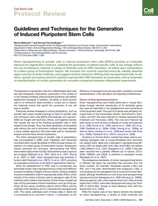
  • 7. selection was key to generating fully reprogrammed cells, ulti- mately leading to the discovery that selection methods were un- necessary and actually counterproductive (Blelloch et al., 2007; Maherali et al., 2007; Meissner et al., 2007). The identification of iPSC colonies based solely upon morpho- logical criteria requires a considerable degree of ESC expertise. In general, mouse ESC colonies can be distinguished by their re- fractive, or ‘‘shiny,’’ appearance and tight, well-defined borders, while human ESC colonies display a cobblestone appearance with prominent nucleoli and pronounced individual cell borders. The stepwise morphological changes that occur during reprog- ramming have been depicted in both systems (Figure 2). Of importance to note is that morphologically similar but non- iPSC colonies also arise during the course of human fibroblast reprogramming. These colonies are often mistaken for iPSCs, particularly by novices in the field, but are distinguishable from iPSCs becuase they are loose and granular in appearance, contain phase-bright cells, and appear earlier in the process (after 1–2 weeks) (Lowry et al., 2008; Takahashi et al., 2007b). Additional methods to identify iPSCs have been described; these techniques become useful when one is dealing with cell types that provide a high background of non-iPSC colonies (for example, those formed during human fibroblast reprogramming), or when ESC expertise is lacking. Two such methods have used ESC-specific surface antigen expression and loss of transgene dependence as strategies to identify reprogrammed cells. For example, isolation of the Thy-1-SSEA-1+ population during the course of mouse fibroblast reprogramming greatly enriches for cells poised to become iPSCs (Stadtfeld et al., 2008b), and live staining of cultures for the hESC-specific surface antigen Tra- 1-81 has aided in the identification of genuine hiPSC colonies de- rivedfromhumanfibroblasts(Lowryetal.,2008).Thelossoftrans- gene dependence, which correlates with full reprogramming, can be assessed through the use of reporters that are silenced in the pluripotent state (Stadtfeld et al., 2008b; Zhao et al., 2008), or through factorwithdrawal, whichrequiresaninducibleortransient delivery system. Upon withdrawal, cells that rely on continued factor expression are eliminated, thus permitting selective expan- sion of fully reprogrammed cells (Maherali et al., 2008). Expansion and Characterization of Cells The steps involved in taking a new colony to a fully established iPSC line are identical to those for ESC derivation, which have been described in detail elsewhere (Akutsu et al., 2006; Lerou et al., 2008; Nagy et al., 2003). Of particular importance to note, however, are the methods used to passage mouse and human iPSCs, as well as those used to ensure purity of the resulting iPSC lines. Mouse iPSCs/ESCs can withstand single-cell dissociation, and newly derived colonies can be immediately subjected to enzymatic passaging, thus facilitating their quick expansion into lines. Human iPSCs/ESCs, however, survive poorly as single cells, and initial passaging of new colonies must be done me- chanically; several passages (approximately five to ten) are required before the cells can be adapted to enzymatic dissocia- tion (Lerou et al., 2008). As hiPSCs/ESCs are highly prone to differentiation, especially within the first few passages, it is im- portant to continually remove differentiated structures to prevent 0d 6d3d 9d BA DC 17d16d14d 8d 12d10d 18d 19d 20d i ii Figure 2. Progressive Formation of iPSC Colonies (A) Time course of reprogramming in primary infected mouse fibroblasts. The arrow at 3d indi- cates a nascent colony. At day 9, two adjacent colonies are shown; only one has reactivated the endogenous Oct4-GFP reporter allele, represent- ing a true iPSC colony. Bottom right image de- picts iPSC colonies from an established line (Stadtfeld et al., 2008b). (B) Two representative colonies derived from pri- mary infected mouse fibroblasts. (Bi) iPSC colony, characterized by a tight well-defined border and Oct4-GFP expression; (Bii) differentiated colony, consisting of loosely packed cells and lacking expression of Oct4-GFP (Stadtfeld et al., 2008b). (C) Colony tracking during the reprogramming of secondary human fibroblasts with a doxycy- cline-inducible system. After doxycycline with- drawal at 14 days, colony regression occurs, and at day 17, a hESC-like colony is seen to emerge (indicated by arrow). This colony contin- ued to develop (denoted by arrows) and, by day 20, showed signs of differentiation typical for hESCs (Maherali et al., 2008). (D) Primary human iPSC colony, depicting the flat colony structure with cobblestone morphology (top panel). Lower panel illustrates the character- istic morphology with pronounced individual cell borders. Cell Stem Cell 3, December 4, 2008 ª2008 Elsevier Inc. 601 Cell Stem Cell Protocol Review
  • 8. them from being carried forward in the expansion. While use of a ROCK inhibitor can greatly facilitate hiPSC/hESC line expansion (Watanabe et al., 2007), it is not known to safeguard against differentiation and may result in carryover of differenti- ated cells; thus, its use is left to the discretion of the experi- menter. Both mouse and human iPSC cultures can harbor initial contamination with improperly reprogrammed or differentiated cells, and subcloning may be necessary to ensure the quality of newly derived lines (Maherali et al., 2007; Wernig et al., 2007). This has been of particular importance in iPSC clones that maintain transgene expression, for instance, in partially reprogrammed cells (Mikkelsen et al., 2008). Several criteria have been set forth to ascertain whether a fully reprogrammed state has been achieved, which include an array of unique features associated with pluripotency, encompassing morphological, molecular, and functional attributes (Figure 3). Morphologically, iPSCs must appear identical to ESCs and dem- onstrate unlimited self-renewal. Weeks 1-2 (106 cells available) - Analysis of pluripotency gene expression - RT-PCR,immunostaining - DNA methylation analysis - Bisulfite sequencing - Begin in vitro differentiation - Perform blastocyst injections (also tetraploid complementation) Weeks 14-16 - Determine germline transmission Weeks 6-8 - Analyze in vivo differentiation: Teratomas Chimeras (Tetraploid complementation) Weeks 2-3 - Analyze in vitro differentiation - Inject teratomas (~106 cells) - Other molecular assays: - Transcriptional profiling - Karyotype - ESC-like histone modifications (ChIP) - X chromosome reactivation (FISH) Weeks 10-12 - Mate to test germline transmission ~2 weeks 3-4 weeks Weeks 16-20 - Analyze teratomas Weeks 2-3 (20 colonies available) - Analysis of pluripotency gene expression - RT-PCR,immunostaining - DNA methylation analysis - Bisulfite sequencing - Begin in vitro differentiation Weeks 6-8 - Inject teratomas (~107 cells) - Other molecular assays: - Transcriptional profiling - Karyotype - ESC-like histone modifications (ChIP) - X chromosome reactivation (FISH) Weeks 4-6 - Analyze in vitro differentiation Picking of primary iPSC colonies (time = 0) 3 wks 6 wks 12 wks 18 wks Morphological analysis Continued - Analysis of self-renewal Continued - Analysis of self-renewal Figure 3. Timeline for the Characterization of Mouse and Human iPSC Lines Suggested time points for key assays are indi- cated, providing a rough outline for a general characterization of iPSCs. Red text indicates morphological assays, green indicates molecular assays, and blue indicates functional character- ization. ChIP, chromatin immunoprecipitation; FISH, fluorescent in situ hybridization. On a molecular level, iPSCs must dis- play gene expression profiles that are indistinguishable from ESCs, which ex- tends to the display of other associated features, including (1) protein-level ex- pression of key pluripotency factors (e.g., Oct4, Nanog) and ESC-specific surface antigens (e.g., SSEA-1 in mouse; SSEA- 3/-4,Tra-1-60/-81inhuman);(2)functional telomeraseexpression;and(3)expression of genes involved in retroviral silencing, such as de novo methyltransferases and Trim28 (Lei et al., 1996; Wolf and Goff, 2007). Accordingly, genuine iPSCs must be independent of transgene expression and thus lack expression of the delivered factors. iPSCs must also be epigenetically similar to ESCs, demonstrating DNA de- methylation at the promoters of pluripo- tency genes, X chromosome reactivation in female cells (Maherali et al., 2007; Ride- out et al., 2001), and the presence of bivalentdomainsatdevelopmentalgenes, consisting of overlapping histone modifi- cations that have opposing roles (Bern- stein et al., 2006; Maherali et al., 2007; Wernig et al., 2007). At a functional level, iPSCs must dem- onstrate the ability to differentiate into lin- eages from all three embryonic germ layers. A hierarchy of criteria has been put forth, and in order of increasing levels of stringency, these in- clude: (1) in vitro differentiation, (2) teratoma formation, (3) chi- mera contribution, (4) germline transmission, and (5) tetraploid complementation (direct generation of entirely ESC/iPSC-de- rived mice) (Jaenisch and Young, 2008). As performing all available assays for the demonstration of pluripotency is infeasible, a suggested minimal set of criteria should be fulfilled in order to ascertain that a genuine iPSC has been obtained. Accordingly, these include (1) all morphological attributes, including unlimited self-renewal; (2) expression of key pluripotency genes with a concomitant downregulation of lineage-specific genes associated with the cell of origin; (3) transgene independence; and (4) proof of functional differentia- tion through the highest-stringency test acceptable. In mouse, the accepted standard is germline transmission, demonstrating the competence of iPSCs to contribute to all line- ages including germ cells and ultimately giving rise to offspring. While tetraploid complementation remains the most stringent 602 Cell Stem Cell 3, December 4, 2008 ª2008 Elsevier Inc. Cell Stem Cell Protocol Review
  • 9. functional assay, iPSCs, unlike ESCs, have not been reported to autonomously generate full-term mice. It is not yet clear whether this reflects a fundamental issue in reprogramming, such as an inability to fully reset the epigenome of a somatic cell, or whether it is a technical issue that remains to be addressed, such as the presence of proviral integrations, the identity of the starting cell type, or the passage number of the iPSCs. Further experiments are therefore needed to truly ascertain whether iPSCs are capable of fulfilling this criterion. For a functional assessment of pluripotency in human cells, teratoma formation should be demonstrated and include both histological and immunohistochemical analysis to confirm the presence of structures derived from all three germ layers (Gertow et al., 2007). While the ability to form teratomas is con- sidered the most stringent assay for human cells, it is not as rigorous as the assays available for mouse cells. For instance, first-generation mouse iPSCs, though able to form teratomas, could not give rise to live-born chimeras (Takahashi and Yama- naka, 2006), indicating that additional assays would be benefi- cial in testing the functional differentiation capacity of hiPSCs. As such, directed differentiation efforts have introduced robust in vitro assays coupled with transplant models to assess the function of specific cell types derived in vitro from hiPSCs/ hESCs (Kroon et al., 2008; Lee et al., 2007; Mummery et al., 2003). In addition to the demonstration of pluripotency, it is crucial to ensure that the resulting iPSCs are free from genetic aberrations. Cells cultured for long periods of time can become genetically unstable, particularly human ESCs in that they have a tendency to acquire abnormal karyotypes (Draper et al., 2004; Lerou et al., 2008). Thus, testing iPSC lines periodically for genetic lesions is important for proper maintenance of the line. Calculation and Reporting of Reprogramming Efficiencies An accurate assessment of reprogramming efficiency is critical for proper comparison between individual experiments, particu- larly in the interpretation of optimization procedures and the reprogramming of different cell types. Most iPSC derivations have reported the frequency of colony formation, which is an indication of the number of colonies formed per number of cells seeded, yet the large range in reported values indicates that this measure portrays an incomplete view of derivation effi- ciency, thus requiring other variables to be factored into the calculations. Differences in factor delivery strongly contribute to discrep- ancies between independent experiments. For example, variabil- ity in viral titer leads to quantitative differences in factor delivery, thus altering the proportion of cells that receive all factors and ultimately leading to a change in the frequency of colony forma- tion. Other contributing factors include the plating efficiency of cells, cell survival (encompassing proliferation and cell death/ apoptosis), and the counting of sister clones (multiple iPSC colonies derived from an individual cell). The specific methods used to quantify colony number often differ between groups, which also strongly contributes to varia- tion in the reported efficiencies. Such methods include morphol- ogy-based counts, alkaline phosphatase (AP) activity, immunos- taining, transgenic and knockin reporter allele expression, and the ability of a colony to form an iPSC line; all methods vary in their accuracy to reflect genuine iPSC colonies. While the ability to form an iPSC line is the most stringent quantification method, it is laborious and not feasible for large colony numbers. The use of knockin reporter alleles or immunostaining for endogenous pluripotency gene expression are suitable surrogate methods; however, morphology-based identification or AP activity is not sufficient for denoting true pluripotent cell colonies (Brambrink et al., 2008). Transgene-based reporter methods should also be used cautiously, as expression of such alleles is not subject to the same regulation as endogenous knockin reporter alleles. This has been exemplified by the kinetics of Oct4 reporter gene reactivation during cell fusion, which occurs much faster with a transgenic allele than an endogenous knockin allele (Do and Scholer, 2004; Maherali et al., 2007). A number of steps can be taken to reduce variability in the reported frequencies and gain the most accurate and reliable estimate of the true reprogramming efficiency. These include (1) controlling for factor delivery through assessment of expres- sion and coinfectivity and to report values as a fraction of the cells expressing all factors, rather than the total number of input cells; (2) calculation of plating efficiency, which can be done via cell counts or single-cell plating; (3) eliminating the count of sister clones through single-cell plating or retrospective analysis of integration patterns; and (4) use of a reliable and stringent method to identify and quantify iPSC colonies. Such standardi- zation between methods will greatly facilitate the interpretation and comparison of independent experiments and, in turn, accel- erate progress in the field. Concluding Remarks The generation of iPSCs represents a major advance in the field of regenerative medicine and provides a powerful tool for the study of cell-fate transitions. While new techniques and insights are continually unveiled, the foundation of iPSC derivation rests upon successful manipulation of a core set of transcription factors. The key steps involved in this process consist of the choice of factors and molecules used, their delivery method, and the choice of target cell type, as well as the parameters of factor expression, culture conditions, methods to identify cells, and the assays used to verify pluripotency. To fully exploit the abundance of new information requires a standardization of certain parameters of the reprogramming process, such as the calculation of reprogramming efficiency and qualification of the pluripotent state. As such, this review has attempted to present a comprehensive comparison of the currently available technologies for iPSC derivation and put forth standards to minimize variability between independent experiments, thus providing a framework to aid in the designing and conducting of future experiments, as well in the evaluation of existing iPSC literature. ACKNOWLEDGMENTS We thank Todd Meyerrose, Jose Polo, Matthias Stadtfeld, and Jochen Utikal for discussion and critical reading of the manuscript, and Tom DeCesare for figure contributions. N.M. is supported by the Natural Sciences and Engineer- ing Research Council of Canada and Alberta Scholarship Programs. K.H. is supported by an NIH Director’s Innovator Award, the Harvard Stem Cell Institute, the Kimmel Foundation, and the V Foundation. Cell Stem Cell 3, December 4, 2008 ª2008 Elsevier Inc. 603 Cell Stem Cell Protocol Review
  • 10. REFERENCES Aasen, T., Raya, A., Barrero, M.J., Garreta, E., Consiglio, A., Gonzalez, F., Vassena, R., Billic, J., Pekarik, V., Tiscornia, G., et al. (2008). Efficient and rapid generation of induced pluripotent stem cells from human keratinocytes. Nat. Biotechnol. 26, 1276–1284. Akutsu, H., Cowan, C.A., and Melton, D. (2006). Human embryonic stem cells. Methods Enzymol. 418, 78–92. Amit, M., and Itskovitz-Eldor, J. (2006). Maintenance of human embryonic stem cells in animal serum- and feeder layer-free culture conditions. Methods Mol. Biol. 331, 105–113. Aoi, T., Yae, K., Nakagawa, M., Ichisaka, T., Okita, K., Takahashi, K., Chiba, T., and Yamanaka, S. (2008). Generation of pluripotent stem cells from adult mouse liver and stomach cells. Science 321, 699–702. Bernstein, B.E., Mikkelsen, T.S., Xie, X., Kamal, M., Huebert, D.J., Cuff, J., Fry, B., Meissner, A., Wernig, M., Plath, K., et al. (2006). A bivalent chromatin structure marks key developmental genes in embryonic stem cells. Cell 125, 315–326. Blelloch, R., Venere, M., Yen, J., and Ramalho-Santos, M. (2007). Generation of induced pluripotent stem cells in the absence of drug selection. Cell Stem Cell 1, 245–247. Bosnali, M., and Edenhofer, F. (2008). Generation of transducible versions of transcription factors Oct4 and Sox2. Biol. Chem. 389, 851–861. Brambrink, T., Foreman, R., Welstead, G.G., Lengner, C.J., Wernig, M., Suh, H., and Jaenisch, R. (2008). Sequential expression of pluripotency markers during direct reprogramming of mouse somatic cells. Cell Stem Cell 2, 151–159. Cheng, J., Dutra, A., Takesono, A., Garrett-Beal, L., and Schwartzberg, P.L. (2004). Improved generation of C57BL/6J mouse embryonic stem cells in a defined serum-free media. Genesis 39, 100–104. Cowan, C.A., Klimanskaya, I., McMahon, J., Atienza, J., Witmyer, J., Zucker, J.P., Wang, S., Morton, C.C., McMahon, A.P., Powers, D., et al. (2004). Deri- vation of embryonic stem-cell lines from human blastocysts. N. Engl. J. Med. 350, 1353–1356. Cowan, C.A., Atienza, J., Melton, D.A., and Eggan, K. (2005). Nuclear reprog- ramming of somatic cells after fusion with human embryonic stem cells. Science 309, 1369–1373. Dimos, J.T., Rodolfa, K.T., Niakan, K.K., Weisenthal, L.M., Mitsumoto, H., Chung, W., Croft, G.F., Saphier, G., Leibel, R., Goland, R., et al. (2008). Induced pluripotent stem cells generated from patients with ALS can be differentiated into motor neurons. Science 321, 1218–1221. Do, J.T., and Scholer, H.R. (2004). Nuclei of embryonic stem cells reprogram somatic cells. Stem Cells 22, 941–949. Draper, J.S., Smith, K., Gokhale, P., Moore, H.D., Maltby, E., Johnson, J., Meisner, L., Zwaka, T.P., Thomson, J.A., and Andrews, P.W. (2004). Recurrent gain of chromosomes 17q and 12 in cultured human embryonic stem cells. Nat. Biotechnol. 22, 53–54. Eminli, S., Utikal, J.S., Arnold, K., Jaenisch, R., and Hochedlinger, K. (2008). Reprogramming of Neural Progenitor Cells into iPS Cells in the Absence of Exogenous Sox2 Expression. Stem Cells 26, 2467–2474. Gaudet, F., Hodgson, J.G., Eden, A., Jackson-Grusby, L., Dausman, J., Gray, J.W., Leonhardt, H., and Jaenisch, R. (2003). Induction of tumors in mice by genomic hypomethylation. Science 300, 489–492. Gertow, K., Przyborski, S., Loring, J.F., Auerbach, J.M., Epifano, O., Oton- koski, T., Damjanov, I., and Ahrlund-Richter, L. (2007). Isolation of human embryonic stem cell-derived teratomas for the assessment of pluripotency. Curr. Protoc. Stem Cell Biol. Chapter 1, Unit1B.4. Gump, J.M., and Dowdy, S.F. (2007). TAT transduction: the molecular mech- anism and therapeutic prospects. Trends Mol. Med. 13, 443–448. Hanna, J., Markoulaki, S., Schorderet, P., Carey, B.W., Beard, C., Wernig, M., Creyghton, M.P., Steine, E.J., Cassady, J.P., Foreman, R., et al. (2008). Direct reprogramming of terminally differentiated mature B lymphocytes to pluripo- tency. Cell 133, 250–264. Hochedlinger, K., and Jaenisch, R. (2006). Nuclear reprogramming and pluripotency. Nature 441, 1061–1067. Hochedlinger, K., Yamada, Y., Beard, C., and Jaenisch, R. (2005). Ectopic ex- pression of Oct-4 blocks progenitor-cell differentiation and causes dysplasia in epithelial tissues. Cell 121, 465–477. Hockemeyer, D., Soldner, F., Cook, E.G., Gao, Q., Mitalipova, M., and Jaenisch, R. (2008). A drug-inducible system for direct reprogramming of human somatic cells to pluripotency. Cell Stem Cell 3, 346–353. Huangfu, D., Maehr, R., Guo, W., Eijkelenboom, A., Snitow, M., Chen, A.E., and Melton, D.A. (2008a). Induction of pluripotent stem cells by defined factors is greatly improved by small-molecule compounds. Nat. Biotechnol. 26, 795–797. Huangfu, D., Osafune, K., Maehr, R., Guo, W., Eijkelenboom, A., Chen, S., Muhlestein, W., and Melton, D.A. (2008b). Induction of pluripotent stem cells from primary human fibroblasts with only Oct4 and Sox2. Nat. Biotechnol. 26, 1269–1275. Jackson-Grusby, L., Laird, P.W., Magge, S.N., Moeller, B.J., and Jaenisch, R. (1997). Mutagenicity of 5-aza-20 -deoxycytidine is mediated by the mammalian DNA methyltransferase. Proc. Natl. Acad. Sci. USA 94, 4681–4685. Jaenisch, R., and Young, R. (2008). Stem cells, the molecular circuitry of pluripotency and nuclear reprogramming. Cell 132, 567–582. Jahner, D., Stuhlmann, H., Stewart, C.L., Harbers, K., Lohler, J., Simon, I., and Jaenisch, R. (1982). De novo methylation and expression of retroviral genomes during mouse embryogenesis. Nature 298, 623–628. Kim, J.B., Zaehres, H., Wu, G., Gentile, L., Ko, K., Sebastiano, V., Arauzo- Bravo, M.J., Ruau, D., Han, D.W., Zenke, M., et al. (2008). Pluripotent stem cells induced from adult neural stem cells by reprogramming with two factors. Nature 454, 646–650. Kroon, E., Martinson, L.A., Kadoya, K., Bang, A.G., Kelly, O.G., Eliazer, S., Young, H., Richardson, M., Smart, N.G., Cunningham, J., et al. (2008). Pancre- atic endoderm derived from human embryonic stem cells generates glucose- responsive insulin-secreting cells in vivo. Nat. Biotechnol 26, 443–452. Kustikova, O., Fehse, B., Modlich, U., Yang, M., Dullmann, J., Kamino, K., von Neuhoff, N., Schlegelberger, B., Li, Z., and Baum, C. (2005). Clonal dominance of hematopoietic stem cells triggered by retroviral gene marking. Science 308, 1171–1174. Lee, H., Shamy, G.A., Elkabetz, Y., Schofield, C.M., Harrsion, N.L., Panagiota- kos, G., Socci, N.D., Tabar, V., and Studer, L. (2007). Directed differentiation and transplantation of human embryonic stem cell-derived motoneurons. Stem Cells 25, 1931–1939. Lei, H., Oh, S.P., Okano, M., Juttermann, R., Goss, K.A., Jaenisch, R., and Li, E. (1996). De novo DNA cytosine methyltransferase activities in mouse embry- onic stem cells. Development 122, 3195–3205. Lerou, P.H., Yabuuchi, A., Huo, H., Miller, J.D., Boyer, L.F., Schlaeger, T.M., and Daley, G.Q. (2008). Derivation and maintenance of human embryonic stem cells from poor-quality in vitro fertilization embryos. Nat. Protocols 3, 923–933. Liu, H., Zhu, F., Yong, J., Zhang, P., Hou, P., Li, H., Jiang, W., Cai, J., Liu, M., Cui, K., et al. (2008). Generation of induced pluripotent stem cells from adult rhesus monkey fibroblasts. Cell Stem Cell 3, this issue, 587–590. Lois, C., Hong, E.J., Pease, S., Brown, E.J., and Baltimore, D. (2002). Germline transmission and tissue-specific expression of transgenes delivered by lentivi- ral vectors. Science 295, 868–872. Lowry, W.E., Richter, L., Yachechko, R., Pyle, A.D., Tchieu, J., Sridharan, R., Clark, A.T., and Plath, K. (2008). Generation of human induced pluripotent stem cells from dermal fibroblasts. Proc. Natl. Acad. Sci. USA 105, 2883–2888. Maherali, N., Sridharan, R., Xie, W., Utikal, J., Eminli, S., Arnold, K., Stadtfeld, M., Yachechko, R., Tchieu, J., Jaenisch, R., et al. (2007). Directly reprog- rammed fibroblasts show global epigenetic remodeling and widespread tissue contribution. Cell Stem Cell 1, 55–70. Maherali, N., Ahfeldt, T., Rigamonti, A., Utikal, J., Cowan, C., and Hochedlin- ger, K. (2008). A high-efficiency system for the generation and study of human induced pluripotent stem cells. Cell Stem Cell 3, 340–345. 604 Cell Stem Cell 3, December 4, 2008 ª2008 Elsevier Inc. Cell Stem Cell Protocol Review
  • 11. Marson, A., Levine, S.S., Cole, M.F., Frampton, G.M., Brambrink, T., John- stone, S., Guenther, M.G., Johnston, W.K., Wernig, M., Newman, J., et al. (2008). Connecting microRNA genes to the core transcriptional regulatory circuitry of embryonic stem cells. Cell 134, 521–533. Meissner, A., Wernig, M., and Jaenisch, R. (2007). Direct reprogramming of genetically unmodified fibroblasts into pluripotent stem cells. Nat. Biotechnol. 25, 1177–1181. Mikkelsen, T.S., Hanna, J., Zhang, X., Ku, M., Wernig, M., Schorderet, P., Bernstein, B.E., Jaenisch, R., Lander, E.S., and Meissner, A. (2008). Dissecting direct reprogramming through integrative genomic analysis. Nature 454, 49–55. Miller, D.G., Adam, M.A., and Miller, A.D. (1990). Gene transfer by retrovirus vectors occurs only in cells that are actively replicating at the time of infection. Mol. Cell. Biol. 10, 4239–4242. Mummery, C., Ward-van Oostwaard, D., Doevendans, P., Spijker, R., van den Brink, S., Hassink, R., van der Heyden, M., Opthof, T., Pera, M., de la Riviere, A.B., et al. (2003). Differentiation of human embryonic stem cells to cardiomyo- cytes: role of coculture with visceral endoderm-like cells. Circulation 107, 2733–2740. Nagy, A., Gertsenstein, M., and Vintersten, K. (2003). Manipulating the Mouse Embryo: A Laboratory Manual. 764. Nakagawa, M., Koyanagi, M., Tanabe, K., Takahashi, K., Ichisaka, T., Aoi, T., Okita, K., Mochiduki, Y., Takizawa, N., and Yamanaka, S. (2008). Generation of induced pluripotent stem cells without Myc from mouse and human fibro- blasts. Nat. Biotechnol. 26, 101–106. Naldini, L., Blomer, U., Gallay, P., Ory, D., Mulligan, R., Gage, F.H., Verma, I.M., and Trono, D. (1996). In vivo gene delivery and stable transduction of nondividing cells by a lentiviral vector. Science 272, 263–267. Nightingale, S.J., Hollis, R.P., Pepper, K.A., Petersen, D., Yu, X.J., Yang, C., Bahner, I., and Kohn, D.B. (2006). Transient gene expression by nonintegrating lentiviral vectors. Mol. Ther. 13, 1121–1132. Okita, K., Ichisaka, T., and Yamanaka, S. (2007). Generation of germline- competent induced pluripotent stem cells. Nature 448, 313–317. Okita, K., Nakagawa, M., Hyenjong, H., Ichisaka, T., and Yamanaka, S. (2008). Generation of Mouse Induced Pluripotent Stem Cells Without Viral Vectors. Science 322, 949–953. Park, I.H., Arora, N., Huo, H., Maherali, N., Ahfeldt, T., Shimamura, A., Lensch, M.W., Cowan, C., Hochedlinger, K., and Daley, G.Q. (2008a). Disease-specific induced pluripotent stem cells. Cell 134, 877–886. Park, I.H., Lerou, P.H., Zhao, R., Huo, H., and Daley, G.Q. (2008b). Generation of human-induced pluripotent stem cells. Nat. Protocols 3, 1180–1186. Park, I.H., Zhao, R., West, J.A., Yabuuchi, A., Huo, H., Ince, T.A., Lerou, P.H., Lensch, M.W., and Daley, G.Q. (2008c). Reprogramming of human somatic cells to pluripotency with defined factors. Nature 451, 141–146. Ramezani, A., and Hawley, R.G. (2002). Overview of the HIV-1 lentiviral vector system. Curr. Protoc. Stem Cell Biol. Chapter 16, Unit 16.21. Rideout, W.M., III, Eggan, K., and Jaenisch, R. (2001). Nuclear cloning and epigenetic reprogramming of the genome. Science 293, 1093–1098. Rosenzweig, A. (2007). Vectors for gene therapy. Curr. Protoc. Hum. Genet. Chapter 12. Shi, Y., Desponts, C., Do, J.T., Hahm, H.S., Scholer, H.R., and Ding, S. (2008a). Induction of pluripotent stem cells from mouse embryonic fibroblasts by Oct4 and Klf4 with small-molecule compounds. Cell Stem Cell 3, 568–574. Shi, Y., Do, J.T., Desponts, C., Hahm, H.S., Scholer, H.R., and Ding, S. (2008b). A combined chemical and genetic approach for the generation of induced pluripotent stem cells. Cell Stem Cell 2, 525–528. Silva, J., Barrandon, O., Nichols, J., Kawaguchi, J., Theunissen, T.W., and Smith, A. (2008). Promotion of reprogramming to ground state pluripotency by signal inhibition. PLoS Biol. 6, e253. Stadtfeld, M., Brennand, K., and Hochedlinger, K. (2008a). Reprogramming of pancreatic beta cells into induced pluripotent stem cells. Curr. Biol. 18, 890–894. Stadtfeld, M., Maherali, N., Breault, D.T., and Hochedlinger, K. (2008b). Defin- ing molecular cornerstones during fibroblast to iPS cell reprogramming in mouse. Cell Stem Cell 2, 230–240. Stadtfeld, M., Nagaya, M., Utikal, J., Weir, G., and Hochedlinger, K. (2008c). Induced Pluripotent Stem Cells Generated Without Viral Integration. Science 322, 945–949. Tada, M., Takahama, Y., Abe, K., Nakatsuji, N., and Tada, T. (2001). Nuclear reprogramming of somatic cells by in vitro hybridization with ES cells. Curr. Biol. 11, 1553–1558. Takahashi, K., and Yamanaka, S. (2006). Induction of pluripotent stem cells from mouse embryonic and adult fibroblast cultures by defined factors. Cell 126, 663–676. Takahashi, K., Okita, K., Nakagawa, M., and Yamanaka, S. (2007a). Induction of pluripotent stem cells from fibroblast cultures. Nat. Protocols 2, 3081–3089. Takahashi, K., Tanabe, K., Ohnuki, M., Narita, M., Ichisaka, T., Tomoda, K., and Yamanaka, S. (2007b). Induction of pluripotent stem cells from adult human fibroblasts by defined factors. Cell 131, 861–872. Tiscornia, G., Singer, O., and Verma, I.M. (2006). Production and purification of lentiviral vectors. Nat. Protocols 1, 241–245. Varas, F., Stadtfeld, M., De Andres-Aguayo, L., Maherali, N., di Tullio, A., Pantano, L., Notredame, C., Hochedlinger, K., and Graf, T. (2008). Fibroblast derived induced pluripotent stem cells show no common retroviral vector insertions. Stem Cells. Published online November 13, 2008. 10.1634/stem- cells.2008-0696. Wakayama, T., Perry, A.C., Zuccotti, M., Johnson, K.R., and Yanagimachi, R. (1998). Full-term development of mice from enucleated oocytes injected with cumulus cell nuclei. Nature 394, 369–374. Watanabe, K., Ueno, M., Kamiya, D., Nishiyama, A., Matsumura, M., Wataya, T., Takahashi, J.B., Nishikawa, S., Nishikawa, S., Muguruma, K., et al. (2007). A ROCK inhibitor permits survival of dissociated human embryonic stem cells. Nat. Biotechnol. 25, 681–686. Wernig, M., Meissner, A., Foreman, R., Brambrink, T., Ku, M., Hochedlinger, K., Bernstein, B.E., and Jaenisch, R. (2007). In vitro reprogramming of fibroblasts into a pluripotent ES-cell-like state. Nature 448, 318–324. Wernig, M., Lengner, C.J., Hanna, J., Lodato, M.A., Steine, E., Foreman, R., Staerk, J., Markoulaki, S., and Jaenisch, R. (2008a). A drug-inducible trans- genic system for direct reprogramming of multiple somatic cell types. Nat. Biotechnol. 26, 916–924. Wernig, M., Meissner, A., Cassady, J.P., and Jaenisch, R. (2008b). c-Myc is dispensable for direct reprogramming of mouse fibroblasts. Cell Stem Cell 2, 10–12. Wolf, D., and Goff, S.P. (2007). TRIM28 mediates primer binding site-targeted silencing of murine leukemia virus in embryonic cells. Cell 131, 46–57. Ying, Q.L., Wray, J., Nichols, J., Batlle-Morera, L., Doble, B., Woodgett, J., Cohen, P., and Smith, A. (2008). The ground state of embryonic stem cell self-renewal. Nature 453, 519–523. Yu, J., Vodyanik, M.A., Smuga-Otto, K., Antosiewicz-Bourget, J., Frane, J.L., Tian, S., Nie, J., Jonsdottir, G.A., Ruotti, V., Stewart, R., et al. (2007). Induced pluripotent stem cell lines derived from human somatic cells. Science 318, 1917–1920. Zhao, Y., Yin, X., Qin, H., Zhu, F., Liu, H., Yang, W., Zhang, Q., Xiang, C., Hou, P., Song, Z., et al. (2008). Two supporting factors greatly improve the efficiency of human iPSC generation. Cell Stem Cell 3, 475–479. Cell Stem Cell 3, December 4, 2008 ª2008 Elsevier Inc. 605 Cell Stem Cell Protocol Review