1. Cell Stem Cell
Review
Interspecies Nuclear Transfer:
Implications for Embryonic Stem Cell Biology
Zeki Beyhan,1 Amy E. Iager,1 and Jose B. Cibelli1,2,3,*
1Cellular Reprogramming Laboratory, Department of Animal Science
2Department of Physiology
Michigan State University, B270 Anthony Hall, East Lansing, MI 48824, USA
3Programa Andaluz de Terapia Celular y Medicina Regenerativa, Avenida Ame´ rico Vespucio 5, Bloque 2, 2a Planta 41092 Isla
de la Cartuja, Sevilla, Andalucı´a, Spain
*Correspondence: cibelli@msu.edu
DOI 10.1016/j.stem.2007.10.009
Accessibility of human oocytes for research poses a serious ethical challenge to society. This fact
categorically holds true when pursuing some of the most promising areas of research, such as
somatic cell nuclear transfer and embryonic stem cell studies. One approach to overcoming this
limitation is to use an oocyte from one species and a somatic cell from another. Recently, several
attempts to capture the promises of this approach have met with varying success, ranging from es-
tablishing human embryonic stem cells to obtaining live offspring in animals. This review focuses on
the challenges and opportunities presented by the formidable task of overcoming biological differ-
ences among species.
Introduction
The oocyte is invaluable. This is an unmistakable fact
underscored by the scarcity of oocytes available from
species where they could be most useful, i.e., human,
for biomedical applications, and endangered or extinct
species, for their conservation and rescue. One of the pos-
sibilities presented by somatic cell nuclear transfer (SCNT)
is so-called interspecies cloning, where the recipient
ooplast and donor nucleus are derived from different spe-
cies. Hypothetically, having the means to take advantage
of readily available recipient oocytes to reprogram the do-
nor nucleus of a different species holds tremendous
promise.
Perhaps the most important prospective application of
interspecies SCNT (iSCNT) lies in its potential to facilitate
reprogramming of human somatic cells without many of
the significant ethical challenges surrounding the use of
human oocytes. Ethical considerations aside, the ques-
tion of availability makes SCNT using human oocytes
unfeasible as a long-term solution for cellular reprogram-
ming. While several alternative strategies such as egg
sharing and egg donation programs are suggested as
the source of human oocytes for assisted reproductive
technologies, neither of these approaches has been
implemented broadly enough to overcome the shortage
of human oocytes facing the research and therapeutic
development community. As an alternative approach,
producing competent human oocytes from human ESCs
has not been realized yet either (Hubner et al., 2003).
These constraints draw attention to iSCNT as a workable
strategy to address human oocyte shortage for ESC stud-
ies. The recent ruling by the Human Fertilization Embryol-
ogy Authority (HFEA)—the government body in charge of
overseeing IVF treatments and research using human
embryos in the UK—to allow iSCNT experiments using
human somatic cells and nonhuman oocytes offers scien-
tists the long-awaited legal framework to explore the po-
tential of the iSCNT procedure to its full extent. This bold
measure taken by the HEFA, and upheld by a large group
of scientists, implicitly recognizes the irreplaceable nature
of the mammalian oocyte (http://www.hfea.gov.uk/en/
1581.html).
The use of nonhuman oocytes for reprogramming could
be of immediate value as a tool for the production of
human-nuclear-transfer-derived embryonic stem cells
(NTESCs) from individuals suffering from such late onset
diseases as diabetes, Parkinson’s disease, and Alz-
heimer’s disease, among others. In turn, these cells could
be used to develop new treatments in vitro.
We reported the first iSCNT experiments shortly after
the cloning of a lamb from somatic cells (Wilmut et al.,
1997; Dominko et al., 1999). Since then, the feasibility of
interspecies cloning has been addressed by several
researchers employing various model systems. More
than 40 articles have been published in which oocytes
and somatic cells from a number of species have been
used to generate embryos via interspecies nuclear trans-
fer (Table 1). Live offspring have been obtained by com-
bining closely related species, such as cattle/gaur (Bos
taurus/Bos grunensis) (Lanza et al., 2000) and domestic
sheep/argali sheep (O. aries/O. musimon) (White et al.,
1999). In some of the reported experiments, however,
genetic distance between donor and recipient species
spanned taxonomic classes, such as cattle/chicken (Bos
taurus/Gallus gallus) (Liu et al., 2004) and rabbit/panda
(Oryctolagus cuniculus/A. melanoleuca) (Chen et al.,
2002). The majority of these experiments have failed to
produce viable embryos. A common limitation in making
comparisons between these iSCNT reports is that the
definition of experimental endpoints and criteria for
502 Cell Stem Cell 1, November 2007 ª2007 Elsevier Inc.
2. successful reprogramming was often ill-defined, except
for those resulting in live offspring. Nevertheless, the
potential impact of a successful iSCNT scheme is suffi-
ciently attractive to maintain ample scientific interest in
this subject. In this review, we will summarize the recent
literature on iSCNT and address a number of technical
and theoretical concerns regarding these experiments.
We also propose an outline for the evaluation of iSCNT
experiments, given that many reports in the literature to
date lack a common framework to gauge and compare
developmental outcomes.
What Is Interspecies SCNT?
A ‘‘species,’’ the basic unit of taxonomic classification, is
defined as a group of organisms that share certain pheno-
typical characteristics, forming a reproductively isolated
entity. In practice, however, it is not always easy to draw
lines between different species, and researchers have
had to define subgroups and transitory groups like sub-
species and breeds. For the sake of simplicity, we will
define the term species as a group of organisms that could
interbreed naturally and produce fertile offspring. We will
also use an abridged hierarchy of taxonomic units in our
discussions.
As previously mentioned, nuclear transfer (NT) experi-
ments that employ oocytes and donor cells from two
different species are defined as interspecies or interspe-
cies nuclear transfers (iSCNT) (Figure 1). The two main
assumptions required for iSCNT are that early develop-
mental events and mechanisms are evolutionarily con-
served among mammals and that molecules that regu-
late these events in mammalian oocytes are capable of
interacting with nuclei from another species. The validity
of these assumptions, however, deserves vigorous scru-
tiny. Although most mammalian embryos follow a very
similar pattern of ontogenic development, significant dif-
ferences in many aspects of the process do exist among
evolutionarily divergent taxonomic groups (Gilbert and
Bolker, 2001). Temporal regulation of developmental
events—such as cell-cycle progression, embryonic
genome activation (EGA), blastocyst formation, implan-
tation, and organogenesis—differs from species to spe-
cies. One wonders, therefore, how these developmental
processes are regulated in an embryo reconstructed us-
ing an oocyte and a donor cell from different species.
What constituent of this unusual embryo drives the devel-
opment? Is there crosstalk between the donor nucleus
and the recipient cytoplasm? What developmental pro-
gram is executed—the oocyte’s, the donor’s, or both?
Are the interspecies cybrid cells functional and viable?
Is the resulting embryo/fetus viable? Are any live
offspring produced, and, if so, are they fertile? These
are some of the most important questions that need to
be, and can be, addressed by interspecies cloning
experiments.
Potential Applications of iSCNT
Theoretically, an iSCNT approach would be beneficial in
any situation in which an alternative source of ooplasm
is needed, due to either ethical or technical consider-
ations, such as establishing primate ESCs from iSCNT
embryos or cloning endangered species, respectively.
The ultimate endpoints of these applications are either
(1) to generate a preimplantation embryo to be used as
a source of ESCs or (2) to produce live offspring in all
animals with the exception of human.
Embryos cloned for ESC establishment need not prog-
ress through all developmental stages. Instead, a few
functional equivalents of inner cell mass (ICM) cells or sin-
gle blastomeres in an SCNT embryo could be adequate to
establish an ESC line (Klimanskaya et al., 2006, 2007;
Wakayama et al., 2001). This concept could be applied
to iSCNT (Figure 1), assuming that the pattern of gene
expression of the interspecies embryos approximates
that of same-species SCNTs or of fertilized preimplanta-
tion embryos at the same stage of development. iSCNT
has been proposed for creating human ESC lines in an ef-
fort to avoid the ethically charged issue of soliciting human
oocytes for research purposes (Fulka and Mrazek, 2004).
While this alternative may alleviate some ethical and prac-
tical concerns, the use of such cells for therapeutic pur-
poses is in doubt, due to potential risks of transmission
of infectious diseases. Further, ECSs isolated from cybrids
may maintain mitochondria derived from the nonhuman
recipient oocyte, a result that is likely to have deleteri-
ous long-term physiological and immunological conse-
quences to human recipients (Hall et al., 2006). Nonethe-
less, the ability to produce ESCs from iSCNT embryos
could facilitate the creation of new cellular models of
human disease and could significantly advance our under-
standing of basic nuclear-cytoplasmic interactions be-
tween a somatic cell and an oocyte.
Using iSCNT to produce live offspring, a goal of some
of the earliest iSCNT experiments, focuses primarily on
applications involving the preservation/rescue of endan-
gered species (Dominko et al., 1998). Although the main
procedures to construct cloned embryos for any purpose
are essentially the same, embryos cloned to produce live
offspring require a more comprehensive and complete
reprogramming of the somatic genome, since they need
to progress through all developmental milestones and
survive a rigorous in vivo selection process. A few studies
have successfully employed iSCNT in cloning endangered
species, such as gaur (Lanza et al., 2000), mouflon (Loi
et al., 2001), and African wild cat (Hidalgo et al., 2003),
using oocytes from closely related species.
Preimplantation and Postimplantation
Development of iSCNT Embryos
The majority of iSCNT experiments published to date have
reported the production of at least blastocyst-stage
embryos, although with varying degrees of efficiency (Ta-
ble 1). Development of blastocyst-stage iSCNT embryos
was reported even with some crossclass NT experiments
in which the donor species exhibits no functional equiva-
lent of the blastocyst stage during normal embryonic
development (Liu et al., 2004). Depending on the species
and the experiment, the frequency of blastocyst
Cell Stem Cell 1, November 2007 ª2007 Elsevier Inc. 503
Cell Stem Cell
Review
3. Table 1. Comprehensive List of Interspecies Cloning Experiments Reported to Date
Taxonomic
Relationship Recipient Oocyte Donor Cell Blastocyst Implantation
Live
Offspring Reference
Interclass Cow (B. taurus) Chicken (G. gallus) YES NET NA Liu et al., 2004
Cow (B. taurus) Rat (R. norvegicus) NAa
NO NA Dominko et al., 1999
Rabbit (O. cuniculus) Panda
(A. melanoleuca)
YES YES NO Chen et al., 2002
Interorder Cow (B. taurus) Whale
(B. acutorostrata)
NO NA NA Ikumi et al., 2004
Cow (B. taurus) Dog (C. familiaris) YES NET NA Murakami et al., 2005
Cow (B. taurus) Human
(H. sapiens)
YES NET NA Chang et al., 2003;
Illmensee et al., 2006
Cow (B. taurus) Rhesus monkey
(M. mulatta)
YES NET NA Dominko et al., 1999
Cow (B. taurus) Mouse
(M. musculus)
NO NA NA Arat et al., 2003
Cow (B. taurus) Pig (S. sucrofa) YES NO NA Dominko et al., 1999
Rabbit (O. cuniculus) Ibex (C. ibex) YES NET NA Jiang et al., 2005
Rabbit (O. cuniculus) Domestic cat
(F. catus)
YES YES NO Wen et al., 2005
Rabbit (O. cuniculus) Marbled cat
(P. marmorata)
YES NET NA Thongphakdee
et al., 2006
Rabbit (O. cuniculus) Human (H. sapiens) YES NET NA Chen et al., 2003
Rabbit (O. cuniculus) Rhesus monkey
(M. mulatta)
YES NET NA Yang et al., 2003
Rabbit (O. cuniculus) Camel
(C. dromedaries)
YES NET NA Zhao et al., 2006
Rabbit (O. cuniculus) Pig (S. sucrofa) YES NET NA Chen et al., 2006
Rabbit (O. cuniculus) Tbetan antelope
(P. hodgsonii)
YES NET NA Zhao et al., 2006
Pig (S. sucrofa) Whale
(B. acutorostrata)
NO NA NA Ikumi et al., 2004
Pig (S. sucrofa) Tiger (P. tigris) YES NET NA Hashem et al., 2007
Interfamily Cow (B. taurus) Takin (B. taxicolor) YES NET NA Li et al., 2006a
Cow (B. taurus) Sheep (O. aries) YES YES NO Dominko et al., 1999;
Hua et al., 2007
Goat (C. hirus) Tibetan antelope
(P. hodgsonii)
YES NET NA Zhao et al., 2007
Intergenus Cow (B. taurus) Buffalo (B. bubalis) YES NET NA Kitiyanant et al., 2001
Cow (B. taurus) Goral (N. goral) YES NET NA Oh et al., 2006
Wild cat
(F. silvestris)
Leopard cat
(P. bengalensis)
YES YES NO Yin et al., 2006
Interspecies Cow (B. taurus) Gaur (B. gaurus) YES YES YES Lanza et al., 2000;
Mastromonaco
et al., 2007
Cow (B. taurus) Gaur/Cow hybrid YES YES NA Dindot et al., 2004
Cow (B. taurus) Yak (B. grunniens) YES YES YES Li et al., 2006a, 2006b
Cow (B. taurus) Zebu (B. indicus) YES YES YES Meirelles et al., 2001
Cow (B. taurus) Banteng
(Bos javanicus)
YES YES NO Sansinena et al., 2005
504 Cell Stem Cell 1, November 2007 ª2007 Elsevier Inc.
Cell Stem Cell
Review
4. development varied between 4% and 44%. Overall, the
ability of an iSCNT embryo to develop to the blastocyst
stage decreases as the taxonomic distance between do-
nor and recipient species increases. Several reports—in
addition to our own experience with iSCNT embryos—
suggest that major barriers to the development of such
embryos are first manifested at the time of EGA, i.e., the
time when the genome of the zygote—in this case, that
of the somatic cell—becomes independent from the ma-
ternal transcripts and initiates transcription on its own.
These findings reveal that early preimplantation develop-
ment of iSCNT embryos is controlled by the oocyte and,
further, suggest that developmental arrest appears to be
imposed just before the time when high-level EGA should
take place in bovine embryos (Latham, 2005).
In the majority of experiments, resulting iSCNT blasto-
cysts were not transferred to surrogate animals, and their
capacity to implant and develop further was not investi-
gated. As indicated in Table 1, and as discussed previ-
ously, in the few instances in which blastocyst implanta-
tion and development were assessed, full-term cloned
offspring was a rare event and was only observed in
iSCNT between closely related species, underscoring
the importance of compatibility between donor-cell and
recipient-oocyte species.
Rabbit Oocytes as Highly Efficient
iSCNT Recipients
More than a quarter of iSCNT studies reported were per-
formed using rabbit oocytes. The resulting iSCNT embryos
developed to the blastocyst stage with remarkably high
efficiency, a phenomenon replicated using donor nuclei
from multiple species. Depending on the donor spe-
cies—these include cat, ibex, panda, camel, antelope,
Table 1. Continued
Taxonomic
Relationship Recipient Oocyte Donor Cell Blastocyst Implantation
Live
Offspring Reference
Goat (C. hirus) Ibex (C. ibex) YES NET NA Jiang et al., 2005
Domestic cat
(F. catus)
Wild cat
(F. silvestris)
YES YES YES Gomez et al., 2003, 2004
Sheep (O. aries) Muflon (O. orientalis
musimon)
YES YES YES Loi et al., 2001
NET, no embryo transfer; NA, not applicable.
a
Embryos were transferred at two cell stage.
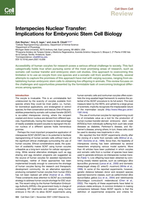
Figure 1. Schematic Representation of
Interspecies Nuclear Transfer for
Derivation of NTESCs
Cell Stem Cell 1, November 2007 ª2007 Elsevier Inc. 505
Cell Stem Cell
Review
5. macaque, and human—6%–33% of iSCNT embryos de-
veloped into blastocysts (Chen et al., 2003; Wen et al.,
2005). These numbers put the efficiency of iSCNT using
rabbit oocytes within or above the range of successful
intraspecies SCNT blastocyst development frequencies
(Dinnyes et al., 2001; Chesne et al., 2002). Assuming that
none of the blastocysts reported were of parthenogenetic
origin—not determined by the authors—it is intriguing to
speculate that the rabbit oocyte may be more efficient at
supporting preimplantation development than other spe-
cies. Unfortunately, none of the reported rabbit iSCNT ex-
periments addressed molecular and physiologic aspects
of preimplantation development, leaving the mechanism
behind the observed high efficiencies of rabbit iSCNT
open to question. One can speculate, however, that the
high success rate could be due to intrinsic characteristics
of rabbit oocytes that make them more effective in driving
early mammalian developmental events. We cannot rule
out the possibility that the high efficiency could also be at-
tributed to the technical expertise of the group responsible
for the bulk of the rabbit iSCNT experiments. Taken
together, a detailed examination of rabbit oocytes at a mo-
lecular and functional level in the context of iSCNT may
provide interesting insights into somatic cell reprogram-
ming forces that operate during the cloning process.
Challenges Faced by the iSCNT Model
Although the number of interspecies cloning experiments
is not large enough to definitively answer fundamental
biological questions, studies reported to date imply fairly
constricted species barriers that prevent an iSCNT em-
bryo from developing into a viable fetus and offspring. In
addition to the failures of iSCNT embryos during preim-
plantation development, notably around EGA, those that
can develop into blastocysts are likely to fail to implant
in the uterus. This is reflected in the observation that
many different recipient-oocyte/donor-cell combinations
were able to develop to the blastocyst stage (albeit at
reduced frequencies compared to their intraspecies coun-
terparts) but failed thereafter (Table 1).
The majority of reported iSCNT experiments were de-
signed to address the preimplantation development of re-
constructed embryos only at the morphological level and
generally did not address physiological aspects of devel-
opment in great depth. There is, to date, a distinct lack of
reports examining interactions and compatibility between
nuclei and the cytoplasm. It is not yet clear whether the
failure to produce a high percentage of iSCNT-derived
blastocysts—as a percentage of fused oocytes—should
be attributed to incompatibility between oocyte proteins
and the transplanted nuclei, between the mitochondria
and the transplanted nuclei, or both (Figure 2). Not surpris-
ingly, due to the few iSCNT embryos that have implanted,
attempts to explore fetal-maternal interactions between
the embryo and the recipient uterus are simply absent.
The theory of reproductive isolation proposes that the
emergence of two species occurs when recombination,
through either mutations or hybridizations, renders two
populations unable to interbreed and to continue to repro-
duce. To the extent that a fertilization using sperm from
one species and oocytes from another is equivalent to
iSCNT in the context of evolutionary biology, the pub-
lished findings are consistent with this model, in that
only closely related species seem capable of generating
iSCNT offspring. Although we do not know the identity
of the particular genes that cause postzygotic reproduc-
tive isolation, it is reasonable to speculate that this pool
of genes could contain good candidates for factors that
might sabotage the successful development of iSCNT
embryos (Orr et al., 2004).
iSCNT for Embryonic Stem Cell Isolation
In addition to providing a great model to study nuclear
cytoplasmic interactions, iSCNT embryos can potentially
Figure 2. Developmental Milestones Required to Establish Interspecies Nuclear Transfer Embryonic Stem Cells
Yellow text boxes indicate potential problems associated with the model. TE, trophoectoderm; ICM, inner cell mass.
506 Cell Stem Cell 1, November 2007 ª2007 Elsevier Inc.
Cell Stem Cell
Review
6. be used for the isolation of ESCs. While many intraspecies
SCNT blastocysts fail to develop into live offspring, a sub-
stantial number of ESCs or embryonic stem-like cells have
been derived from SCNT embryos in cattle and mouse
models (Cibelli et al., 1998; Wakayama et al., 2001,
2006; Wang et al., 2005).
Earlier reports indicated that the functional characteris-
tics of ESCs derived by NT (ntESCs) may not be compro-
mised by the aberrations observed in cloned fetuses and
embryos, including chromosomal, genetic, and epigenetic
abnormalities (Renard et al., 1999; Hill et al., 2000;
Humpherys et al., 2001, 2002). Live, healthy offspring
have been obtained by using ntESCs as nuclear donors
in a second round of NT, indicating that at least some of
the ntESCs are competent to support full-term develop-
ment (Wakayama et al., 2005b). In addition, mouse chi-
meras generated with ntESCs resulted in germline trans-
mission of the injected cells, strongly supporting the
idea that they are functionally similar to conventional
ESCs (Wakayama et al., 2005a, 2005b, 2005c). A recent
study by Wakayama et al., employing 150 mouse ntESC
lines, reported that these ntESCs are comparable to their
in vivo-derived counterparts in terms of their differentia-
tion capacity, pluripotency marker expression profile,
global gene expression profile, and select methylation
characteristics (Wakayama et al., 2006). These observa-
tions are not only limited to the mouse. Recently, rabbit
ESCs have also been established from fertilized, parthe-
nogenetic and NT embryos with high efficiency (Fang
et al., 2006). Conventional and NT rabbit ESCs exhibit
similar characteristics in their ESC marker expression
and in their in vivo and in vitro differentiation abilities (Fang
et al., 2006). Taken together, these data demonstrate
that the developmental problems observed for SCNT
embryos do not seem to affect their ability to form ESCs,
reinforcing the fact that iSCNT could be used to address
questions related to interspecies nucleocytoplasmic com-
patibility and even to provide another source of cytoplasm
for isolating human ESCs, albeit with the limitations out-
lined previously.
To date, only one report (Chen et al., 2003) has estab-
lished ESCs from rabbit/human iSCNT blastocysts. The
authors produced 158 blastocysts, employing rabbit
oocytes and human fibroblasts from four individuals rang-
ing in age from 5 to 60 years old. They isolated NTESCs
from 14 iSCNT blastocysts, 4 of which were expanded
for more than 25 passages (Chen et al., 2003). The re-
ported frequency of blastocyst development (14.5%)
and the frequency of ESC establishment (13% and 4%
up to passage 10 and passage 25, respectively) are very
encouraging, considering the fact that many more intra-
species SCNT attempts have produced only a handful of
blastocysts and no human embryonic stem cell line. In
this report, the ESC lines produced by iSCNT were posi-
tive for various protein markers known to be expressed
by human ESC lines, such as alkaline phosphatase,
SSEA-3, SSEA-4, TRA-1-10, and TRA-1-85. Upon differ-
entiation, embryoid bodies and outgrowths expressed
marker proteins specific for several somatic cell line-
ages—such as myoglobin, a-fetoprotein, a-1-antitrypsin,
VEGF receptor-2, Tie-2, nestin, and MyoD1—consistent
with their presumed pluripotency. These ntESCs pheno-
typically resembled human ESCs isolated from fertilized
embryos. While the report indicates that the ESCs carried
both human and rabbit mitochondrial DNA, quantitative
and functional analyses testing the compatibility of the
donor nuclei and recipient mitochondria were not per-
formed. However, the successful, prolonged culture of
ntESCs derived in this way suggests at least some degree
of compatibility. This study is interesting, since it implies
that rabbit oocytes could enable human somatic nuclei
to go through not only several landmarks of preimplanta-
tion development, including EGA and compaction, but
also could overcome the possible nucleocytoplasmic
conflict during establishment and differentiation of
ESCs. These results have yet to be replicated by an inde-
pendent group; nonetheless, in the event that the results
are consistent in other hands, future iSCNT studies using
rabbit oocytes are warranted.
Is SCNT the only way to obtain bona fide ESCs from
adult individuals? Recently, three different independent
groups reported that mouse somatic cells can be forced
to dedifferentiate by exogenously inserting four genes,
Oct4, Klf4, c-myc, and Sox2 (Okita et al., 2007; Takahashi
and Yamanaka, 2006; Wernig et al., 2007; Maherali et al.,
2007). These findings imply that the requirement for
oocytes to reprogram somatic cells could be overcome
one day. While true, considerable additional research
must be conducted to understand and subsequently
mimic the epigenetic mechanisms that take place during
early embryonic development. Among the limitations of
these induced pluripotent stem cells (iPS) is the low
efficiency of their derivation. As reported by Takahashi
and Yamanaka (Takahashi and Yamanaka, 2006), in order
to obtain one pluripotent colony of cells, 5000 cells must
be transfected, although this efficiency has been im-
proved, at least to some degree (Maherali et al., 2007).
Nevertheless, murine iPS cell generation remains 100–
500 times less efficient than NTESC derivation (Wa-
kayama et al., 2005c). Furthermore, two of the four genes
required to obtain iPS are proto-oncogenes, casting seri-
ous doubts about the safety of the cells if they are ever
produced in human for therapeutic purposes. Nonethe-
less, it would be unwise not to recognize the intrinsic value
of iPS cells as a biological tool, reinforcing the notion that
all avenues should be explored when trying to understand
the gene reprogramming processes necessary to dedif-
ferentiate somatic cells, including iSCNT. Indeed, the au-
thors of these studies have collaborated to contribute
a Correspondence article in the October issue of Cell
Stem Cell (Hyun et al., 2007) underscoring the necessity
of pursuing all avenues toward the generation of human
pluripotent cells.
Theoretical Considerations for iSCNT Models
SCNT is an inefficient technique, even in the context of
intraspecies cloning. The best frequencies reported for
offspring born after embryos are transferred into surrogate
Cell Stem Cell 1, November 2007 ª2007 Elsevier Inc. 507
Cell Stem Cell
Review
7. mothers hover around 5%. It is generally accepted that
the litmus test for functional genome reprogramming is
having produced healthy and fertile adult offspring. For
SCNT embryos, failing this test is the norm. These failures
have been associated with early embryonic losses and
various developmental abnormalities, including an en-
larged, less-vascularized placenta; pulmonary immaturity;
and liver, renal, and endocrine failure (Chavatte-Palmer
et al., 2002; Hill, 2002; Hill et al., 1999, 2000, 2002). Nu-
merous factors have been investigated as the potential
culprits of failed reprogramming in SCNT embryos. Failed
epigenetic reorganization of the genome has emerged as
one likely source of problems associated with SCNT.
These problems include deregulation of chromatin struc-
ture through alterations in DNA and histone methylation/
acetylation patterns (Chung et al., 2003; Santos et al.,
2003) and aberrant expression of imprinted genes
(Humpherys et al., 2001; Mann et al., 2003). Unfortunately,
our understanding of the regulation of epigenetic changes
is still limited, and as a consequence, we are at a disadvan-
tage when trying to discover their role in the context of
SCNT (see reviews, Hochedlinger and Jaenisch, 2003;
Jaenisch et al., 2005; Kishigami et al., 2006).
Nuclear-Mitochondrial Compatibility
The fact that iSCNT experiments yield live offspring only
when the oocyte and donor-cell sources used are derived
from related species may be the result of incompatibilities
in mitochondrial physiology.
Earlier studies using mouse blastomere NTs have sug-
gested that the typical pattern of maternal inheritance
observed in many mammalian species does not apply in
SCNT, and varying degrees of heteroplasmy were ob-
served in most of the resulting embryos, fetuses, and
live offspring (Hiendleder et al., 2003). Moreover, even tis-
sue-specific selection of certain haplotypes was observed
in cloned mice. With few exceptions, neutral segregation
of mtDNA in most bovine SCNT embryos, fetuses, and
animals appears to be the dominant pattern of inheritance
in which the amount of donor-cell mtDNA is not more than
the original amount contributed during reconstruction of
the embryo (i.e., the donor-cell mitochondria are not
selectively replicated over the recipient mitochondria)
(Hiendleder et al., 2003).
Mitochondria are semiautonomous organelles, with
their own genomes and transcriptional machinery. This
compact genome encodes 13 protein subunits of oxida-
tive phosphorylation, 22 tRNAs, and 2 rRNAs. To be
operational, however, the mitochondria also rely on ‘‘im-
ported’’ proteins encoded in the nucleus. Hence, close
coordination between the nuclear and mitochondrial ge-
nomes is essential (Brenner et al., 2004; St John et al.,
2004). Coevolution of nuclear and mitochondrial genomes
and the transfer of genetic information from mitochondria
to the nucleus have resulted in a very specific and unique
complementation of mitochondrial and nuclear function
within an individual species. This specific interaction has
also been proposed to contribute to the speciation pro-
cess (Herrmann et al., 2003). This assumption would
lead us to conclude that the compatibility of nuclear and
mitochondrial genomes may be of paramount importance
to iSCNT experiments.
In all eukaryotic cells, mitochondria play a major role in
many biological processes. They are known to be involved
in ATP production, regulation of intracellular calcium
levels, apoptosis, and cellular aging. During mammalian
gametogenesis, fertilization, and embryogenesis, mito-
chondria have an unusual morphology and pattern of
transmission from one generation to another. Mitochon-
dria in oocytes and preimplantation embryos are spherical
in shape and bear fewer, less-prominent cristae. More-
over, a subset of the mitochondrion pool in primordial
germ cells serves as the founders of the mitochondria
population in the oocyte and in the offspring. This genetic
bottleneck is thought to ensure mitochondrial homo-
plasmy (defined as having mitochondria derived from a
single source—in this case, the oocyte), which is important
to the maintenance of proper mitochondrial function.
During SCNT, a relatively small number of the donor-cell
mitochondria are inserted into the reconstructed embryo,
resulting in mtDNA heteroplasmy. Studies on ovine,
mouse, and bovine SCNT embryos indicate a high degree
of variability in mitochondrial distribution, with some ani-
mals displaying complete homoplasmy (Evans et al.,
1999; Hua et al., 2007; Meirelles et al., 2001) and others
displaying heteroplasmy to varying degrees (Han et al.,
2003; Hiendleder et al., 2003; Inoue et al., 2004; Takeda
et al., 2003). In heteroplasmic animals, the level of contri-
bution from donor-cell mitochondria is also highly variable
between subjects and within tissues of the same subject.
It has been reported that the level of heteroplasmy in-
creases when iSCNT is performed (St John et al., 2004).
Although healthy, live offspring have been obtained by
both intraspecies and iSCNT, possible negative effects
of heteroplasmy introduced in these animals may be re-
sponsible, in part, for many of the failures of iSCNT. For
example, incompatibility of mitochondrial and nuclear
genomes could impair mitochondrial function, leading to
suboptimal respiration (St John et al., 2004). Most of the
available SCNT studies have not provided information
about the mitochondrial DNA and the metabolic status
of SCNT embryos and animals.
The study by Chen et al. (2003) describes the presence
of rabbit mitochondria in human cells. However, it is un-
known whether those mitochondria were able to fully re-
store functional nucleocytoplasmic crosstalk. In human-
monkey cybrids, for instance, mitochondria from common
chimpanzee, pigmy chimpanzee, and gorilla were defi-
cient in mitochondrial complex I activity, and the cybrids
displayed reduced cellular respiration (Barrientos et al.,
1998; McKenzie et al., 2003).
To date, NT studies addressing mitochondrial transmis-
sion have limited their scope to the detection of mtDNA
and have provided no information about indicators of mi-
tochonrial function. Two recent studies have investigated
the amount of mtDNA, ATP production, and gene expres-
sion in bovine SCNT embryos with different haplotypes
(Jiao et al., 2007) and mtRNA expression in sheep goat
508 Cell Stem Cell 1, November 2007 ª2007 Elsevier Inc.
Cell Stem Cell
Review
8. SCNT embryos (Ma et al., 2007). The results of these stud-
ies suggest that the haplotype of recipient oocyte affects
the ATP output, and developmental competence of
SCNT embryos and that of the donor cell’s mitochondria
are selectively eliminated in iSCNT embryos during preim-
plantation development. Although metabolic pathways
are well conserved among mammals, the proper activity
of respiratory chain complexes (i.e., involving nuclei-
mitochondrial compatibility, as discussed above) has
never been directly studied in iSCNT embryos, leaving
a significant gap in our understanding of the potential
role that metabolic insufficiencies may play in the success
of both iSCNT and SCNT developmental competency.
Taken together, these studies suggest that more data
are necessary to determine whether or not cell-donor/
recipient-oocyte mitochondrial incompatibilities are the
cause of the problems in iSCNT.
Activation of the Embryonic Genome
Early development in mammals is controlled by proteins
and mRNAs stored in the oocyte during oogenesis. After
fertilization, these factors direct early cleavage divisions
and are gradually depleted. Depletion of these molecules
seems to coincide with the activation of the embryonic ge-
nome. Experiments using RNA polymerase II inhibitors
have established that the embryo can develop up to the
two cell, four cell, eight cell, and 16 cell stages in mouse,
pig, human, rabbit, and sheep and cattle embryos, re-
spectively, using maternal transcripts. These experiments
demonstrated that the stage at which the embryo gains
full control of transcription is a species-specific occur-
rence (Latham and Schultz, 2001; Schultz, 2002). How-
ever, recent studies have established that this process is
more dynamic than previously thought. In fact, transcrip-
tion can start as early as the one cell stage and can grad-
ually increase until the embryonic genome gains control,
reaching the ‘‘tipping point’’ at the stages previously de-
scribed in the RNA polymerase II inhibitor experiments.
Since timely and orderly expression of developmental
genes is very critical for embryos to develop properly
(Latham, 2005), the process of EGA plays a major role
by providing control over spatial and temporal patterns
of gene expression during preimplantation development.
Considering that live offspring have been obtained using
SCNT, it is easy to assume that donor somatic-genome-
initiated transcription can either adapt to or be adapted
to a developmentally compatible program, even though
resulting gene expression patterns are reportedly altered
in some of these animals (Latham, 2005; Latham and
Schultz, 2001). In the context of SCNT, regulation of
EGA deserves special attention, because the depletion
of maternal messages stored in the ooplasm and chroma-
tin modification appear to be concurrent events and could
affect the timing of transcriptional initiation. For an iSCNT
embryo to develop successfully into a blastocyst and be-
yond, it needs to coordinate both the donor and recipient
components of EGA.
Data on reactivation of endogenous genes from the do-
nor cell are scarce. Only one study directly addresses
questions of EGA in iSCNT embryos; in this study, bo-
vine/mouse iSCNT embryos reactivated a stable trans-
gene (EGFP under CMV promoter) by the eight cell stage,
while several selected endogenous genes failed to be
transcribed by the same stage (Arat et al., 2003). In this
study, no bovine/mouse iSCNT embryos developed fur-
ther than the eight cell stage. Despite these limited data
in gene expression, morphologic and developmental ob-
servations provide indirect evidence suggesting that, in
the case of closely related species, at least some iSCNT
embryos are able to activate the donor-cell genome, de-
velop into blastocysts, and complete fetal development.
Our experience with iSCNT between distant species,
however, suggests that the majority of failed embryos ex-
perience a developmental block when the genome of the
recipient oocyte is required to undergo a major activation
event. Collectively, these results suggest that EGA could
manifest itself as the culprit in the failure to overcome
one of the species-specific developmental blocks and
also deserves more rigorous attention in the context of
iSCNT.
Minimal Standards for Future iSCNT Studies
In light of the arguments we have made, there are only two
sound approaches for assessing whether reprogramming
has occurred in iSCNT embryos: (1) establishing pluripo-
tent embryonic cells that can be later analyzed in detail
or (2) obtaining live offspring in all animals with the excep-
tion of human. The majority of published studies using
iSCNT are difficult to interpret, in part due to a lack of com-
pelling evidence showing that cells can or cannot be re-
programmed by a given oocyte. We would like to put for-
ward specific methodological approaches that may yield
more meaningful results: (1) PCR-based methods should
be used to identify genomic and mitochondrial DNA, using
primers specific to both species. (2) Greater emphasis
should be placed on quantification, and not just the detec-
tion, of mtDNA. (3) If enough cells are generated, karyo-
typing should be performed to determine the relative sta-
bility of resulting cell genomes and to confirm their origin.
(4) Endpoints for reprogramming should be either the es-
tablishment of pluripotent embryonic cell lines or in vivo
development. (5) If preimplantation development is the
focus of the study, basic physiological aspects of embryo
biology should be addressed, such as timing of EGA,
expression of the donor cell’s specific genes, and embryo
metabolism.
Although studies focusing on preimplantation stages of
development in iSCNT embryos can and have uncovered
many interesting facts about developmental physiology,
future research in this field will undoubtedly shift from
purely descriptive reports to more complex quantitative
analyses that seek to better define the relationship
between molecular events and developmental success.
Conclusions
With few exceptions, available iSCNT data suggest that
species-specific barriers stand in the way of reprogram-
ming somatic cells into embryonic stem cells. These
Cell Stem Cell 1, November 2007 ª2007 Elsevier Inc. 509
Cell Stem Cell
Review
9. barriers appear to be overcome, or disappear, only when
the two species are sufficiently closely related as to be
able to crossbreed. In the event that studies aimed at the
isolation of human ESCs using iSCNT prove successful,
they could present an invaluable experimental model to in-
vestigate causes and potential treatments for late-onset
diseases in human. Meanwhile, SCNT preimplantation
embryos could be used to provide information about phys-
iological problems associated with iSCNT, such as cell-cy-
cle kinetics, EGA, and embryo metabolism. A detailed un-
derstanding of the limitations imposed on these embryos
by species differences might lead to our being able to ma-
nipulate the limiting mechanisms and to exploit the iSCNT
model in more practical ways. Although it presents a chal-
lenging task, the application of iSCNT could offer benefits
not only for practical purposes but also for understanding
fundamental aspects of biology.
ACKNOWLEDGMENTS
The authors thank Steve Suhr for critically reading the manuscript.
Funding was provided by the Naylor Family Foundation, the Michigan
State University Foundation, and the Michigan Agriculture Experiment
Station.
REFERENCES
Arat, S., Rzucidlo, J., and Stice, S. (2003). Gene expression and in vitro
development of inter-species nuclear transfer embryos. Mol. Reprod.
Dev. 66, 334–342.
Barrientos, A., Kenyon, L., and Moraes, C.T. (1998). Human xenomito-
chondrial cybrids. Cellular models of mitochondrial complex I defi-
ciency. J. Biol. Chem. 273, 14210–14217.
Brenner, C.A., Kubisch, H.M., and Pierce, K.E. (2004). Role of the
mitochondrial genome in assisted reproductive technologies and em-
bryonic stem cell-based therapeutic cloning. Reprod. Fertil. Dev. 16,
743–751.
Chang, K.H., Lim, J.M., Kang, S.K., Lee, B.C., Moon, S.Y., and Hwang,
W.S. (2003). Blastocyst formation, karyotype, and mitochondrial DNA
of interspecies embryos derived from nuclear transfer of human cord
fibroblasts into enucleated bovine oocytes. Fertil. Steril. 80, 1380–
1387.
Chavatte-Palmer, P., LeBourhis, D., Camous, S., Vignon, X., Renard,
J.P., and Enright, B.P. (2002). Reproductive characteristics of cloned
heifers derived from adult somatic cells. Biol. Reprod. 66, 6–13.
Chen, D.Y., Wen, D.C., Zhang, Y.P., Sun, Q.Y., Han, Z.M., Liu, Z.H.,
Shi, P., Li, J.S., Xiangyu, J.G., Lian, L., et al. (2002). Interspecies im-
plantation and mitochondria fate of panda-rabbit cloned embryos.
Biol. Reprod. 67, 637–642.
Chen, Y., He, Z.X., Liu, A., Wang, K., Mao, W.W., Chu, J.X., Lu, Y.,
Fang, Z.F., Shi, Y.T., Yang, Q.Z., et al. (2003). Embryonic stem cells
generated by nuclear transfer of human somatic nuclei into rabbit oo-
cytes. Cell Res. 13, 251–263.
Chen, T., Zhang, Y.L., Jiang, Y., Liu, J.H., Schatten, H., Chen, D.Y., and
Sun, Q.Y. (2006). Interspecies nuclear transfer reveals that demethyla-
tion of specific repetitive sequences is determined by recipient oo-
plasm but not by donor intrinsic property in cloned embryos. Mol. Re-
prod. Dev. 73, 313–317.
Chesne, P., Adenot, P.G., Viglietta, C., Baratte, M., Boulanger, L., and
Renard, J.P. (2002). Cloned rabbits produced by nuclear transfer from
adult somatic cells. Nat. Biotechnol. 20, 366–369.
Chung, Y.G., Ratnam, S., Chaillet, J.R., and Latham, K.E. (2003).
Abnormal regulation of DNA methyltransferase expression in cloned
mouse embryos. Biol. Reprod. 69, 146–153.
Cibelli, J.B., Stice, S.L., Golueke, P.J., Kane, J.J., Jerry, J., Blackwell,
C., Ponce de Leon, F.A., and Robl, J.M. (1998). Transgenic bovine chi-
meric offspring produced from somatic cell-derived stem-like cells.
Nat. Biotechnol. 16, 642–646.
Dindot, S.V., Farin, P.W., Farin, C.E., Romano, J., Walker, S., Long, C.,
and Piedrahita, J.A. (2004). Epigenetic and genomic imprinting analy-
sis in nuclear transfer derived Bos gaurus/Bos taurus hybrid fetuses.
Biol. Reprod. 71, 470–478.
Dinnyes, A., Dal, Y.P., Barber, M., Liu, L., Xu, J., Zhou, P.L., and Yang,
X.Z. (2001). Development of cloned embryos from adult rabbit fibro-
blasts: effect of activation treatment and donor cell preparation. Biol.
Reprod. 64, 257–263.
Dominko, T., Mitalipova, M., Haley, B., Beyhan, Z., Memili, E., and
First, N. (1998). Bovine oocyte as a universal recipient cytoplasm in
mammalian nuclear transfer. Theriogenology 49, 385.
Dominko, T., Mitalipova, M., Haley, B., Beyhan, Z., Memili, E., McKusick,
B., and First, N.L. (1999). Bovine oocyte cytoplasm supports develop-
ment of embryos produced by nuclear transfer of somatic cell nuclei
from various mammalian species. Biol. Reprod. 60, 1496–1502.
Evans, M.J., Gurer, C., Loike, J.D., Wilmut, I., Schnieke, A.E., and
Schon, E.A. (1999). Mitochondrial DNA genotypes in nuclear trans-
fer-derived cloned sheep. Nat. Genet. 23, 90–93.
Fang, Z.F., Gai, H., Huang, Y.Z., Li, S.G., Chen, X.J., Shi, J.J., Wu, L.,
Liu, A., Xu, P., and Sheng, H.Z. (2006). Rabbit embryonic stem cell lines
derived from fertilized, parthenogenetic or somatic cell nuclear transfer
embryos. Exp. Cell Res. 312, 3669–3682.
Fulka, J., Jr., and Mrazek, M. (2004). Cloning of mammals-biological
aspects. Cas. Lek. Cesk. 143, 295–298.
Gilbert, S.F., and Bolker, J.A. (2001). Homologies of process and mod-
ular elements of embryonic construction. J. Exp. Zool. 291, 1–12.
Gomez, M.C., Jenkins, J.A., Giraldo, A., Harris, R.F., King, A., Dresser,
B.L., and Pope, C.E. (2003). Nuclear transfer of synchronized african
wild cat somatic cells into enucleated domestic cat oocytes. Biol.
Reprod. 69, 1032–1041.
Gomez, M.C., Pope, C.E., Giraldo, A., Lyons, L.A., Harris, R.F., King,
A.L., Cole, A., Godke, R.A., and Dresser, B.L. (2004). Birth of African
Wildcat cloned kittens born from domestic cats. Cloning Stem Cells
6, 247–258.
Hall, V.J., Stojkovic, P., and Stojkovic, M. (2006). Using therapeutic
cloning to fight human disease: A conundrum or reality? Stem Cells
24, 1628–1637.
Han, Z.M., Chen, D.Y., Li, J.S., Sun, Q.Y., Wan, Q.H., Kou, Z.H., Rao,
G., Lei, L., Liu, Z.H., and Fang, S.G. (2003). Mitochondrial DNA heter-
oplasmy in calves cloned by using adult somatic cell. Mol. Reprod.
Dev. 67, 207–214.
Hashem, M.A., Bhandari, D.P., Kang, S.K., and Lee, B.C. (2007). Cell
cycle analysis and interspecies nuclear transfer of in vitro cultured
skin fibroblasts of the Siberian tiger (Panthera tigris Altaica). Mol.
Reprod. Dev. 74, 403–411.
Herrmann, R.G., Maier, R.M., and Schmitz-Linneweber, C. (2003).
Eukaryotic genome evolution: rearrangement and coevolution of com-
partmentalized genetic information. Philos. Trans. R. Soc. Lond. B
Biol. Sci. 358, 87–97.
Hidalgo, C.O., Diez, C., Duque, P., Facal, N., and Gomez, E. (2003).
Pregnancies and improved early embryonic development with bovine
oocytes matured in vitro with 9-cis-retinoic acid. Reproduction 125,
409–416.
Hiendleder, S., Zakhartchenko, V., Wenigerkind, H., Reichenbach,
H.D., Bruggerhoff, K., Prelle, K., Brem, G., Stojkovic, M., and Wolf,
E. (2003). Heteroplasmy in bovine fetuses produced by intra- and in-
ter-subspecific somatic cell nuclear transfer: neutral segregation of
nuclear donor mitochondrial DNA in various tissues and evidence for
recipient cow mitochondria in fetal blood. Biol. Reprod. 68, 159–166.
Hill, J.R. (2002). Abnormal in utero development of cloned animals: im-
plications for human cloning. Differentiation 69, 174–178.
510 Cell Stem Cell 1, November 2007 ª2007 Elsevier Inc.
Cell Stem Cell
Review
10. Hill, J.R., Roussel, A.J., Cibelli, J.B., Edwards, J.F., Hooper, N.L.,
Miller, M.W., Thompson, J.A., Looney, C.R., Westhusin, M.E., Robl,
J.M., et al. (1999). Clinical and pathologic features of cloned transgenic
calves and fetuses (13 case studies). Theriogenology 51, 1451–1465.
Hill, J.R., Burghardt, R.C., Jones, K., Long, C.R., Looney, C.R., Shin,
T., Spencer, T.E., Thompson, J.A., Winger, Q.A., and Westhusin,
M.E. (2000). Evidence for placental abnormality as the major cause
of mortality in first-trimester somatic cell cloned bovine fetuses. Biol.
Reprod. 63, 1787–1794.
Hill, J.R., Schlafer, D.H., Fisher, P.J., and Davies, C.J. (2002). Abnor-
mal expression of trophoblast major histocompatibility complex class
I antigens in cloned bovine pregnancies is associated with a pro-
nounced endometrial lymphocytic response. Biol. Reprod. 67, 55–63.
Hochedlinger, K., and Jaenisch, R. (2003). Nuclear transplantation,
embryonic stem cells, and the potential for cell therapy. N. Engl. J.
Med. 349, 275–286.
Hua, S., Zhang, Y., Song, K., Song, J., Zhang, Z., Zhang, L., Zhang, C.,
Cao, J., and Ma, L. (2007). Development of bovine-ovine interspecies
cloned embryos and mitochondria segregation in blastomeres during
preimplantation. Anim. Reprod. Sci. Published online March 6, 2007.
10.1016/j.anireprosci.2007.03.002.
Hubner, K., Fuhrmann, G., Christenson, L.K., Kehler, J., Reinbold, R.,
De La Fuente, R., Wood, J., Strauss, J.F., III, Boiani, M., and Scholer,
H.R. (2003). Derivation of oocytes from mouse embryonic stem cells.
Science 300, 1251–1256.
Humpherys, D., Eggan, K., Akutsu, H., Hochedlinger, K., Rideout,
W.M., 3rd, Biniszkiewicz, D., Yanagimachi, R., and Jaenisch, R.
(2001). Epigenetic instability in ES cells and cloned mice. Science
293, 95–97.
Humpherys, D., Eggan, K., Akutsu, H., Friedman, A., Hochedlinger, K.,
Yanagimachi, R., Lander, E.S., Golub, T.R., and Jaenisch, R. (2002).
Abnormal gene expression in cloned mice derived from embryonic
stem cell and cumulus cell nuclei. Proc. Natl. Acad. Sci. USA 99,
12889–12894.
Hyun, I., Hochedlinger, K., Jaenisch, R., and Yamanaka, S. (2007).
New advances in iPS cell research do not obviate the need for human
embryonic stem cells. Cell Stem Cell 1, 367–368.
Ikumi, S., Sawai, K., Takeuchi, Y., Iwayama, H., Ishikawa, H., Ohsumi,
S., and Fukui, Y. (2004). Interspecies somatic cell nuclear transfer for in
vitro production of Antarctic minke whale (Balaenoptera bonaerensis)
embryos. Cloning Stem Cells 6, 284–293.
Illmensee, K., Levanduski, M., and Zavos, P.M. (2006). Evaluation of
the embryonic preimplantation potential of human adult somatic cells
via an embryo interspecies bioassay using bovine oocytes. Fertil.
Steril. 85 (Suppl 1), 1248–1260.
Inoue, K., Ogonuki, N., Yamamoto, Y., Takano, K., Miki, H., Mochida,
K., and Ogura, A. (2004). Tissue-specific distribution of donor mito-
chondrial DNA in cloned mice produced by somatic cell nuclear trans-
fer. Genesis 39, 79–83.
Jaenisch, R., Hochedlinger, K., and Eggan, K. (2005). Nuclear cloning,
epigenetic reprogramming and cellular differentiation. Novartis Found.
Symp. 265, 107–118.
Jiang, Y., Chen, T., Nan, C.L., Ouyang, Y.C., Sun, Q.Y., and Chen, D.Y.
(2005). In vitro culture and mtDNA fate of ibex-rabbit nuclear transfer
embryos. Zygote 13, 233–240.
Jiao, F., Yan, J.B., Yang, X.Y., Li, H., Wang, Q., Huang, S.Z., Zeng, F.,
and Zeng, Y.T. (2007). Effect of oocyte mitochondrial DNA haplotype
on bovine somatic cell nuclear transfer efficiency. Mol. Reprod. Dev.
74, 1278–1286.
Kishigami, S., Wakayama, S., van Thuan, N., and Wakayama, T.
(2006). Cloned mice and embryonic stem cell establishment from adult
somatic cell. Hum. Cell 19, 2–10.
Kitiyanant, Y., Saikhun, J., Chaisalee, B., White, K.L., and Pavasuthi-
paisit, K. (2001). Somatic cell cloning in Buffalo (Bubalus bubalis): ef-
fects of interspecies cytoplasmic recipients and activation proce-
dures. Cloning Stem Cells 3, 97–104.
Klimanskaya, I., Chung, Y., Becker, S., Lu, S.J., and Lanza, R. (2006).
Human embryonic stem cell lines derived from single blastomeres.
Nature 444, 481–485.
Klimanskaya, I., Chung, Y., Becker, S., Lu, S.J., and Lanza, R. (2007).
Derivation of human embryonic stem cells from single blastomeres.
Nat. Protoc. 2, 1963–1972.
Lanza, R.P., Cibelli, J.B., Diaz, F., Moraes, C.T., Farin, P.W., Farin,
C.E., Hammer, C.J., West, M.D., and Damiani, P. (2000). Cloning of
an endangered species (Bos gaurus) using interspecies nuclear trans-
fer. Cloning 2, 79–90.
Latham, K.E. (2005). Early and delayed aspects of nuclear reprogram-
ming during cloning. Biol. Cell 97, 119–132.
Latham, K.E., and Schultz, R.M. (2001). Embryonic genome activation.
Front. Biosci. 6, D748–D759.
Li, Y., Dai, Y., Du, W., Zhao, C., Wang, H., Wang, L., Li, R., Liu, Y., Wan,
R., and Li, N. (2006a). Cloned endangered species takin (Budorcas
taxicolor) by inter-species nuclear transfer and comparison of the blas-
tocyst development with yak (Bos grunniens) and bovine. Mol. Reprod.
Dev. 73, 189–195.
Li, Y., Dai, Y., Du, W., Zhao, C., Wang, L., Wang, H., Liu, Y., Li, R., and
Li, N. (2006b). In vitro development of yak (Bos grunniens) embryos
generated by interspecies nuclear transfer. Anim. Reprod. Sci. 101,
45–59.
Liu, S.Z., Zhou, Z.M., Chen, T., Zhang, Y.L., Wen, D.C., Kou, Z.H., Li,
Z.D., Sun, Q.Y., and Chen, D.Y. (2004). Blastocysts produced by nu-
clear transfer between chicken blastodermal cells and rabbit oocytes.
Mol. Reprod. Dev. 69, 296–302.
Loi, P., Ptak, G., Barboni, B., Fulka, J., Jr., Cappai, P., and Clinton, M.
(2001). Genetic rescue of an endangered mammal by cross-species
nuclear transfer using post-mortem somatic cells. Nat. Biotechnol.
19, 962–964.
Ma, L.B., Yang, L., Zhang, Y., Cao, J.W., Hua, S., and Li, J.X. (2007).
Quantitative analysis of mitochondrial RNA in goat-sheep cloned em-
bryos. Mol. Reprod. Dev. 75, 33–39. Published online June 14, 2007.
10.1002/mrd.20736.
Maherali, N., Sridharan, R., Xie, W., Utikal, J., Eminli, S., Arnold, K.,
Stadtfeld, M., Yachechko, R., Tchieu, J., Jaenisch, R., et al. (2007).
Directly reprogrammed fibroblasts show global epigenetic remodeling
and widespread tissue contribution. Cell Stem Cell 1, 55–70.
Mann, M.R.W., Chung, Y.G., Nolen, L.D., Verona, R.I., Latham, K.E.,
and Bartolomei, M.S. (2003). Disruption of imprinted gene methylation
and expression in cloned preimplantation stage mouse embryos. Biol.
Reprod. 69, 902–914.
Mastromonaco, G.F., Favetta, L.A., Smith, L.C., Filion, F., and King,
W.A. (2007). The influence of nuclear content on developmental com-
petence of gaur x cattle hybrid in vitro fertilized and somatic cell
nuclear transfer embryos. Biol. Reprod. 76, 514–523.
McKenzie, M., Chiotis, M., Pinkert, C.A., and Trounce, I.A. (2003).
Functional respiratory chain analyses in murid xenomitochondrial cy-
brids expose coevolutionary constraints of cytochrome b and nuclear
subunits of complex III. Mol. Biol. Evol. 20, 1117–1124.
Meirelles, F.V., Bordignon, V., Watanabe, Y., Watanabe, M., Dayan, A.,
Lobo, R.B., Garcia, J.M., and Smith, L.C. (2001). Complete replace-
ment of the mitochondrial genotype in a Bos indicus calf reconstructed
by nuclear transfer to a Bos taurus oocyte. Genetics 158, 351–356.
Murakami, M., Otoi, T., Wongsrikeao, P., Agung, B., Sambuu, R., and
Suzuki, T. (2005). Development of interspecies cloned embryos in yak
and dog. Cloning Stem Cells 7, 77–81.
Oh, B.C., Kim, J.T., Shin, N.S., Kwon, S.W., Kang, S.K., Lee, B.C., and
Hwang, W.S. (2006). Production of blastocysts after intergeneric nu-
clear transfer of goral (Naemorhedus goral) somatic cells into bovine
oocytes. J. Vet. Med. Sci. 68, 1167–1171.
Okita, K., Ichisaka, T., and Yamanaka, S. (2007). Generation of germ-
line-competent induced pluripotent stem cells. Nature 448, 313–317.
Cell Stem Cell 1, November 2007 ª2007 Elsevier Inc. 511
Cell Stem Cell
Review
11. Orr, H.A., Masly, J.P., and Presgraves, D.C. (2004). Speciation genes.
Curr. Opin. Genet. Dev. 14, 675–679.
Renard, J.P., Chastant, S., Chesne, P., Richard, C., Marchal, J.,
Cordonnier, N., Chavatte, P., and Vignon, X. (1999). Lymphoid hypo-
plasia and somatic cloning. Lancet 353, 1489–1491.
Sansinena, M.J., Hylan, D., Hebert, K., Denniston, R.S., and Godke,
R.A. (2005). Banteng (Bos javanicus) embryos and pregnancies pro-
duced by interspecies nuclear transfer. Theriogenology 63, 1081–
1091.
Santos, F., Zakhartchenko, V., Stojkovic, M., Peters, A., Jenuwein, T.,
Wolf, E., Reik, W., and Dean, W. (2003). Epigenetic marking correlates
with developmental potential in cloned bovine preimplantation em-
bryos. Curr. Biol. 13, 1116–1121.
Schultz, R.M. (2002). The molecular foundations of the maternal to
zygotic transition in the preimplantation embryo. Hum. Reprod. Up-
date 8, 323–331.
St John, J.C., Lloyd, R.E., Bowles, E.J., Thomas, E.C., and El Shour-
bagy, S. (2004). The consequences of nuclear transfer for mammalian
foetal development and offspring survival. A mitochondrial DNA per-
spective. Reproduction 127, 631–641.
Takahashi, K., and Yamanaka, S. (2006). Induction of pluripotent stem
cells from mouse embryonic and adult fibroblast cultures by defined
factors. Cell 126, 663–676.
Takeda, K., Akagi, S., Kaneyama, K., Kojima, T., Takahashi, S., Imai,
H., Yamanaka, M., Onishi, A., and Hanada, H. (2003). Proliferation of
donor mitochondrial DNA in nuclear transfer calves (Bos taurus) de-
rived from cumulus cells. Mol. Reprod. Dev. 64, 429–437.
Thongphakdee, A., Numchaisrika, P., Omsongkram, S., Chatdarong,
K., Kamolnorranath, S., Dumnui, S., and Techakumphu, M. (2006).
In vitro development of marbled cat embryos derived from interspecies
somatic cell nuclear transfer. Reprod. Domest. Anim. 41, 219–226.
Wakayama, T., Tabar, V., Rodriguez, I., Perry, A.C., Studer, L., and
Mombaerts, P. (2001). Differentiation of embryonic stem cell lines gen-
erated from adult somatic cells by nuclear transfer. Science 292, 740–
743.
Wakayama, S., Kishigami, S., Van Thuan, N., Ohta, H., Hikichi, T., Miz-
utani, E., Yanagimachi, R., and Wakayama, T. (2005a). Propagation of
an infertile hermaphrodite mouse lacking germ cells by using nuclear
transfer and embryonic stem cell technology. Proc. Natl. Acad. Sci.
USA 102, 29–33.
Wakayama, S., Mizutani, E., Kishigami, S., Thuan, N.V., Ohta, H., Hikichi,
T., Bui, H.T., Miyake, M., and Wakayama, T. (2005b). Mice cloned by nu-
clear transfer from somatic and ntES cells derived from the same individ-
uals. J. Reprod. Dev. 51, 765–772.
Wakayama, S., Ohta, H., Kishigami, S., Thuan, N.V., Hikichi, T., Mizutani,
E., Miyake, M., and Wakayama, T. (2005c). Establishment of male and
female nuclear transfer embryonic stem cell lines from different mouse
strains and tissues. Biol. Reprod. 72, 932–936.
Wakayama, S., Jakt, M.L., Suzuki, M., Araki, R., Hikichi, T., Kishigami,
S., Ohta, H., Van Thuan, N., Mizutani, E., Sakaide, Y., et al. (2006).
Equivalency of nuclear transfer-derived embryonic stem cells to those
derived from fertilized mouse blastocyst. Stem Cells 24, 2023–2033.
Wang, L., Duan, E., Sung, L.Y., Jeong, B.S., Yang, X., and Tian, X.C.
(2005). Generation and characterization of pluripotent stem cells
from cloned bovine embryos. Biol. Reprod. 73, 149–155.
Wen, D.C., Bi, C.M., Xu, Y., Yang, C.X., Zhu, Z.Y., Sun, Q.Y., and Chen,
D.Y. (2005). Hybrid embryos produced by transferring panda or cat so-
matic nuclei into rabbit MII oocytes can develop to blastocyst in vitro.
J. Exp. Zoolog. A Comp. Exp. Biol. 303, 689–697.
Wernig, M., Meissner, A., Foreman, R., Brambrink, T., Ku, M., Hoched-
linger, K., Bernstein, B.E., and Jaenisch, R. (2007). In vitro reprogram-
ming of fibroblasts into a pluripotent ES-cell-like state. Nature 448,
318–324.
White, K.L., Bunch, T.D., Mitalipov, S., and Reed, W.A. (1999). Estab-
lishment of pregnancy after the transfer of nuclear transfer embryos
produced from the fusion of argali (Ovis ammon) nuclei into domestic
sheep (Ovis aries) enucleated oocytes. Cloning 1, 47–54.
Wilmut, I., Schnieke, A.E., McWhir, J., Kind, A.J., and Campbell, K.H.S.
(1997). Viable offspring derived from fetal and adult mammalian cells.
Nature 385, 810–813.
Yang, C.X., Han, Z.M., Wen, D.C., Sun, Q.Y., Zhang, K.Y., Zhang, L.S.,
Wu, Y.Q., Kou, Z.H., and Chen, D.Y. (2003). In vitro development and
mitochondrial fate of macaca-rabbit cloned embryos. Mol. Reprod.
Dev. 65, 396–401.
Yin, X., Lee, Y., Lee, H., Kim, N., Kim, L., Shin, H., and Kong, I. (2006).
In vitro production and initiation of pregnancies in inter-genus nuclear
transfer embryos derived from leopard cat (Prionailurus bengalensis)
nuclei fused with domestic cat (Felis silverstris catus) enucleated oo-
cytes. Theriogenology 66, 275–282.
Zhao, Z.J., Ouyang, Y.C., Nan, C.L., Lei, Z.L., Song, X.F., Sun, Q.Y.,
and Chen, D.Y. (2006). Rabbit oocyte cytoplasm supports develop-
ment of nuclear transfer embryos derived from the somatic cells of
the camel and Tibetan antelope. J. Reprod. Dev. 52, 449–459.
Zhao, Z.J., Li, R.C., Cao, H.H., Zhang, Q.J., Jiang, M.X., Ouyang, Y.C.,
Nan, C.L., Lei, Z.L., Song, X.F., Sun, Q.Y., and Chen, D.Y. (2007). Inter-
species nuclear transfer of Tibetan antelope using caprine oocyte as
recipient. Mol. Reprod. Dev. 74, 412–419.
512 Cell Stem Cell 1, November 2007 ª2007 Elsevier Inc.
Cell Stem Cell
Review