SlideShare a Scribd company logo
1 of 14
Download to read offline
RESEARCH ARTICLE
Autocrine/Paracrine Sphingosine-
1-Phosphate Fuels Proliferative and Stemness
Qualities of Glioblastoma Stem Cells
Giovanni Marfia,1
Rolando Campanella,2
Stefania Elena Navone,1
Clara Di Vito,3
Elena Riccitelli,3
Loubna Abdel Hadi,3
Andrea Bornati,4
Gisele de Rezende,4
Paola Giussani,3
Cristina Tringali,3
Paola Viani,3
Paolo Rampini,1
Giulio Alessandri,5
Eugenio Parati,5
and Laura Riboni3
Accumulating reports suggest that human glioblastoma contains glioma stem-like cells (GSCs) which act as key determinants
driving tumor growth, angiogenesis, and contributing to therapeutic resistance. The proliferative signals involved in GSC pro-
liferation and progression remain unclear. Using GSC lines derived from human glioblastoma specimens with different prolif-
erative index and stemness marker expression, we assessed the hypothesis that sphingosine-1-phosphate (S1P) affects the
proliferative and stemness properties of GSCs. The results of metabolic studies demonstrated that GSCs rapidly consume
newly synthesized ceramide, and export S1P in the extracellular environment, both processes being enhanced in the cells
exhibiting high proliferative index and stemness markers. Extracellular S1P levels reached nM concentrations in response to
increased extracellular sphingosine. In addition, the presence of EGF and bFGF potentiated the constitutive capacity of GSCs
to rapidly secrete newly synthesized S1P, suggesting that cooperation between S1P and these growth factors is of central
importance in the maintenance and proliferation of GSCs. We also report for the first time that S1P is able to act as a prolifer-
ative and pro-stemness autocrine factor for GSCs, promoting both their cell cycle progression and stemness phenotypic pro-
file. These results suggest for the first time that the GSC population is critically modulated by microenvironmental S1P, this
bioactive lipid acting as an autocrine signal to maintain a pro-stemness environment and favoring GSC proliferation, survival
and stem properties.
GLIA 2014;62:1968–1981
Key words: stem cell niche, sphingolipid metabolism, ceramide, CD133, growth factors
Introduction
The eradication of glioblastoma multiforme (GBM)
(WHO grade 4) remains a tremendous clinical challenge
in human oncology. Indeed, this tumor accounts for the most
common, aggressive and lethal primary brain cancer in adults,
exhibiting a dismal prognosis, despite extensive surgical resec-
tion, and adjuvant radiotherapy and/or chemotherapy
(Schwartzbaum et al., 2006; Stupp et al., 2005). The finding
that GBM contains functional subsets of cells with stem-like
properties named glioma stem-like cells (GSCs) (Ignatova
et al., 2002; Singh et al., 2004) has opened up novel oppor-
tunities and promises for the development of new therapies
for this devastating cancer.
GSCs are self-renewing, multipotent cells, with the
capacity to establish and maintain glioma tumors at the clo-
nal level, leading to the hypothesis that they are tumor-
initiating cells (Bao et al., 2006; Lathia et al., 2011a). More-
over, these rare subpopulations of cells possess an elevated
View this article online at wileyonlinelibrary.com. DOI: 10.1002/glia.22718
Published online July 5, 2014 in Wiley Online Library (wileyonlinelibrary.com). Received Mar 22, 2014, Accepted for publication June 20, 2014.
Address correspondence to Laura Riboni, Department of Medical Biotechnology and Translational Medicine, LITA-Segrate, University of Milan, via Fratelli Cervi,
93, 20090 Segrate, Milan, Italy. E-mail: laura.riboni@unimi.it
From the 1
Laboratory of Experimental Neurosurgery and Cell Therapy, Neurosurgery Unit, Fondazione IRCCS Ca Granda Ospedale Maggiore Policlinico Milan,
University of Milan, Italy; 2
Neurosurgical Unit, San Carlo Borromeo Hospital, Milan, Italy; 3
Department of Medical Biotechnology and Translational Medicine, LITA-
Segrate, University of Milan, Italy; 4
Department of Pathology, San Carlo Borromeo Hospital, Milan, Italy; 5
Cellular Neurobiology Laboratory, Cerebrovascular Dis-
eases Unit, IRCCS Foundation, Neurological Institute “C. Besta”, Milan, Italy.
Giovanni Marfia and Rolando Campanella contributed equally to this work.
1968 VC 2014 Wiley Periodicals, Inc.
proliferative potential, and intrinsic resistance to therapy,
being thus considered a key determinant driving tumor
growth, and relapse after resection and therapy (Hadjipanayis
and Van Meir, 2009; Laks et al., 2009). In appropriate cul-
ture conditions, GSCs form free-floating cell aggregates with
a spheroid morphology, named neurospheres. These cancer
neurospheres preserve many of the fundamental characteristics
of the parent tumor, including cell heterogeneity, and the
ability to recapitulate the parent tumor’s cell composition and
invasiveness when transplanted in murine brain (Lee et al.,
2006). In addition, the capacity of the original tumor cells to
form neurospheres is by itself an indicator of the in vivo
aggressiveness of the tumor (Pallini et al., 2008).
A key role in the regulation of GSCs behavior is played
by their microenvironment. Indeed, different studies demon-
strated that GSCs reside in a particular tumor microenviron-
ment that provides essential cues to their maintenance, and is
necessary to support their behavior (Charles et al., 2012;
Heddleston et al., 2011).
Different molecules, including lipid mediators appear to
play a key role in the GSC microenvironment (Filatova et al.,
2013; Krishnamoorthy and Honn, 2011). Among lipid medi-
ators involved in GSC properties, sphingosine-1-phosphate
(S1P) has recently emerged as a key bioactive factor. This
simple sphingoid is produced during sphingolipid metabolism
via the conversion of ceramide (Cer) to sphingosine (Sph),
which is then phosphorylated to S1P by kinase (SK) 1 and 2
(Le Stunff et al., 2004; Liu et al., 2012). S1P exerts its effects
predominantly in the extracellular milieu, after interaction
with five specific G protein-coupled receptors (S1PR1–5),
located on the cell surface (Rosen et al., 2009). Accumulating
literature supports that S1P acts as an onco-promoter signal,
favoring growth, invasion, and therapy resistance in various
human cancers, including GBMs (Maceyka et al., 2012; Pyne
and Pyne, 2010; Takuwa et al., 2012). Of relevance, the pres-
ence of S1P in both glioma cell lines and human gliomas is
critical for glioma cell proliferation and survival, a down-
regulation of SK1 suppressed growth of human GBM cells
and xenografts, and a higher expression of S1PR1 in GBM
has been correlated with poor prognosis (Kapitonov et al.,
2009; Van Brocklyn et al., 2005; Yoshida et al., 2010).
It has been shown that the effects of S1P on cell prolif-
eration, and its ability to block apoptotic pathways are antag-
onized by its metabolic precursor Cer, an anti-proliferative
and pro-apoptotic intracellular messenger (Pettus et al.,
2002), suggesting that the altered balance between these two
lipids has been implicated in tumorigenesis, tumor progres-
sion, and chemo-resistance (Furuya et al., 2011; Pyne and
Pyne, 2010). However, little is known on the role of S1P in
GSCs. Recent studies reported that S1P acts as an invasive
signal in GSCs (Annabi et al., 2009; Mora et al., 2010), and
that the inhibition of SK, or the administration of a S1PR
antagonist, results in GSC death (Estrada-Bernal et al., 2012;
Mora et al., 2010). Very recently, we reported that GSCs
derived from U87 GBM cells and those isolated from a
human GBM specimen can release S1P extracellularly, and
that S1P acts as a first messenger to enhance GSC chemore-
sistance (Riccitelli et al., 2013). Although these studies have
provided insight into the S1P-related pathways integral to
GSC invasivity and chemoresistance, we know very little
about autocrine machinery controlling GSC proliferation,
and particularly on the significance of S1P in this event. In
order to expand our investigation on SIP and to better under-
stand its role on GSC activity, in this study we focused on
the potential role of S1P on the proliferative and stemness
properties of the GSCs.
Materials and Methods
Clinical Data and Tumor Collection
GBM tissue samples were collected following surgical resection from
two patients (pts), aging 56 (pt 1), and 59 years (pt 2), under insti-
tutional review board-approved protocols. The preoperative Karnof-
sky performance status (KPS) was normal (100%) in pt 1 and low
(55%) in pt 2. The samples were donated by patients who signed
the free and informed consent form.
Histopathologic Analyses
Histology was performed on specimens fixed in a 4% paraformalde-
hyde (PFA) solution (Sigma-Aldrich, Basel, Switzerland) in Dulbec-
co’s phosphate-buffered saline (D-PBS) (Euroclone, Milan, Italy) and
subsequently embedded in paraffin, cut at 30 mm, and stained with
hematoxylin-eosin. Both GBM samples were histologically confirmed
and classified as grade IV GBMs in accordance with WHO estab-
lished guidelines (Louis et al., 2007).
Immunohistochemistry
The sections were deparaffinized and then dehydrated. In the next
step, blocks were incubated in fresh citrate/HCl buffer solution for
antigen recovery and, after 60 min incubation, were washed with
D-PBS. Samples were then incubated with biotinylated monoclonal
Ki-67/MIB1 antibody (1:100; Ventana, Tucson, AZ, USA) and
rabbit anti-glial fibrillary acidic protein (GFAP; 1:200, Chemicon,
Temecula, CA, USA). Then the slides were exposed to peroxidase-
labeled streptavidin followed by washing with D-PBS, and finally
exposed to diaminobenzidine hydrochloride chromogen (BD Bio-
sciences, San Jose, CA). Afterwards, they were covered with cover-
slips following dehumidification. Stained slides were observed by a
pathologist using an Olympus BX41 (Olympus, Tokyo, Japan)
light microscope.
One thousand tumor cells were counted in several areas of tis-
sue where positively stained nuclei were evenly distributed. In those
cases with uneven distribution of positive nuclei, the tumor cells
were counted in the areas with highest density of positive nuclei by
visual analysis (Ralte et al., 2001). The MIB-1 labelling index was
calculated as a percentage of labelled nuclei.
Marfia et al.: S1P Prompts Growth and Stemness of GSCs
December 2014 1969
Establishment of GSCs and Primary Cultures of
GBM Cells
After surgical removal, tumor samples were placed in D-PBS supple-
mented with 1% penicillin/streptomycin (Sigma-Aldrich, Basel, Swit-
zerland). GSCs were isolated essentially as previously described
(Riccitelli et al., 2013; Salmaggi et al., 2006), with some modifica-
tions. In particular, samples were mechanically dissociated in a Petri
dish with a scalpel, and suspension was collected with D-PBS and
centrifuged (300g for 10 min). The pellet was enzymatically digested
by treatment with 0.25% Liberase Blendzyme2 (Roche Diagnostics,
Indianapolis, USA) in D-PBS for 2 h. After dilution with D-PBS,
the suspension was centrifuged as above. Cells were then resus-
pended in Stem Cell Medium (SCM), consisting of Dulbecco’s
modified Eagle’s medium (DMEM)/F12 (1:1) containing 10 ng/mL
fibroblast growth factor-2 (bFGF) (Peprotech Inc. Rocky Hill, NY,
USA), 20 ng/mL epidermal growth factor (EGF) (RD Systems,
Minneapolis, USA), and 1% penicillin/streptomycin. Cells were then
seeded in 75-cm2
flasks, and grown in a humidified incubator con-
taining 5% CO2 and 5% O2 at 37
C.
Primary cultures of GBM cells were obtained from an aliquot
of the pellet obtained from tumor specimens as described above.
Cells were plated in DMEM supplemented with 10% fetal bovine
serum (FBS) (v/v), 2 mM L-glutamine, 100 units/mL penicillin,
100 lg/mL streptomycin, and 0.25 lg/mL amphotericin B and
incubated at 37
C in 5% CO2 humidified atmosphere.
Culture medium of both GSCs and GBM cells was refreshed
twice per week to guarantee constant supply of required growth fac-
tors and nutrients. Both cell types were detached and dissociated
weekly using Tryple Select (Gibco, Grand Island, NY, USA).
Cells were observed with an inverted phase-contrast micro-
scope (Nikon Eclipse TE300, Nikon, Shinjuku, Tokyo, Japan), and
images were acquired with a digital camera (Zeiss Axiovision, Zeiss,
Oberkochen, Germany) using Axion Vision software (Zeiss). Cell
viability was evaluated by Trypan Blue dye-exclusion assay, and cells
counted in a Fuchs-Rosenthal chamber.
Intracranial Xenograft Studies
To assess in vivo tumor formation and growth, adult 6-week old
NOD-SCID mice (Charles River, Calco, Lecco, Italy) were used for
transplants. We minimized the number of animals used in experi-
ments and did great efforts to reduce their sufferings. All surgical
procedures were performed under sterile conditions and aseptic man-
ner and were performed according to NIH and institutional guide-
lines for animal care and handling. Animals (n 5 5 for each cell line)
were deeply anaesthetized, and cells were injected stereotactically
into the frontal lobe, essentially as we previously described (Salmaggi
et al., 2006). In detail, a burr hole was made in the skull 1.2 mm
lateral to the bregma using a drill, and 2.0 3 105
cells in 2 mL of
PBS were stereotactically injected over 3 min at a depth of 4 mm.
At 5 weeks, mice were anesthetized and transcardially perfused with
4% paraformaldehyde in saline. The brains were removed, post-
fixed, and prepared for histology (Salmaggi et al., 2006). Tumor vol-
ume was calculated by measuring the diameter and length of each
tumor in the respective slide with the maximum tumor size
(Schmidt et al., 2004). Maximal and minimal diameter of the tumor
was measured in mm on hematoxylin and eosin stained sections and
the approximate tumor volume was calculated using the following
formula: tumor volume (mm3
) 5 (max. diameter) 3 (min. diame-
ter)2
3 0.5.
To identify human cells, slides were labeled with a specific
mouse anti-human nuclei antibody (MAB 1281, Chemicon) (1:50,
overnight at 4
C). Next they were washed and incubated with sec-
ondary antibody Cy-3-conjugated anti-mouse IgG (1:1,000, Jackson,
Bar Harbor, Maine, USA) for 1 h at room temperature and then
washed again and counter stained with 4’-6-diamidino-2-
phenylindole (DAPI) to identify cell nuclei. Immunofluorescence
measurements were made using a confocal microscopy (Leica Sys-
tem, Wetzlar, Germany).
Immunophenotypic Analyses
Immunophenotypic analyses of GSCs were performed by FACS,
using single cell suspensions. For each analysis, 5.0 3 104
cells were
incubated with the appropriate phycoerytrine- (PE) or fluorescein
isothiocyanate- (FITC) conjugated antibody to evaluate the expres-
sion of the following pattern of stemness/progenitor cell markers:
CD15, CD31, CD34, CD45, CD133 (Miltenyi Biotec, Bisley, Sur-
rey, UK), and CD90 (Millipore Temecula, CA, USA). Isotype-
matched mouse immunoglobulins were used as control. After 30
min at 4
C, cells were washed once with D-PBS, centrifuged at
300g for 10 min, and finally fixed with 4% PFA. Cell analyses were
performed on a FACS flow cytometer and CellQuest software (BD
Biosciences, San Jose, CA). A Forward Scatter vs. Side Scatter dot
plot was used to include only viable nucleated cells. Post-processing
data analyses were performed with FlowJo software (Treestar, Inc.
Ashland). At least 2 3 104
events per sample were acquired.
GCS Proliferation Assays
GSCs were plated in 12-well multiplates (3.0 3 105
cells/well) in
SCM, and incubated in 5% CO2 and 5% O2 at 37
C for 7 days.
At the end, cells were collected and centrifuged (300g, 10 min). The
supernatant was discarded, and the pellet was re-suspended in 400
lL of TrypLE Select (Gibco) and incubated at 37
C for 5 min. The
suspension was then mechanically dissociated by pipetting up and
down for 2 min at room temperature, then neutralized with an equal
volume of SCM, and centrifuged as above. Viable cells were then
counted by Trypan Blue dye-exclusion assay, using a Fuchs-
Rosenthal chamber. Growth curves were obtained by counting the
number of viable cells at each passage one week after plating.
To evaluate the proliferation index, GSCs were plated in SCM
as above, and the index was calculated by dividing the cell number
at specific time points (2 and 7 days) and the number of input cells
at time 0.
Cell Cycle Analysis
Cells (5.0 3 105
) were fixed in one mL of cold ethanol (70% vol/
vol in D-PBS) with gentle vortexing (5 sec) to obtain a mono-
dispersed cell suspension, and maintained in ethanol, at 4
C, for at
least 2 h. Cells were then centrifuged (300g, 10 min) and the etha-
nol thoroughly decanted. The pellet was re-suspended in 500 mL of
D-PBS and incubated with 10 mg/mL RNAse A, at 37
C for
1970 Volume 62, No. 12
15 min. After centrifugation as above, the new pellet was resus-
pended in 500 mL of D-PBS containing 50 mg/mL propidium
iodide, and incubated at 37
C, 20 min in the dark. Cell cycle distri-
bution was measured on a FACSCalibur flow cytometer, and data
analyzed using Cell Quest software (BD Biosciences, San Jose, CA).
Metabolic Studies by Cell Labeling
Metabolic studies using [C3-3
H]-D-erythro-sphingosine (3
H-Sph)
(PerkinElmer, Boston, MA, USA) were performed as previously
reported (Riboni et al., 2000), with some modifications. In detail, at
the time of experiment, cells were plated in 6-multiwell plates (5.0
3 105
cells/well), and incubated for a given period of time in com-
plete SCM containing 20 nM–1mM 3
H-Sph (0.4 mCi/mL) and
phosphatase inhibitors (Riccitelli et al., 2013). After exposure to 3
H-
Sph for different times, GSCs were centrifuged (500g, 5 min at
4
C), and media and cells were collected. Cellular internalization of
Sph was confirmed by radioactivity counting, and possible cytotoxic
effects were excluded by a cell viability assay.
Sphingolipid and S1P Extraction and Purification
Total lipids were extracted from cells with chloroform/methanol at
4
C as previously reported (Anelli et al., 2005). Briefly, after parti-
tioning of the total lipid extract, the upper alkaline aqueous phase
(containing S1P) was evaporated under a nitrogen stream and
counted for radioactivity. The lower organic phase (containing Cer,
sphingomyelin, and neutral glycosphingolipids) was subjected to a
mild alkaline hydrolysis by the addition of 0.15 volumes of 0.1 M
NH4OH, and the phases were then separated by centrifugation.
Extracellular S1P was extracted and partially purified from cell
medium by a two-step partitioning, at first in alkaline conditions;
then a back extraction of the aqueous phase was carried out in acidic
conditions (Anelli et al., 2005). The final organic phase, containing
extracellular S1P, was then evaporated under a nitrogen stream.
Separation and Quantification of Sph Metabolites
Sphingolipids in the methanolysed organic phase and aqueous phases
were counted for radioactivity by liquid scintillation, and applied to
silica gel HPTLC plates (Merck, Darmstadt, Germany). The plates
were developed in chloroform/methanol/water (55:20:3, by vol.) for
the organic phase. For the quantification of 3
H-SlP, the fractions
containing cellular and extracellular S1P were applied to HPTLC
plates and developed in n-butanol/acetic acid/water (3:1:1, by vol.).
Standard 3
H-S1P, obtained from 3
H-Sph by enzymatic phosphoryla-
tion (Anelli et al., 2005), was chromatographed on the same plate,
and used as internal standard.
At the end of the chromatographic separation, HPTLC plates
were submitted to digital autoradiography (Beta-Imager 2000, Bio-
space, Paris, France). The radioactivity associated with individual
sphingolipids was determined with the b-Vision analysis software
provided by Biospace. The recognition and identification of radioac-
tive Cer, sphingomyelin, glucosyl-ceramide (Glc-Cer), and S1P were
performed as previously reported (Anelli et al., 2005; Riboni et al.,
2000). 3
H-S1P degradation was quantified as tritiated water in the
final aqueous phase of medium partitioning. To this purpose, ali-
quots of the aqueous phase of the partitioned media were submitted
to fractional distillation (Anelli et al., 2005), and counted for radio-
activity as above.
Cell Treatments
For S1P treatment, stock solution of S1P (100 mM) (Enzo Life Sci-
ences, Farmingdale, NY, USA) was prepared in fatty acid free bovine
serum albumin (4 mg/mL in D-PBS). For FTY720 treatment,
FTY720 (Cayman Chemicals, Ann Arbor, MI, USA) was dissolved
in absolute ethanol at a concentration of 5 mM. At the time of the
experiments, stock solutions were diluted at 200 nM (S1P) and
100 nM (FTY720) final concentrations in fresh SCM and adminis-
tered to cells. In the case of GBM cells treatment, stock solution of
S1P was diluted at 200 nM in supplemented DMEM without FBS.
After incubation for 48h, the number of viable cells, the apoptotic
and necrotic events, the proliferation index and the cell cycle were
analyzed.
Assessment of Apoptosis
To evaluate apoptosis, GSCs were processed by human Annexin V-
FITC kit (BMS306FI, Bender MedSystems, Vienna, Austria).
Briefly, cells were collected, washed in cold D-PBS and resuspended
in 200 mL of binding buffer (10 mM Hepes/NaOH, pH 7.4,
140 mM NaCl, 2.5 mM CaCl2) containing 5 mL of Annexin V-
FITC solution. The mixture was incubated for 10 min in the dark
at room temperature, and at the end, one mL of binding buffer was
added. After washing with D-PBS, the final cell pellet was resus-
pended in 200 mL of binding buffer, containing 1 mg/mL propidium
iodide, and samples were immediately analysed by flow cytometry.
This double staining procedure allowed us to distinguish early stage
apoptotic cells (Annexin V1
/Propidium Iodide-
), from late stage
apoptotic cells (Annexin V1
/Propidium Iodide1
), and necrotic cells
(Annexin V-
/Propidium Iodide1
). Data were acquired by a FACS
flow cytometer and analyzed by CellQuest software (BD Bioscence,
San Jose, CA).
Statistical Analyses
The data are shown as mean 6 standard deviation (SD) of at least
three independent experiments, performed in triplicate using separate
culture preparations. The statistical significance was determined by
the two-tailed Student’s t test. Differences were considered statisti-
cally significant at P  0.05.
Results
Histological Analysis and Proliferative Features of
the Human GBMs
Microscopically, GBM specimens obtained from pt 1 and 2
were composed of poorly differentiated glial cells, with
numerous multinucleated giant cells. Confluent areas of
necrosis and parenchymal destruction with serpiginous foci
were present. Vascular proliferation was seen throughout the
lesions, often around necrotic areas and in peripheral zones of
infiltration. Diffuse reactivity for GFAP, regularly distributed,
was seen in both cases. Proliferative activity was prominent,
with detectable mitosis in nearly every field. Areas with large
Marfia et al.: S1P Prompts Growth and Stemness of GSCs
December 2014 1971
number of MIB-1-stained nuclei in brown, reflecting active
cellular proliferation, were evident in SC02 (Fig. 1A). The
mean value of MIB-1 labeling index, determined by the Ki-
67/MIB-1 antibody, was 2063.6% in pt 1 and 6067.1%
in pt 2.
SC01 and SC02 Grow as Neurospheres and Exhibit
In Vivo Tumorigenic Potential
GSCs obtained from pt 1 and 2 GBM specimens were
named SC01 and SC02, respectively. In appropriate culture
conditions that favor GSC survival and proliferation, both
GBM-derived cancer cell lines formed GSCs that grew as free
floating clusters, namely neurospheres (Fig. 1B).
GSCs were next validated in vivo for their tumorigenic
potential in immunodeficient mice. After intracranial injec-
tion, both GSC lines formed tumors in all injected mice,
with histological appearance similar to human GBMs (Fig.
1C). The mean tumor volume 6 SD at sacrifice was
9.1 6 4.8 mm3
and 12.9 6 5.5 mm3
in mice injected with
SC01 and SC02, respectively. The human origin of the tumor
was demonstrated by the positive staining with an anti-
human nuclei antibody, which identified cells positive to
human marker in the slides (Salmaggi et al., 2006). These
results, showing that both cell lines exhibit the capacity for
sphere formation, and are tumorigenic in brains of immuno-
compromised mice, demonstrated that SC01 and SC02
expressed functional features of GSCs (Singh et al., 2004).
SC01 and SC02 Neurospheres Differ in the
Expression of Stemness Markers
In order to phenotype SC01 and SC02, we determined the
expression of different putative stem/progenitor cell markers.
As shown in Fig. 2, all the investigated markers were highly
expressed in both GSCs, with CD90 as the highest (90%)
expressed one. It should be noted that CD90 has been
recently identified as a novel marker for GSCs in high-grade
gliomas and as a more general marker than the recognized
GSC marker CD133 (He et al., 2012). The expression of all
other markers, including CD133 and CD15, was significantly
higher in SC02 than SC01.
SC01 and SC02 Differ in the Proliferation Rate and
Cell Cycle Parameters
In the used culture condition, the analyses of the cellular
growth curves showed that, along with passages, the cell num-
ber gradually increased, and this increase was much more
pronounced in the SC02 line (Fig. 3A). Moreover, at both 2
and 7 days in culture, the proliferation index was significantly
higher in SC02 than SC01 (Fig. 3B).
The results of the cell cycle evaluation by flow cytome-
try revealed differences in the number of cells undergoing
mitosis between the two cell lines. In fact, when compared
with SC01, the SC02 cell line exhibited a significant increase
in the percentage of cells in the mitotic phases and a decrease
in that of cells in G0/G1 phase (Fig. 3C).
FIGURE 2: Slow- and fast-proliferating GSCs growing as neuro-
sphere differ in stemness marker expression. The expression of
different stemness and precursor markers in SC01 (grey) and SC02
(blue) cells, obtained by cytofluorimetric analyses is shown. Data
are expressed as % of total cells and are the mean 6 SD of 3–5
independent experiments. *P 0.05; **P  0.01; ***P  0.001 SC02
versus SC01. Similar data were obtained at different cell passages.
[Color figure can be viewed in the online issue, which is available
at wileyonlinelibrary.com.]
FIGURE 1: Ki-67 immunoreactivity of GBMs and tumor-derived neurospheres. A: Tumor tissue from pt 2 stained with anti-Ki-67 antibody.
The image shows positive nuclear expression of Ki-67 in brown and negative nuclei counterstained in blue (magnification, 40x). B: Repre-
sentative microscopic image of GSCs grown as neurosphere (magnification, 20x). C: Representative histological image (hematoxylin and
eosin staining) of a brain tumor derived from GSCs after intracranial orthotopic xenograft of nude mice. [Color figure can be viewed in
the online issue, which is available at wileyonlinelibrary.com.]
1972 Volume 62, No. 12
SC01 and SC02 Differ in Sph Metabolism and
Extracellular Release of S1P
To follow S1P metabolism and its breakdown product, we
used Sph labeled with tritium at the C3 position of the long
chain base backbone. This label remains in the lipid through-
out the phosphorylation and recycling steps of the sphingoid
base, and thus allows tracking of all metabolites as long as
the sphingoid backbone remains intact, as well as S1P irre-
versible degradation as tritiated water. After administration,
tritiated Sph was rapidly incorporated into the two GSC lines
in a time-dependent fashion, and, after 120 min, total incor-
porated radioactivity accounted for 197.9 6 20.7 in SC01,
and 214.2 6 19.8 nCi/dish in SC02, that is approximately
half of the administered radioactivity. At all investigated
times, the level of intracellular 3
H-Sph was found low, repre-
senting less than 10% of the total incorporated radioactivity
in both cell lines, this level being 3-, 4-fold higher in SC01
than that of SC02 cells (Fig. 4). These data indicate that
both GSC types are able to rapidly incorporate and metabo-
lize exogenous Sph, and that the SC02 cells are particularly
efficient in this metabolization.
At different pulse times, the bulk of incorporated radio-
activity was associated to the N-acylated derivatives of Sph,
and, in less amounts, to the 1-phosphorylation products (Fig.
4). It is worth noting that the ratio between N-acylated and
1-phosphorylated metabolites was 21.3 and 15.5 in SC01,
and 5.2 and 4.9 in SC02 at 15 and 120 min, respectively.
The main N-acylated metabolites produced during pulse
in both GSC lines were represented by Cer and complex
sphingolipids, including sphingomyelin, and, in much lower
amounts, GlcCer. All over pulse, Cer was by far the major
3
H-metabolite, its anabolic derivatives increasing with time
(Fig. 5). A significant decrease in 3
H-Cer in SC02 compared
with SC01 was observed after 120 min pulse (Fig. 5, upper
panel). All over pulse, in SC02 cells a marked elevation of
sphingomyelin and Glc-Cer was also evident; at 120 min, in
SC02 these sphingolipids increased by about 110% and
250%, respectively, relative to SC01 (Fig. 5, lower panels).
As reported above, the metabolites derived from the 1-
phosphorylation process represented the second class of Sph
derivatives (Fig. 4). Among them, intracellular S1P repre-
sented only a minor fraction of 1-phosphorylated Sph metab-
olites and, at 15 min pulse, its level was significantly lower in
SC01 than SC02 (Fig. 6A). Thereafter, there was no relevant
change in the cellular S1P level between SC01 and SC02. At
all investigated times, tritiated water, represented the vast
majority of Sph 1-phosphorylation products, its content being
significantly higher (4-, 6-fold) in SC02 than in SC01 (Fig.
6B). After pulse with [C3-3
H]Sph, tritiated water derives
from the oxidation of tritiated hexadecenal produced from
3
H-S1P by S1P lyase (Anelli et al., 2005). Our results
FIGURE 3: Slow- and fast-proliferating GSCs differ in proliferation
and cell cycle parameters. A: SC01 and SC02 were plated at the
same initial density, and at each passage viable cells were counted;
(B) GSCs at the 5th
passage were plated, and after 2 and 7 days in
culture, the proliferation index (PI) was calculated as described in
“Materials and Methods”; (C) GSCs were plated as above, and cell
cycle distribution was analyzed by flow cytometry. Similar data
were obtained at different cell passages. *P 0.05; **P 0.01;
***P 0.001, SC02 versus SC01. [Color figure can be viewed in the
online issue, which is available at wileyonlinelibrary.com.]
Marfia et al.: S1P Prompts Growth and Stemness of GSCs
December 2014 1973
indicate that a rapid and efficient degradation of S1P, derived
from the phosphorylation of exogenous Sph, occurs in GSCs.
Besides tritiated water, the analyses of the culture media
from both cell lines revealed the presence of labeled S1P (Fig.
6C,D), which increased in a time-dependent fashion in both
cell lines. Remarkably, at all investigated times, the S1P levels
in the extracellular milieu of SC02 were significantly higher
than those of SC01 (Fig. 6C). At 120 min pulse extracellular
S1P from SC02 accounted for about 4-fold that of intracellu-
lar S1P, and about 9-fold that of SC01 (P  0.001).
Extracellular Secretion of S1P by SC Cells is Highly
Efficient and Stimulated by Growth Factors
To better define the S1P export from GSCs, we next
addressed the possibility that elevated levels of Sph might
influence its metabolism and S1P export. After pulse of GSCs
with 1 mM Sph, the percentage of phosphorylated metabo-
lites, including both intracellular S1P and its catabolic prod-
uct water, increased in respect with the N-acylated ones in
both cell lines. In the presence of 1 mM Sph, the amount of
FIGURE 4: Slow- and fast-proliferating GSCs differ in Sph metab-
olism. SC01 (left panels) and SC02 (right panels) cells were
pulsed with 20 nM 3
H-Sph for 15–120 min. At the end, cells and
media were processed and analyzed as described in “Materials
and Methods”. The radioactivity distribution (as % of total incor-
porated radioactivity) among unmetabolized Sph (green), and its
N-acylation (light yellow), and 1-phosphorylation (blue) products
is shown. Data are the mean of three experiments, performed in
duplicate. *P  0.05; ***P  0.001, SC02 versus SC01. [Color fig-
ure can be viewed in the online issue, which is available at
wileyonlinelibrary.com.]
FIGURE 5: Cer consumption to SM and Glc-Cer is enhanced in
fast-proliferating GSCs. Content of labeled Cer (A), sphingomy-
elin (B), and Glc-Cer (C) in SC01 (light grey) and SC02 (blue) at
15–120 min pulse with 3
H-Sph. Data are expressed as percent of
N-acylated metabolites, and are the mean 6 SD of 3 experi-
ments. **P  0.01; ***P  0.001, SC02 versus SC01. [Color figure
can be viewed in the online issue, which is available at
wileyonlinelibrary.com.]
1974 Volume 62, No. 12
released S1P was relevant, reaching on average the concentra-
tion of 2 nM in both cell lines (data not shown).
Since EGF and bFGF are recognized as autocrine signals
in GSCs, we then evaluated the possible influence of these
growth factors on S1P release. The results demonstrated that
either in the absence or presence of EGF and bFGF, GSCs
can release S1P, reflecting a constitutive S1P secretion by the
cells. It is relevant to note that this secretion was significantly
enhanced by the presence of EGF and bFGF (Fig. 7A,B),
supporting an autocrine loop induced by these growth
factors.
Extracellular S1P Prompts GSC Proliferation and
Stemness
On the basis of the above results, it was of interest to investi-
gate whether extracellular S1P can influence the GSC prolif-
eration index and/or stemness. In this scenario, GSCs were
cultured in S1P-enriched SCM medium for 48 h, and the
number of viable cells was determined. As shown in Fig. 8A,
the addition of S1P resulted in a significant increase in viable
cells compared with the control group (P  0.01). These
results suggest that extracellular S1P promotes GSC survival
and/or proliferation. Trypan blue exclusion test revealed that
few cells (less than 5%) were stained in control and S1P-
treated cells (data not shown). Cytofluorimetric studies were
performed. These studies, besides confirming the low level of
dead cells in both control and S1P-treated cells, demonstrated
that the number of cells in the early and late apoptosis, as
well as the necrotic one was significantly reduced after S1P
administration (Fig. 8B). However, due to the low levels of
death cells, it is unlikely that the increase in cell number in
S1P-treated cells results from reduced apoptosis/necrosis, and
FIGURE 6: Slow- and fast-proliferating GSCs differ in S1P degradation and export. The radioactivity associated to (A) intracellular S1P
(S1Pin), (B) water, and (C) extracellular S1P (S1Pout) in SC01 (dotted line) and SC02 (straight line) is shown at 15–120 min pulse with 3
H-Sph.
Results are expressed as nCi/well, and are the mean 6 SD of three experiments in duplicate. **P  0.01; ***P  0.001, SC02 versus SC01. D:
Representative autoradiographic image of the extracellular S1P containing fractions from SC01 and SC02, after 15–120 min pulse. [Color fig-
ure can be viewed in the online issue, which is available at wileyonlinelibrary.com.]
Marfia et al.: S1P Prompts Growth and Stemness of GSCs
December 2014 1975
it may indicate an effect on cell cycle progression. We then
performed FACS analyses to determine the cell cycle profile
in S1P-treated cells. After culturing in S1P-containing
medium, quantitative analysis showed that cells in the G0/G1
phase decreased and those in the S and G2/M phases signifi-
cantly increased (Fig. 8C), suggesting that GSCs cultured in
S1P-containing medium transitioned from the G1 phase to
the S and G2/M phases of the cell cycle. In agreement, we
found that the proliferation index of S1P-treated cells was
increased significantly in comparison with cells that did not
receive the sphingoid molecule, and this effect was inhibited
by FTY720 (Fig. 8D). Taken together, these results point out
that extracellular S1P, after binding to S1PRs, promotes GSC
survival and proliferation. In order to evaluate if the prolifer-
ating effect of S1P was selective for GSCs or shared with
non-stem GBM cells, we prepared primary cultures of GBM
cells from tumor samples from pt 1 and 2, and evaluated the
effect of S1P on their proliferation. These experiments dem-
onstrated that 200 nM S1P significantly increased the prolif-
eration of GBM cells from pt 1 and pt 2, by 26.8% and
38.2%, respectively (P  0.01 in both cases).
Finally, we evaluated the effect of nanomolar concentra-
tions of S1P on cell stemness. Remarkably, the results of the
phenotypic characterization by FACS analyses demonstrated
that S1P significantly increased the expression of different
stemness and precursor markers in GSCs (Fig. 9, upper
panel), including, among others, CD133 and CD15. This
S1P effect could be due to either an induction of stem cell
marker expression or a selective expansion of cells that are
already expressing stem cell markers. In order to clarify this
point, we evaluated the effect of S1P on stem marker expres-
sion in primary cultures of GBM cells prepared from pt 1
and 2. We first found that primary GBM cells express very
low levels of stemness markers. Indeed, as shown in Fig. 9
(lower panel), the percentages of CD1331
and CD151
cells
in GBM cells from pt 2 were by far lower (15- and 25-fold)
the corresponding value of SC02, their corresponding GSCs
(Fig. 9, upper panel). We found similar differences in the
stem marker expression by GBM cells from pt 1 and SC01.
Of relevance, and differently from what we observed in
GSCs, S1P exerted only modest, if any, effect on the stemness
profile of primary GBM cells (Fig. 9, lower panel).
Discussion
A central unresolved issue with GSCs is their growth signal-
ing and cell cycle control. This lack of progress is especially
surprising given that the histopathologic assessment in the
diagnosis of GBM includes the identification of a high tumor
cell mitotic index (Persano et al., 2013; Pojo and Costa,
2011), as well as that a key determinant of GSCs is their
capacity for extensive proliferation and self-renewing (Al-Hajj
et al., 2004; Oliver and Wechsler-Reya, 2004).
S1P is increasingly recognized as a critical growth factor
for a number of cancer cell types and, among others, glioma
cells (Yester et al., 2011). A role for S1P as a potential mito-
genic factor in GSCs, however, remains uncharacterized. In
this study we investigated the origin and role for S1P in GSC
properties and specifically in GSC proliferation and stemness.
To the best of our knowledge, this work is the first to address
this issue.
Among different GSC lines prepared from GBM speci-
mens, on the bases of proliferation index and cell cycle
parameters, we selected two lines, named SC01 and SC02, as
representative of slow- and fast-proliferating cells, respectively.
These GSC populations demonstrated heterogeneity not only
FIGURE 7: Growth factors stimulate S1P export by GSCs. GSCs
were pulsed with 20 nM 3
H-Sph in SCM without (- GFs) or with
(1 GFs) EGF and bFGF for 120 min. A: Representative autoradio-
graphic image of the extracellular S1P containing fractions from
SC01 and SC02, separated by HPTLC. B: Radioactivity associated
to extracellular S1P (S1Pout) is expressed as nCi/well and is the
mean 6 SD of three experiments. **P  0.01; ***P  0.001, 1 GFs
versus - GFs. [Color figure can be viewed in the online issue,
which is available at wileyonlinelibrary.com.]
1976 Volume 62, No. 12
in terms of proliferative potential, but also of expression of
stem cell markers, the fast proliferative status of SC02 being
paralleled by a higher expression of stemness and progenitor
cell parameters.
Metabolic studies revealed that both GSC lines were
able to efficiently take up and metabolize Sph, but with sig-
nificant differences between slow-proliferating and fast-
proliferating cells. In particular, SC02 exhibited an extremely
rapid Sph processing, and a significant higher ratio between
1-phosphorylated and N-acylated metabolites than SC01,
indicating that in the two cell lines a different propensity
exists in the use of Sph by the two different metabolic routes.
Moreover, we show that the two cell types had different
kinetics of Cer consumption. Both types of Cer consumption,
which convert Cer to SM and GlcCer were potentiated in
SC02, and appear functional to avoid Cer accumulation in
these cells.
A further result of the metabolic experiments was that
GSCs constitutively exhibit the property to efficiently release
S1P in the extracellular microenvironment, in line with our
recent study (Riccitelli et al., 2013). The novel finding is that
the proliferative properties of GSCs appeared as related to
efficient and rapid release of newly produced S1P. Indeed
extracellular S1P level was up to 10-fold higher in SC02 than
SC01, suggesting that the high extent of S1P release by SC02
cells reflects, and most probably participates, in their
FIGURE 8: Extracellular S1P promotes GSC survival and proliferation. SC02 cells were treated in the absence (Ct) or presence of 200 nM
S1P alone (S1P) or with 100 nM FTY720 (S1P1FTY) for 48 hours. At the end, cell viability (A), apoptosis (B), cell cycle parameters (C),
and proliferation index (PI) (D) were assessed as reported in “Materials and Methods”. *P  0.05; **P  0.01; ***P  0.001, S1P-treated
versus Ct. Similar effects of S1P were obtained on SC01 cells. [Color figure can be viewed in the online issue, which is available at
wileyonlinelibrary.com.]
Marfia et al.: S1P Prompts Growth and Stemness of GSCs
December 2014 1977
proliferation and stemness features. In spite representative of
different GSC lines, the limited number of GSC lines here
used and the heterogeneity of GBMs warrant further studies
to confirm these findings.
Overall, these metabolic experiments demonstrate that
fast proliferating cells exhibit an increased flux through the
pathway converting Sph to S1P, paralleled by an increase of
Cer to complex sphingolipids, the most relevant changes
being gain of extracellular S1P and loss of intracellular Sph
and Cer. Since the balance between S1P and Cer/Sph levels is
believed to provide a rheostat mechanism determinant for cell
proliferation and fate (Spiegel and Merrill, 1996), our results
suggest that altering this rheostat in favor of S1P provides
SC02 an advantage in their proliferative and stemness qual-
ities. In agreement, a decrease in total ceramides and an
increase in S1P content with increasing glioma grade were
reported (Abuhusain et al., 2013; Riboni et al., 2002).
It is becoming increasingly clear that GSC properties
involve a complex interplay between GSCs and their micro-
environment, and the GSC microenvironment provides essen-
tial cues to their maintenance (Charles et al., 2012; Filatova
et al., 2013; Heddleston et al., 2011). Further results here
obtained underscore the relevance of the extracellular environ-
ment in affecting the extent of S1P export. Indeed, as dis-
cussed below, we found that the availability of Sph, as well as
that of growth factors are critical factors influencing the
extracellular levels of S1P in GSCs.
When GSCs were fed with increased concentrations of
Sph, released S1P was found in the nanomolar range, i.e. in
the range of Kd for S1P binding to its receptors (Rosen
et al., 2009). Free Sph originates from the intracellular degra-
dation of complex sphingolipids, and high Sph may also
result from its release from necrotic cells (Cavalli et al.,
2002), particularly abundant in aggressive GBMs (Lacroix
et al., 2001). The observed high capacity of S1P export sug-
gests that, in circumstances of high sphingolipid degradation,
or in perinecrotic regions where GSCs are enriched in (Seidel
et al., 2010), S1P biosynthesis and release occur very rapidly
and to a high extent in GSCs, providing these cells a favor-
able environment.
Extrinsically, GSCs are regulated by growth factors and,
among them, EGF and bFGF, which play a crucial role as
autocrine factors sustaining GSC self-renewal and prolifera-
tion (Li et al., 2009; Soeda et al., 2008), and support the
growth of GSC in culture (Lee et al., 2006). We provide evi-
dence that the presence of bFGF and EGF significantly pro-
moted the S1P export by GSCs into their extracellular milieu.
Notably, in spite that S1P is a potent chemoattractant for
neural stem cells, these cells are not able to export S1P in
their external environment, even in the presence of the
growth factors (Kimura et al., 2007). Our study supports that
GSCs not only receive S1P signal from extracellular sources
but, differently from neural stem cells, also efficiently trans-
mit S1P as signal to manipulate their environment. Evolving
data in GBMs suggest that multiple GSC pools exist within
these tumors with different proliferative state or control
mechanism, and that these pools may replenish each other
(Piccirillo et al., 2009). Whether S1P originates from and/or
participates to the dynamic heterogeneous nature of GSCs
remains a future challenge.
We continued by analyzing the response of GSCs to
S1P, and found that GSCs switched to a faster growth mode
when exposed to exogenous S1P, implying that S1P can act as
an autocrine signal for GSCs not only to invade and survive,
but also to grow and proliferate. Indeed, here we show for
the first time that the extracellular S1P sends a proliferative
FIGURE 9: Extracellular S1P enhances stemness in GSCs but not
in GBM cells. SC02 cells (upper panel) and GBM cells from pt 2
(lower panel) were incubated in the absence (Ct) or presence of
200 nM S1P (S1P) for 48 h. At the end, the immunophenotypic
profile was assessed by cytofluorimetric analyses. Results are
expressed as percent of total cells and are the mean 6 SD of
three independent experiments. **P  0.01; ***P  0.001, S1P-
treated versus Ct. Similar effects of S1P were observed on SC01
and GBM cells from pt 1. [Color figure can be viewed in the
online issue, which is available at wileyonlinelibrary.com.]
1978 Volume 62, No. 12
signal to GSCs that is strong enough to promote cell cycle
and progression into G2/M phase of GSCs. Further experi-
ments showed that S1P was able to exert a proliferative effect
also in primary GBM cells from both pt 1 and pt 2, in agree-
ment with previous findings (Van Brocklyn et al., 2002;
Yoshida et al., 2010). Thus, it appears that GSCs share with
non-stem GBM cells the ability to respond to S1P with
increased proliferation.
We found that the proliferative action of S1P in GSCs
was inhibited by FTY720, a Sph analogue that, after cellular
internalization and phosphorylation, is released extracellularly
and binds to S1P receptors. It has been demonstrated that
phosphorylated FTY720 induces internalization and
degradation of the S1P1 receptor, resulting in prolonged
down-regulation of this receptor (Oo et al., 2007). Since
S1P1 expression is increased in CD1331
cells, as well as in
experimental and intracranial glioblastomas (Annabi et al.,
2009), it appears reasonable that S1P1 might be involved in
the proliferating effect of extracellular S1P in CD1331
GSCs.
Of relevance, besides changing the growth pattern of
GSCs, our study provides first evidence that S1P enhanced
the stemness phenotype in GSCs, elevating the level of stem
cell markers. In spite this evidence suggests that S1P may
play a role to maintain GSCs undifferentiated, the
pluripotency potential of S1P in GSCs remains to be
characterized.
The finding that S1P promotes stemness parameters in
GSCs, might reside in its ability to promote either the expres-
sion of different stem markers or the proliferation of GSCs
that are already expressing stem cell markers. In non-stem pri-
mary GBM cells, we observed that S1P did not induce stem-
ness marker expression, thus suggesting that its effect resides
in the selective expansion of GSCs.
We observed that S1P treatment resulted in elevation of
the CD1331
population of GSCs. It is worth noting that
this subpopulation of GSCs was demonstrated to present a
more malignant behavior, and to maintain the GSC pool in
the tumor, also increasing its cellular heterogeneity (Lathia
et al., 2011b; Zeppernick et al., 2008). Although at present
the mechanisms of the stemness-promoting action of S1P are
unknown, it appears reasonable that the S1P might induce
the expansion of CD1331
GSCs, and/or their de-
differentiation to CD1331
GSCs. Of interest, besides
CD1331
cells, S1P increased the CD151
population too,
and this population was reported to complement some part
of CD133 function, as it survives better and proliferates faster
than their negative counterpart (Zeppernick et al., 2008). We
found that S1P also increased the number of cells expressing
the hematopoietic/endothelial progenitor markers CD311
and CD341
. Intriguingly, CD311
and CD341
GSCs were
found to co-express CD1331
, and to be more proliferating
than the negative counterpart (Christensen et al., 2011).
Overall, our study suggests that fast-proliferating GSCs
possess a high propensity for secreting S1P, and that autocrine
S1P acts as microenvironmental signal to increase the GSC
population and to enhance their stem cell properties. Besides
emphasizing the importance of S1P as a proliferative factor,
our study underscores S1P export as a crucial step in connect-
ing GSCs with their niche, and suggests that S1P secretion by
GSCs may also have the effect of targeting functions of non-
stem niche cells in a paracrine manner. In line with this, S1P
has been reported to favor propagation of non-stem cell types
present in the GSC niche, such as astrocytes (Bassi et al.,
2006), endothelial cells (Rosen and Goetzl, 2005), and as
confirmed in our study, in non-stem glioma cells, too (Van
Brocklyn et al., 2002; Yoshida et al., 2010).
In conclusion, this work implicates S1P to be an auto-
crine/paracrine mediator acting as a mitogenic and stemness-
favoring factor through direct effects in GSCs, and possibly
through the induction of their niche. This prompts further
studies to understand the mechanism of S1P secretion and
action in GSC microenvironment, and suggests that the inhi-
bition of S1P release from GSCs, not previously considered
as a GBM therapeutic, could be a valuable strategy to curtail
GBM progression.
Acknowledgment
Grant sponsor: Italian Ministry of University and Scientific
and Technological Research PRIN and FIRST (to LR and
PV) and by LR8 of IRCCS Foundation Neurological Insti-
tute C. Besta.
The authors would like to thank Dr. G. Razionale for
proofreading the manuscript.
References
Abuhusain HJ, Matin A, Qiao Q, Shen H, Kain N, Day BW, Stringer BW,
Daniels B, Laaksonen MA, Teo C, McDonald KL, Don AS. 2013. A metabolic
shift favoring sphingosine 1-phosphate at the expense of ceramide controls
glioblastoma angiogenesis. J Biol Chem 288:37355–37364.
Al-Hajj M, Becker MW, Wicha M, Weissman I, Clarke MF. 2004. Therapeutic
implications of cancer stem cells. Curr Opin Genet Dev 14:43–47.
Anelli V, Bassi R, Tettamanti G, Viani P, Riboni L. 2005. Extracellular release
of newly synthesized sphingosine-1-phosphate by cerebellar granule cells
and astrocytes. J Neurochem 92:1204–1215.
Annabi B, Lachambre MP, Plouffe K, Sartelet H, Beliveau R. 2009. Modulation
of invasive properties of CD1331 glioblastoma stem cells: Arole for
MT1-MMP in bioactive lysophospholipid signaling. Mol Carcinog 48:
910–919.
Bao S, Wu Q, McLendon RE, Hao Y, Shi Q, Hjelmeland AB, Dewhirst MW,
Bigner DD, Rich JN. 2006. Glioma stem cells promote radioresistance by
preferential activation of the DNA damage response. Nature 444:756–760.
Bassi R, Anelli V, Giussani P, Tettamanti G, Viani P, Riboni L. 2006. Sphingo-
sine-1-phosphate is released by cerebellar astrocytes in response to bFGF
Marfia et al.: S1P Prompts Growth and Stemness of GSCs
December 2014 1979
and induces astrocyte proliferation through Gi-protein-coupled receptors.
Glia 53:621–630.
Cavalli AL, Ligutti JA, Gellings NM, Castro E, Page MT, Klepper RE, Palade
PT, McNutt WT, Sabbadini RA. 2002. The Role of TNFa and sphingolipid sig-
naling in cardiac hypoxia: Evidence that cardiomyocytes release TNFa and
sphingosine. Basic Appl Myol 12:167–175.
Charles NA, Holland EC, Gilbertson R, Glass R, Kettenmann H. 2012. The
brain tumor microenvironment. Glia 60:502–514.
Christensen K, Schrïder HD, Kristensen BW. 2011. CD1331
niches and
single cells in glioblastoma have different phenotypes. Neuro Oncol 104:
129–143.
Estrada-Bernal A, Palanichamy K, Ray Chaudhury A, Van Brocklyn JR. 2012.
Induction of brain tumor stem cell apoptosis by FTY720: A potential thera-
peutic agent for glioblastoma. Neuro Oncol 14:405–415.
Filatova A, Acker T, Garvalov BK. 2013. The cancer stem cell niche(s): The
crosstalk between glioma stem cells and their microenvironment. Biochim
Biophys Acta 1830:2496–2508.
Furuya H, Shimizu Y, Kawamori T. 2011. Sphingolipids in cancer. Cancer
Metastasis Rev 30:567–576.
Hadjipanayis CG, Van Meir EG. 2009. Brain cancer propagating cells: Biol-
ogy, genetics and targeted therapies. Trends Mol Med 15:519–530.
He J, Liu Y, Zhu T, Zhu J, DiMeco F, Vescovi AL, Heth JA, Muraszko KM, Fan
X, Lubman DM. 2012. CD90 is identified as a marker for cancer stem cells in
primary high grade gliomas using tissue microarrays. Mol Cell Proteomics 11:
1–8.
Heddleston JM, Hitomi M, Venere M, Flavahan WA, Yan K, Kim Y, Minhas S,
Rich JN, Hjelmeland AB. 2011. Glioma stem cell maintenance: The role of
the microenvironment. Curr Pharm Des 17:2386–2401.
Ignatova TN, Kukekov VG, Laywell ED, Suslov ON, Vrionis FD, Steindler DA.
2002. Human cortical glial tumors contain neural stem-like cells expressing
astroglial and neuronal markers in vitro. Glia 39:193–206.
Kapitonov D, Allegood JC, Mitchell C, Hait NC, Almenara JA, Adams JK,
Zipkin RE, Dent P, Kordula T, Milstien S, Spiegel S. 2009. Targeting sphingo-
sine kinase 1 inhibits AKT signaling, induces apoptosis, and suppresses
growth of human glioblastoma cells and xenografts. Cancer Res 69:6915–
6923.
Kimura A, Ohmori T, Ohkawa R, Madoiwa S, Mimuro J, Murakami T,
Kobayashi CE, Hoshino CY, Yatomi AY, Sakata DY. 2007. Essential roles of
sphingosine 1-phosphate/S1P1 receptor axis in the migration of neural stem
cells toward a site of spinal cord injury. Stem Cells 25:115–124.
Krishnamoorthy S, Honn KV. 2011. Eicosanoids and other lipid mediators and
the tumor hypoxic microenvironment. Cancer Metastasis Rev 30:613–618.
Lacroix M, Abi-Said D, Fourney DR, Gokaslan ZL, Shi W, DeMonte F, Lang
FF, McCutcheon IE, Hassenbusch SJ, Holland E, Hess K, Michael C, Miller D,
Sawaya R. 2001. A multivariate analysis of 416 patients with glioblastoma
multiforme: Prognosis, extent of resection, and survival. J Neurosurg 95:190–
198.
Laks DR, Masterman-Smith M, Visnyei K, Angenieux B, Orozco NM,
Foran I, Yong WH, Vinters HV, Liau LM, Lazareff JA, Mischel PS,
Cloughesy TF, Horvath S, Kornblum HI. 2009. Neurosphere formation is
an independent predictor of clinical outcome in malignant glioma. Stem
Cells 27:980–987.
Lathia JD, Heddleston JM, Venere M, Rich JN. 2011a. Deadly teamwork:
Neural cancer stem cells and the tumor microenvironment. Cell Stem Cell 8:
482–485.
Lathia JD, Hitomi M, Gallagher J, Gadani SP, Adkins J, Vasanji A, Liu L,
Eyler CE, Heddleston JM, Wu Q, Minhas S, Soeda A, Hoeppner DJ, Ravin R,
McKay RD, McLendon RE, Corbeil D, Chenn A, Hjelmeland AB, Park
DM, Rich JN. 2011b. Distribution of CD133 reveals glioma stem cells
self-renew through symmetric and asymmetric cell divisions. Cell Death Dis 2:
e200.
Lee J, Kotliarova S, Kotliarov Y, Li A, Su Q, Donin NM, Pastorino S, Purow
BW, Christopher N, Zhang W, Park JK, Fine HA. 2006. Tumor stem cells
derived from glioblastomas cultured in bFGF and EGF more closely mirror
the phenotype and genotype of primary tumors than do serum-cultured cell
lines. Cancer Cell 9:391–403.
Le Stunff H, Milstien S, Spiegel S. 2004. Generation and metabolism of bio-
active sphingosine-1-phosphate. J Cell Biochem 92:882–899.
Li G, Chen Z, Hu YD, Wei H, Li D, Ji H, Wang DL. 2009. Autocrine factors
sustain glioblastoma stem cell self-renewal. Oncol Rep 21:419–424.
Liu X, Zhang QH, Yi GH. 2012. Regulation of metabolism and transport of
sphingosine-1-phosphate in mammalian cells. Mol Cell Biochem 363:21–33.
Louis DN, Ohgaki H, Wiestler OD, Cavenee WK, Burger PC, Jouvet A,
Scheithauer BW, Kleihues P. 2007. The 2007 WHO classification of tumours
of the central nervous system. Acta Neuropathol 114:97–109.
Maceyka M, Harikumar KB, Milstien, S, Spiegel S. 2012. Sphingosine-1-phos-
phate signaling and its role in disease. Trends Cell Biol 22:50–60.
Mora R, Dokic I, Kees T, H€uber CM, Keitel D, Geibig R, Br€ugge B, Zentgraf
H, Brady NR, Regnier-Vigouroux A. 2010. Sphingolipid rheostat alterations
related to transformation can be exploited for specific induction of lysosomal
cell death in murine and human glioma. Glia 58:1364–1383.
Oliver TG, Wechsler-Reya RJ. 2004. Getting at the root and stem of brain
tumors. Neuron 42:885–888.
Oo ML, Thangada S, Wu MT, Liu CH, Macdonald TL, Lynch KR, Lin CY, Hla
T. 2007. Immunosuppressive and anti-angiogenic sphingosine 1-phosphate
receptor-1 agonists induce ubiquitinylation and proteasomal degradation of
the receptor. J Biol Chem 282:9082–9089.
Pallini R, Ricci-Vitiani L, Banna GL, Signore M, Lombardi D, Todaro M, Stassi
G, Martini M, Maira G, Larocca LM, De Maria R. 2008. Cancer stem cell anal-
ysis and clinical outcome in patients with glioblastoma multiforme. Clin Can-
cer Res 14:8205–8212.
Persano L, Rampazzo E, Basso G, Viola G. 2013. Glioblastoma cancer stem
cells: Role of the microenvironment and therapeutic targeting. Biochem Phar-
macol 85:612–622.
Pettus BJ, Chalfant CE, Hannun YA. 2002. Ceramide in apoptosis: An over-
view and current perspectives. Biochim Biophys Acta 1585:114–125.
Piccirillo SG, Combi R, Cajola L, Patrizi A, Redaelli S, Bentivegna A,
Baronchelli S, Maira G, Pollo B, Mangiola A, DiMeco F, Dalpra L, Vescovi AL.
2009. Distinct pools of cancer stem-like cells coexist within human glioblasto-
mas and display different tumorigenicity and independent genomic evolu-
tion. Oncogene 28:1807–1811.
Pojo M, Costa BM. 2011. Molecular hallmarks of gliomas. In: Garami M, edi-
tor. Molecular targets of CNS tumors. Rijeka: InTech. pp 177–200.
Pyne NJ, Pyne S. 2010. Sphingosine 1-phosphate and cancer. Nat Rev Can-
cer 10:489–503.
Ralte AM, Sharma MC, Karak AK, Mehta VS, Sarkar C. 2001. Clinicopatholog-
ical features, MIB-1 labelling index and apoptotic index in recurrent astrocytic
tumors. Pathol Oncol Res 7:267–278.
Riboni L, Campanella R, Bassi R, Villani R, Gaini SM, Martinelli-Boneschi F,
Viani P, Tettamanti G. 2002. Ceramide levels are inversely associated with
malignant progression of human glial tumors. Glia 39:105–113.
Riboni L, Viani P, Tettamanti G. 2000. Estimating sphingolipid metabolism
and trafficking in cultured cells using radiolabeled compounds. Methods
Enzymol 311:656–682.
Riccitelli E, Giussani P, Di Vito C, Condomitti G, Tringali C, Caroli M, Galli R,
Viani P, Riboni L. 2013. Extracellular sphingosine-1-phosphate: A novel actor
in human glioblastoma stem cell survival. PLoS ONE 8:e68229.
Rosen H, Goetzl EJ. 2005. Sphingosine-1-phosphate and its receptors: An
autocrine and paracrine network. Nat Rev Immunol 5:560–570.
Rosen H, Gonzalez-Cabrera PJ, Sanna MG, Brown S. 2009. Sphingosine 1-
phosphate receptor signaling. Annu Rev Biochem 78:743–768.
Salmaggi A, Boiardi A, Gelati M, Russo A, Calatozzolo C, Ciusani E, Sciacca
FL, Ottolina A, Parati EA, La Porta C, Alessandri G, Marras C, Croci D, De
Rossi M. 2006. Glioblastoma-derived tumorospheres identify a population of
1980 Volume 62, No. 12
tumor stem-like cells with angiogenic potential and enhanced multidrug
resistance phenotype. Glia 54:850–860.
Schmidt KF, Ziu M, Schmidt NO, Vaghasia P, Cargioli TG, Doshi S, Albert
MS, Black PM, Carroll RS, Sun Y. 2004. Volume reconstruction techniques
improve the correlation between histological and in vivo tumor volume meas-
urements in mouse models of human gliomas. J Neurooncol 68:207–215.
Schwartzbaum JA, Fisher JL, Aldape KD, Wrensch M. 2006. Epidemiology
and molecular pathology of glioma. Nat Clin Pract Neurol 2:494–503.
Seidel S, Garvalov BK, Wirta V, von Stechow L, Sch€anzer A, Meletis K, Wolter
M, Sommerlad D, Henze AT, Nister M, Reifenberger G, Lundeberg J, Frisen
J, Acker T. 2010. A hypoxic niche regulates glioblastoma stem cells through
hypoxia inducible factor 2a. Brain 133:983–995.
Singh SK, Hawkins C, Clarke ID, Squire JA, Bayani J, Hide T, Henkelman RM,
Cusimano MD, Dirks PB. 2004. Identification of human brain tumour initiating
cells. Nature 432:396–401.
Soeda A, Inagaki A, Oka N, Ikegame Y, Aoki H, Yoshimura S, Nakashima S,
Kunisada T, Iwama T. 2008. Epidermal growth factor plays a crucial role in
mitogenic regulation of human brain tumor stem cells. J Biol Chem 283:
10958–1866.
Spiegel S, Merrill AH Jr. 1996. Sphingolipid metabolism and cell growth reg-
ulation. FASEB J 10:1388–1397.
Stupp R, Mason WP, van den Bent MJ, Weller M, Fisher B, Taphoorn MJ,
Belanger K, Brandes AA, Marosi C, Bogdahn U, Curschmann J, Janzer RC,
Ludwin SK, Gorlia T, Allgeier A, Lacombe D, Cairncross JG, Eisenhauer E,
Mirimanoff RO. 2005. Radiotherapy plus concomitant and adjuvant temozolo-
mide for glioblastoma. N Engl J Med 352:987–996.
Takuwa Y, Okamoto Y, Yoshioka K, Takuwa N. 2012. Sphingosine-1-phos-
phate signaling in physiology and diseases. Biofactors 38:329–337.
Van Brocklyn JR, Jackson CA, Pearl DK, Kotur MS, Snyder PJ, Prior TW.
2005. Sphingosine kinase-1 expression correlates with poor survival of
patients with glioblastoma multiforme: Roles of sphingosine kinase isoforms
in growth of glioblastoma cell lines. J Neuropathol Exp Neurol 64:695–705.
Van Brocklyn J, Letterle C, Snyder P, Prior T. 2002. Sphingosine-1-phosphate
stimulates human glioma cell proliferation through Gi-coupled receptors:
Role of ERK/MAP kinase and phosphatidylinositol 3-kinase b. Cancer Lett
181:195–204.
Yester JW, Tizazu E, Harikumar KB, Kordula T. 2011. Extracellular and intra-
cellular sphingosine-1-phosphate in cancer. Cancer Metastasis Rev 30:577–
597.
Yoshida Y, Nakada M, Harada T, Tanaka S, Furuta T, Hayashi Y, Kita D,
Uchiyama N, Hayashi Y, Hamada J. 2010. The expression level of
sphingosine-1-phosphate receptor type 1 is related to MIB-1 labelling index
and predicts survival of glioblastoma patients. J Neuro Oncol 9:41–47.
Zeppernick F, Ahmadi R, Campos B, Dictus C, Helmke BM, Becker N, Lichter P,
Unterberg A, Radlwimmer B, Herold-Mende CC. 2008. Stem cell marker
CD133 affects clinical outcome in glioma patients. Clin Cancer Res 14:123–129.
Marfia et al.: S1P Prompts Growth and Stemness of GSCs
December 2014 1981

More Related Content

What's hot

Mutation accumulation in adult stem celles
Mutation accumulation in adult stem cellesMutation accumulation in adult stem celles
Mutation accumulation in adult stem celles06AYDIN
 
Knockdown of long non coding rna tug1 suppresses osteoblast apoptosis in part...
Knockdown of long non coding rna tug1 suppresses osteoblast apoptosis in part...Knockdown of long non coding rna tug1 suppresses osteoblast apoptosis in part...
Knockdown of long non coding rna tug1 suppresses osteoblast apoptosis in part...Clinical Surgery Research Communications
 
Austin Journal of Molecular and Cellular Biology research
Austin Journal of Molecular and Cellular Biology research Austin Journal of Molecular and Cellular Biology research
Austin Journal of Molecular and Cellular Biology research Austin Publishing Group
 
Ovarian_Cancer_Presentation-Tritz_VanLith_WareJoncas_Elwell
Ovarian_Cancer_Presentation-Tritz_VanLith_WareJoncas_ElwellOvarian_Cancer_Presentation-Tritz_VanLith_WareJoncas_Elwell
Ovarian_Cancer_Presentation-Tritz_VanLith_WareJoncas_ElwellZachary WareJoncas
 
FoulkesLi2015-PMS2_founder_mismatch_repair
FoulkesLi2015-PMS2_founder_mismatch_repairFoulkesLi2015-PMS2_founder_mismatch_repair
FoulkesLi2015-PMS2_founder_mismatch_repairKristi Baker
 
Alternative lengthening of telomeres is enriched in, and impacts survival of ...
Alternative lengthening of telomeres is enriched in, and impacts survival of ...Alternative lengthening of telomeres is enriched in, and impacts survival of ...
Alternative lengthening of telomeres is enriched in, and impacts survival of ...Joshua Mangerel
 
Clin Cancer Res-2012-Lechner-4549-59
Clin Cancer Res-2012-Lechner-4549-59Clin Cancer Res-2012-Lechner-4549-59
Clin Cancer Res-2012-Lechner-4549-59Karolina Megiel
 
Osteoblasts remotely supply lung tumors with cancer-promoting SiglecFhigh neu...
Osteoblasts remotely supply lung tumors with cancer-promoting SiglecFhigh neu...Osteoblasts remotely supply lung tumors with cancer-promoting SiglecFhigh neu...
Osteoblasts remotely supply lung tumors with cancer-promoting SiglecFhigh neu...Gul Muneer
 
TiF1-gamma Plays an Essential Role in Murine Hematopoiesis and Regulates Tran...
TiF1-gamma Plays an Essential Role in Murine Hematopoiesis and Regulates Tran...TiF1-gamma Plays an Essential Role in Murine Hematopoiesis and Regulates Tran...
TiF1-gamma Plays an Essential Role in Murine Hematopoiesis and Regulates Tran...Joe Lee
 
Mi r 449b inhibits the migration and invasion of colorectal cancer cells thro...
Mi r 449b inhibits the migration and invasion of colorectal cancer cells thro...Mi r 449b inhibits the migration and invasion of colorectal cancer cells thro...
Mi r 449b inhibits the migration and invasion of colorectal cancer cells thro...Clinical Surgery Research Communications
 
Epigenetic regulation of cardiogenesis
Epigenetic regulation of cardiogenesisEpigenetic regulation of cardiogenesis
Epigenetic regulation of cardiogenesisPasteur_Tunis
 
Search for atoxic cereals: a single blind, cross-over study on the safety of...
Search for atoxic cereals: a single blind, cross-over  study on the safety of...Search for atoxic cereals: a single blind, cross-over  study on the safety of...
Search for atoxic cereals: a single blind, cross-over study on the safety of...Enrique Moreno Gonzalez
 
The Effects of Genetic Alteration on Reprogramming of Fibroblasts into Induc...
The Effects of Genetic Alteration on Reprogramming of  Fibroblasts into Induc...The Effects of Genetic Alteration on Reprogramming of  Fibroblasts into Induc...
The Effects of Genetic Alteration on Reprogramming of Fibroblasts into Induc...remedypublications2
 
Regenerative medicine
Regenerative medicineRegenerative medicine
Regenerative medicineSpringer
 

What's hot (20)

mmc2
mmc2mmc2
mmc2
 
Mutation accumulation in adult stem celles
Mutation accumulation in adult stem cellesMutation accumulation in adult stem celles
Mutation accumulation in adult stem celles
 
Extended Abstract
Extended AbstractExtended Abstract
Extended Abstract
 
Zemfira-March-2015
Zemfira-March-2015Zemfira-March-2015
Zemfira-March-2015
 
Knockdown of long non coding rna tug1 suppresses osteoblast apoptosis in part...
Knockdown of long non coding rna tug1 suppresses osteoblast apoptosis in part...Knockdown of long non coding rna tug1 suppresses osteoblast apoptosis in part...
Knockdown of long non coding rna tug1 suppresses osteoblast apoptosis in part...
 
Austin Journal of Molecular and Cellular Biology research
Austin Journal of Molecular and Cellular Biology research Austin Journal of Molecular and Cellular Biology research
Austin Journal of Molecular and Cellular Biology research
 
Ovarian_Cancer_Presentation-Tritz_VanLith_WareJoncas_Elwell
Ovarian_Cancer_Presentation-Tritz_VanLith_WareJoncas_ElwellOvarian_Cancer_Presentation-Tritz_VanLith_WareJoncas_Elwell
Ovarian_Cancer_Presentation-Tritz_VanLith_WareJoncas_Elwell
 
FoulkesLi2015-PMS2_founder_mismatch_repair
FoulkesLi2015-PMS2_founder_mismatch_repairFoulkesLi2015-PMS2_founder_mismatch_repair
FoulkesLi2015-PMS2_founder_mismatch_repair
 
Alternative lengthening of telomeres is enriched in, and impacts survival of ...
Alternative lengthening of telomeres is enriched in, and impacts survival of ...Alternative lengthening of telomeres is enriched in, and impacts survival of ...
Alternative lengthening of telomeres is enriched in, and impacts survival of ...
 
Clin Cancer Res-2012-Lechner-4549-59
Clin Cancer Res-2012-Lechner-4549-59Clin Cancer Res-2012-Lechner-4549-59
Clin Cancer Res-2012-Lechner-4549-59
 
Osteoblasts remotely supply lung tumors with cancer-promoting SiglecFhigh neu...
Osteoblasts remotely supply lung tumors with cancer-promoting SiglecFhigh neu...Osteoblasts remotely supply lung tumors with cancer-promoting SiglecFhigh neu...
Osteoblasts remotely supply lung tumors with cancer-promoting SiglecFhigh neu...
 
TiF1-gamma Plays an Essential Role in Murine Hematopoiesis and Regulates Tran...
TiF1-gamma Plays an Essential Role in Murine Hematopoiesis and Regulates Tran...TiF1-gamma Plays an Essential Role in Murine Hematopoiesis and Regulates Tran...
TiF1-gamma Plays an Essential Role in Murine Hematopoiesis and Regulates Tran...
 
article
articlearticle
article
 
Mi r 449b inhibits the migration and invasion of colorectal cancer cells thro...
Mi r 449b inhibits the migration and invasion of colorectal cancer cells thro...Mi r 449b inhibits the migration and invasion of colorectal cancer cells thro...
Mi r 449b inhibits the migration and invasion of colorectal cancer cells thro...
 
Epigenetic regulation of cardiogenesis
Epigenetic regulation of cardiogenesisEpigenetic regulation of cardiogenesis
Epigenetic regulation of cardiogenesis
 
440
440440
440
 
oncogene
oncogeneoncogene
oncogene
 
Search for atoxic cereals: a single blind, cross-over study on the safety of...
Search for atoxic cereals: a single blind, cross-over  study on the safety of...Search for atoxic cereals: a single blind, cross-over  study on the safety of...
Search for atoxic cereals: a single blind, cross-over study on the safety of...
 
The Effects of Genetic Alteration on Reprogramming of Fibroblasts into Induc...
The Effects of Genetic Alteration on Reprogramming of  Fibroblasts into Induc...The Effects of Genetic Alteration on Reprogramming of  Fibroblasts into Induc...
The Effects of Genetic Alteration on Reprogramming of Fibroblasts into Induc...
 
Regenerative medicine
Regenerative medicineRegenerative medicine
Regenerative medicine
 

Viewers also liked

CloudValue: +business +produttività +controllo
CloudValue: +business  +produttività  +controlloCloudValue: +business  +produttività  +controllo
CloudValue: +business +produttività +controlloDavide Gazzotti
 
Blog.august2015
Blog.august2015Blog.august2015
Blog.august2015Leon Lim
 
Needleman-Wunsch Algorithm
Needleman-Wunsch AlgorithmNeedleman-Wunsch Algorithm
Needleman-Wunsch AlgorithmProshantaShil
 
WIN WORLD INSIGHTS | ISSUE 18 | YEAR 03
WIN WORLD INSIGHTS | ISSUE 18 | YEAR 03WIN WORLD INSIGHTS | ISSUE 18 | YEAR 03
WIN WORLD INSIGHTS | ISSUE 18 | YEAR 03WIN World
 
Implicaations of information technology for the audit prosess
Implicaations of information technology for the audit prosessImplicaations of information technology for the audit prosess
Implicaations of information technology for the audit prosessAndinie Fatimah
 
Intervento di Fabrizio Vimercati, OTIS
Intervento di Fabrizio Vimercati, OTISIntervento di Fabrizio Vimercati, OTIS
Intervento di Fabrizio Vimercati, OTISinfoprogetto
 
IANCU: Cel mai important moment in secevente foto
IANCU: Cel mai important moment in secevente fotoIANCU: Cel mai important moment in secevente foto
IANCU: Cel mai important moment in secevente fotoDiana Solomon
 
capture_56170178
capture_56170178capture_56170178
capture_56170178Tiew Athit
 

Viewers also liked (14)

CloudValue: +business +produttività +controllo
CloudValue: +business  +produttività  +controlloCloudValue: +business  +produttività  +controllo
CloudValue: +business +produttività +controllo
 
Wedding venue crookston mn
Wedding venue crookston mnWedding venue crookston mn
Wedding venue crookston mn
 
Blog.august2015
Blog.august2015Blog.august2015
Blog.august2015
 
Gypsy lingerie
Gypsy lingerieGypsy lingerie
Gypsy lingerie
 
Rendu Data-painting
Rendu Data-paintingRendu Data-painting
Rendu Data-painting
 
Needleman-Wunsch Algorithm
Needleman-Wunsch AlgorithmNeedleman-Wunsch Algorithm
Needleman-Wunsch Algorithm
 
WIN WORLD INSIGHTS | ISSUE 18 | YEAR 03
WIN WORLD INSIGHTS | ISSUE 18 | YEAR 03WIN WORLD INSIGHTS | ISSUE 18 | YEAR 03
WIN WORLD INSIGHTS | ISSUE 18 | YEAR 03
 
Корпоративный квест
Корпоративный квестКорпоративный квест
Корпоративный квест
 
Implicaations of information technology for the audit prosess
Implicaations of information technology for the audit prosessImplicaations of information technology for the audit prosess
Implicaations of information technology for the audit prosess
 
Christmas 2015 powerpoint
Christmas 2015 powerpointChristmas 2015 powerpoint
Christmas 2015 powerpoint
 
Intervento di Fabrizio Vimercati, OTIS
Intervento di Fabrizio Vimercati, OTISIntervento di Fabrizio Vimercati, OTIS
Intervento di Fabrizio Vimercati, OTIS
 
Disseration_ppt
Disseration_pptDisseration_ppt
Disseration_ppt
 
IANCU: Cel mai important moment in secevente foto
IANCU: Cel mai important moment in secevente fotoIANCU: Cel mai important moment in secevente foto
IANCU: Cel mai important moment in secevente foto
 
capture_56170178
capture_56170178capture_56170178
capture_56170178
 

Similar to Marfia_et_al-2014-Glia

13045 2017 article_455
13045 2017 article_45513045 2017 article_455
13045 2017 article_455beatriz9911
 
Bioinformatics-driven discovery of EGFR mutant Lung Cancer
Bioinformatics-driven discovery of EGFR mutant Lung CancerBioinformatics-driven discovery of EGFR mutant Lung Cancer
Bioinformatics-driven discovery of EGFR mutant Lung CancerPreveenRamamoorthy
 
Insulin resistance and cancer the role of insulin and IGFs
Insulin resistance and cancer the role of insulin and IGFsInsulin resistance and cancer the role of insulin and IGFs
Insulin resistance and cancer the role of insulin and IGFsBladimir Viloria
 
Cell-Replacement Therapy with Stem Cells in Neurodegenerative Diseases
Cell-Replacement Therapy with Stem Cells in Neurodegenerative DiseasesCell-Replacement Therapy with Stem Cells in Neurodegenerative Diseases
Cell-Replacement Therapy with Stem Cells in Neurodegenerative DiseasesSararajputsa
 
Sorafenib
SorafenibSorafenib
Sorafenib3s4num
 
Camryn Romph Senior Thesis
Camryn Romph Senior ThesisCamryn Romph Senior Thesis
Camryn Romph Senior ThesisCamryn Romph
 
Fertility Week - April 2014 Sample Issue
Fertility Week - April 2014 Sample IssueFertility Week - April 2014 Sample Issue
Fertility Week - April 2014 Sample IssueVarun Swamy
 
Surveying double-side effects of hippo pathway in regeneration.pptx
Surveying double-side effects of hippo pathway in regeneration.pptxSurveying double-side effects of hippo pathway in regeneration.pptx
Surveying double-side effects of hippo pathway in regeneration.pptxSimindokht1
 
Inhibition of glutathione by buthionine sulfoximine enhanced the anti-cancer ...
Inhibition of glutathione by buthionine sulfoximine enhanced the anti-cancer ...Inhibition of glutathione by buthionine sulfoximine enhanced the anti-cancer ...
Inhibition of glutathione by buthionine sulfoximine enhanced the anti-cancer ...Ashujit
 
Notch signalling
Notch signallingNotch signalling
Notch signallingSAIMA BARKI
 
2011 - Cellular inhibitor of apoptosis protein-1 (cIAP1) can regulate E2F1 tr...
2011 - Cellular inhibitor of apoptosis protein-1 (cIAP1) can regulate E2F1 tr...2011 - Cellular inhibitor of apoptosis protein-1 (cIAP1) can regulate E2F1 tr...
2011 - Cellular inhibitor of apoptosis protein-1 (cIAP1) can regulate E2F1 tr...Simon Gemble
 
Câncer Cervical (10).pdf
Câncer Cervical  (10).pdfCâncer Cervical  (10).pdf
Câncer Cervical (10).pdfGotaConscincia
 
Seminar summaries Biol. 3095
Seminar summaries Biol. 3095Seminar summaries Biol. 3095
Seminar summaries Biol. 3095nicollearosa
 
Nuclear FABP7 immunoreactivity is preferentially expressed in infiltrated glioma
Nuclear FABP7 immunoreactivity is preferentially expressed in infiltrated gliomaNuclear FABP7 immunoreactivity is preferentially expressed in infiltrated glioma
Nuclear FABP7 immunoreactivity is preferentially expressed in infiltrated gliomaYu Liang
 

Similar to Marfia_et_al-2014-Glia (20)

pone.0143384
pone.0143384pone.0143384
pone.0143384
 
nihms336576
nihms336576nihms336576
nihms336576
 
4640-63316-1-PB
4640-63316-1-PB4640-63316-1-PB
4640-63316-1-PB
 
13045 2017 article_455
13045 2017 article_45513045 2017 article_455
13045 2017 article_455
 
Bioinformatics-driven discovery of EGFR mutant Lung Cancer
Bioinformatics-driven discovery of EGFR mutant Lung CancerBioinformatics-driven discovery of EGFR mutant Lung Cancer
Bioinformatics-driven discovery of EGFR mutant Lung Cancer
 
Insulin resistance and cancer the role of insulin and IGFs
Insulin resistance and cancer the role of insulin and IGFsInsulin resistance and cancer the role of insulin and IGFs
Insulin resistance and cancer the role of insulin and IGFs
 
Cell-Replacement Therapy with Stem Cells in Neurodegenerative Diseases
Cell-Replacement Therapy with Stem Cells in Neurodegenerative DiseasesCell-Replacement Therapy with Stem Cells in Neurodegenerative Diseases
Cell-Replacement Therapy with Stem Cells in Neurodegenerative Diseases
 
Sorafenib
SorafenibSorafenib
Sorafenib
 
Camryn Romph Senior Thesis
Camryn Romph Senior ThesisCamryn Romph Senior Thesis
Camryn Romph Senior Thesis
 
Fertility Week - April 2014 Sample Issue
Fertility Week - April 2014 Sample IssueFertility Week - April 2014 Sample Issue
Fertility Week - April 2014 Sample Issue
 
Surveying double-side effects of hippo pathway in regeneration.pptx
Surveying double-side effects of hippo pathway in regeneration.pptxSurveying double-side effects of hippo pathway in regeneration.pptx
Surveying double-side effects of hippo pathway in regeneration.pptx
 
Inhibition of glutathione by buthionine sulfoximine enhanced the anti-cancer ...
Inhibition of glutathione by buthionine sulfoximine enhanced the anti-cancer ...Inhibition of glutathione by buthionine sulfoximine enhanced the anti-cancer ...
Inhibition of glutathione by buthionine sulfoximine enhanced the anti-cancer ...
 
news and views
news and viewsnews and views
news and views
 
Notch signalling
Notch signallingNotch signalling
Notch signalling
 
Weber-Thesis
Weber-ThesisWeber-Thesis
Weber-Thesis
 
2011 - Cellular inhibitor of apoptosis protein-1 (cIAP1) can regulate E2F1 tr...
2011 - Cellular inhibitor of apoptosis protein-1 (cIAP1) can regulate E2F1 tr...2011 - Cellular inhibitor of apoptosis protein-1 (cIAP1) can regulate E2F1 tr...
2011 - Cellular inhibitor of apoptosis protein-1 (cIAP1) can regulate E2F1 tr...
 
Câncer Cervical (10).pdf
Câncer Cervical  (10).pdfCâncer Cervical  (10).pdf
Câncer Cervical (10).pdf
 
Seminar summaries Biol. 3095
Seminar summaries Biol. 3095Seminar summaries Biol. 3095
Seminar summaries Biol. 3095
 
Nuclear FABP7 immunoreactivity is preferentially expressed in infiltrated glioma
Nuclear FABP7 immunoreactivity is preferentially expressed in infiltrated gliomaNuclear FABP7 immunoreactivity is preferentially expressed in infiltrated glioma
Nuclear FABP7 immunoreactivity is preferentially expressed in infiltrated glioma
 
MasterThesis
MasterThesisMasterThesis
MasterThesis
 

Marfia_et_al-2014-Glia

  • 1. RESEARCH ARTICLE Autocrine/Paracrine Sphingosine- 1-Phosphate Fuels Proliferative and Stemness Qualities of Glioblastoma Stem Cells Giovanni Marfia,1 Rolando Campanella,2 Stefania Elena Navone,1 Clara Di Vito,3 Elena Riccitelli,3 Loubna Abdel Hadi,3 Andrea Bornati,4 Gisele de Rezende,4 Paola Giussani,3 Cristina Tringali,3 Paola Viani,3 Paolo Rampini,1 Giulio Alessandri,5 Eugenio Parati,5 and Laura Riboni3 Accumulating reports suggest that human glioblastoma contains glioma stem-like cells (GSCs) which act as key determinants driving tumor growth, angiogenesis, and contributing to therapeutic resistance. The proliferative signals involved in GSC pro- liferation and progression remain unclear. Using GSC lines derived from human glioblastoma specimens with different prolif- erative index and stemness marker expression, we assessed the hypothesis that sphingosine-1-phosphate (S1P) affects the proliferative and stemness properties of GSCs. The results of metabolic studies demonstrated that GSCs rapidly consume newly synthesized ceramide, and export S1P in the extracellular environment, both processes being enhanced in the cells exhibiting high proliferative index and stemness markers. Extracellular S1P levels reached nM concentrations in response to increased extracellular sphingosine. In addition, the presence of EGF and bFGF potentiated the constitutive capacity of GSCs to rapidly secrete newly synthesized S1P, suggesting that cooperation between S1P and these growth factors is of central importance in the maintenance and proliferation of GSCs. We also report for the first time that S1P is able to act as a prolifer- ative and pro-stemness autocrine factor for GSCs, promoting both their cell cycle progression and stemness phenotypic pro- file. These results suggest for the first time that the GSC population is critically modulated by microenvironmental S1P, this bioactive lipid acting as an autocrine signal to maintain a pro-stemness environment and favoring GSC proliferation, survival and stem properties. GLIA 2014;62:1968–1981 Key words: stem cell niche, sphingolipid metabolism, ceramide, CD133, growth factors Introduction The eradication of glioblastoma multiforme (GBM) (WHO grade 4) remains a tremendous clinical challenge in human oncology. Indeed, this tumor accounts for the most common, aggressive and lethal primary brain cancer in adults, exhibiting a dismal prognosis, despite extensive surgical resec- tion, and adjuvant radiotherapy and/or chemotherapy (Schwartzbaum et al., 2006; Stupp et al., 2005). The finding that GBM contains functional subsets of cells with stem-like properties named glioma stem-like cells (GSCs) (Ignatova et al., 2002; Singh et al., 2004) has opened up novel oppor- tunities and promises for the development of new therapies for this devastating cancer. GSCs are self-renewing, multipotent cells, with the capacity to establish and maintain glioma tumors at the clo- nal level, leading to the hypothesis that they are tumor- initiating cells (Bao et al., 2006; Lathia et al., 2011a). More- over, these rare subpopulations of cells possess an elevated View this article online at wileyonlinelibrary.com. DOI: 10.1002/glia.22718 Published online July 5, 2014 in Wiley Online Library (wileyonlinelibrary.com). Received Mar 22, 2014, Accepted for publication June 20, 2014. Address correspondence to Laura Riboni, Department of Medical Biotechnology and Translational Medicine, LITA-Segrate, University of Milan, via Fratelli Cervi, 93, 20090 Segrate, Milan, Italy. E-mail: laura.riboni@unimi.it From the 1 Laboratory of Experimental Neurosurgery and Cell Therapy, Neurosurgery Unit, Fondazione IRCCS Ca Granda Ospedale Maggiore Policlinico Milan, University of Milan, Italy; 2 Neurosurgical Unit, San Carlo Borromeo Hospital, Milan, Italy; 3 Department of Medical Biotechnology and Translational Medicine, LITA- Segrate, University of Milan, Italy; 4 Department of Pathology, San Carlo Borromeo Hospital, Milan, Italy; 5 Cellular Neurobiology Laboratory, Cerebrovascular Dis- eases Unit, IRCCS Foundation, Neurological Institute “C. Besta”, Milan, Italy. Giovanni Marfia and Rolando Campanella contributed equally to this work. 1968 VC 2014 Wiley Periodicals, Inc.
  • 2. proliferative potential, and intrinsic resistance to therapy, being thus considered a key determinant driving tumor growth, and relapse after resection and therapy (Hadjipanayis and Van Meir, 2009; Laks et al., 2009). In appropriate cul- ture conditions, GSCs form free-floating cell aggregates with a spheroid morphology, named neurospheres. These cancer neurospheres preserve many of the fundamental characteristics of the parent tumor, including cell heterogeneity, and the ability to recapitulate the parent tumor’s cell composition and invasiveness when transplanted in murine brain (Lee et al., 2006). In addition, the capacity of the original tumor cells to form neurospheres is by itself an indicator of the in vivo aggressiveness of the tumor (Pallini et al., 2008). A key role in the regulation of GSCs behavior is played by their microenvironment. Indeed, different studies demon- strated that GSCs reside in a particular tumor microenviron- ment that provides essential cues to their maintenance, and is necessary to support their behavior (Charles et al., 2012; Heddleston et al., 2011). Different molecules, including lipid mediators appear to play a key role in the GSC microenvironment (Filatova et al., 2013; Krishnamoorthy and Honn, 2011). Among lipid medi- ators involved in GSC properties, sphingosine-1-phosphate (S1P) has recently emerged as a key bioactive factor. This simple sphingoid is produced during sphingolipid metabolism via the conversion of ceramide (Cer) to sphingosine (Sph), which is then phosphorylated to S1P by kinase (SK) 1 and 2 (Le Stunff et al., 2004; Liu et al., 2012). S1P exerts its effects predominantly in the extracellular milieu, after interaction with five specific G protein-coupled receptors (S1PR1–5), located on the cell surface (Rosen et al., 2009). Accumulating literature supports that S1P acts as an onco-promoter signal, favoring growth, invasion, and therapy resistance in various human cancers, including GBMs (Maceyka et al., 2012; Pyne and Pyne, 2010; Takuwa et al., 2012). Of relevance, the pres- ence of S1P in both glioma cell lines and human gliomas is critical for glioma cell proliferation and survival, a down- regulation of SK1 suppressed growth of human GBM cells and xenografts, and a higher expression of S1PR1 in GBM has been correlated with poor prognosis (Kapitonov et al., 2009; Van Brocklyn et al., 2005; Yoshida et al., 2010). It has been shown that the effects of S1P on cell prolif- eration, and its ability to block apoptotic pathways are antag- onized by its metabolic precursor Cer, an anti-proliferative and pro-apoptotic intracellular messenger (Pettus et al., 2002), suggesting that the altered balance between these two lipids has been implicated in tumorigenesis, tumor progres- sion, and chemo-resistance (Furuya et al., 2011; Pyne and Pyne, 2010). However, little is known on the role of S1P in GSCs. Recent studies reported that S1P acts as an invasive signal in GSCs (Annabi et al., 2009; Mora et al., 2010), and that the inhibition of SK, or the administration of a S1PR antagonist, results in GSC death (Estrada-Bernal et al., 2012; Mora et al., 2010). Very recently, we reported that GSCs derived from U87 GBM cells and those isolated from a human GBM specimen can release S1P extracellularly, and that S1P acts as a first messenger to enhance GSC chemore- sistance (Riccitelli et al., 2013). Although these studies have provided insight into the S1P-related pathways integral to GSC invasivity and chemoresistance, we know very little about autocrine machinery controlling GSC proliferation, and particularly on the significance of S1P in this event. In order to expand our investigation on SIP and to better under- stand its role on GSC activity, in this study we focused on the potential role of S1P on the proliferative and stemness properties of the GSCs. Materials and Methods Clinical Data and Tumor Collection GBM tissue samples were collected following surgical resection from two patients (pts), aging 56 (pt 1), and 59 years (pt 2), under insti- tutional review board-approved protocols. The preoperative Karnof- sky performance status (KPS) was normal (100%) in pt 1 and low (55%) in pt 2. The samples were donated by patients who signed the free and informed consent form. Histopathologic Analyses Histology was performed on specimens fixed in a 4% paraformalde- hyde (PFA) solution (Sigma-Aldrich, Basel, Switzerland) in Dulbec- co’s phosphate-buffered saline (D-PBS) (Euroclone, Milan, Italy) and subsequently embedded in paraffin, cut at 30 mm, and stained with hematoxylin-eosin. Both GBM samples were histologically confirmed and classified as grade IV GBMs in accordance with WHO estab- lished guidelines (Louis et al., 2007). Immunohistochemistry The sections were deparaffinized and then dehydrated. In the next step, blocks were incubated in fresh citrate/HCl buffer solution for antigen recovery and, after 60 min incubation, were washed with D-PBS. Samples were then incubated with biotinylated monoclonal Ki-67/MIB1 antibody (1:100; Ventana, Tucson, AZ, USA) and rabbit anti-glial fibrillary acidic protein (GFAP; 1:200, Chemicon, Temecula, CA, USA). Then the slides were exposed to peroxidase- labeled streptavidin followed by washing with D-PBS, and finally exposed to diaminobenzidine hydrochloride chromogen (BD Bio- sciences, San Jose, CA). Afterwards, they were covered with cover- slips following dehumidification. Stained slides were observed by a pathologist using an Olympus BX41 (Olympus, Tokyo, Japan) light microscope. One thousand tumor cells were counted in several areas of tis- sue where positively stained nuclei were evenly distributed. In those cases with uneven distribution of positive nuclei, the tumor cells were counted in the areas with highest density of positive nuclei by visual analysis (Ralte et al., 2001). The MIB-1 labelling index was calculated as a percentage of labelled nuclei. Marfia et al.: S1P Prompts Growth and Stemness of GSCs December 2014 1969
  • 3. Establishment of GSCs and Primary Cultures of GBM Cells After surgical removal, tumor samples were placed in D-PBS supple- mented with 1% penicillin/streptomycin (Sigma-Aldrich, Basel, Swit- zerland). GSCs were isolated essentially as previously described (Riccitelli et al., 2013; Salmaggi et al., 2006), with some modifica- tions. In particular, samples were mechanically dissociated in a Petri dish with a scalpel, and suspension was collected with D-PBS and centrifuged (300g for 10 min). The pellet was enzymatically digested by treatment with 0.25% Liberase Blendzyme2 (Roche Diagnostics, Indianapolis, USA) in D-PBS for 2 h. After dilution with D-PBS, the suspension was centrifuged as above. Cells were then resus- pended in Stem Cell Medium (SCM), consisting of Dulbecco’s modified Eagle’s medium (DMEM)/F12 (1:1) containing 10 ng/mL fibroblast growth factor-2 (bFGF) (Peprotech Inc. Rocky Hill, NY, USA), 20 ng/mL epidermal growth factor (EGF) (RD Systems, Minneapolis, USA), and 1% penicillin/streptomycin. Cells were then seeded in 75-cm2 flasks, and grown in a humidified incubator con- taining 5% CO2 and 5% O2 at 37 C. Primary cultures of GBM cells were obtained from an aliquot of the pellet obtained from tumor specimens as described above. Cells were plated in DMEM supplemented with 10% fetal bovine serum (FBS) (v/v), 2 mM L-glutamine, 100 units/mL penicillin, 100 lg/mL streptomycin, and 0.25 lg/mL amphotericin B and incubated at 37 C in 5% CO2 humidified atmosphere. Culture medium of both GSCs and GBM cells was refreshed twice per week to guarantee constant supply of required growth fac- tors and nutrients. Both cell types were detached and dissociated weekly using Tryple Select (Gibco, Grand Island, NY, USA). Cells were observed with an inverted phase-contrast micro- scope (Nikon Eclipse TE300, Nikon, Shinjuku, Tokyo, Japan), and images were acquired with a digital camera (Zeiss Axiovision, Zeiss, Oberkochen, Germany) using Axion Vision software (Zeiss). Cell viability was evaluated by Trypan Blue dye-exclusion assay, and cells counted in a Fuchs-Rosenthal chamber. Intracranial Xenograft Studies To assess in vivo tumor formation and growth, adult 6-week old NOD-SCID mice (Charles River, Calco, Lecco, Italy) were used for transplants. We minimized the number of animals used in experi- ments and did great efforts to reduce their sufferings. All surgical procedures were performed under sterile conditions and aseptic man- ner and were performed according to NIH and institutional guide- lines for animal care and handling. Animals (n 5 5 for each cell line) were deeply anaesthetized, and cells were injected stereotactically into the frontal lobe, essentially as we previously described (Salmaggi et al., 2006). In detail, a burr hole was made in the skull 1.2 mm lateral to the bregma using a drill, and 2.0 3 105 cells in 2 mL of PBS were stereotactically injected over 3 min at a depth of 4 mm. At 5 weeks, mice were anesthetized and transcardially perfused with 4% paraformaldehyde in saline. The brains were removed, post- fixed, and prepared for histology (Salmaggi et al., 2006). Tumor vol- ume was calculated by measuring the diameter and length of each tumor in the respective slide with the maximum tumor size (Schmidt et al., 2004). Maximal and minimal diameter of the tumor was measured in mm on hematoxylin and eosin stained sections and the approximate tumor volume was calculated using the following formula: tumor volume (mm3 ) 5 (max. diameter) 3 (min. diame- ter)2 3 0.5. To identify human cells, slides were labeled with a specific mouse anti-human nuclei antibody (MAB 1281, Chemicon) (1:50, overnight at 4 C). Next they were washed and incubated with sec- ondary antibody Cy-3-conjugated anti-mouse IgG (1:1,000, Jackson, Bar Harbor, Maine, USA) for 1 h at room temperature and then washed again and counter stained with 4’-6-diamidino-2- phenylindole (DAPI) to identify cell nuclei. Immunofluorescence measurements were made using a confocal microscopy (Leica Sys- tem, Wetzlar, Germany). Immunophenotypic Analyses Immunophenotypic analyses of GSCs were performed by FACS, using single cell suspensions. For each analysis, 5.0 3 104 cells were incubated with the appropriate phycoerytrine- (PE) or fluorescein isothiocyanate- (FITC) conjugated antibody to evaluate the expres- sion of the following pattern of stemness/progenitor cell markers: CD15, CD31, CD34, CD45, CD133 (Miltenyi Biotec, Bisley, Sur- rey, UK), and CD90 (Millipore Temecula, CA, USA). Isotype- matched mouse immunoglobulins were used as control. After 30 min at 4 C, cells were washed once with D-PBS, centrifuged at 300g for 10 min, and finally fixed with 4% PFA. Cell analyses were performed on a FACS flow cytometer and CellQuest software (BD Biosciences, San Jose, CA). A Forward Scatter vs. Side Scatter dot plot was used to include only viable nucleated cells. Post-processing data analyses were performed with FlowJo software (Treestar, Inc. Ashland). At least 2 3 104 events per sample were acquired. GCS Proliferation Assays GSCs were plated in 12-well multiplates (3.0 3 105 cells/well) in SCM, and incubated in 5% CO2 and 5% O2 at 37 C for 7 days. At the end, cells were collected and centrifuged (300g, 10 min). The supernatant was discarded, and the pellet was re-suspended in 400 lL of TrypLE Select (Gibco) and incubated at 37 C for 5 min. The suspension was then mechanically dissociated by pipetting up and down for 2 min at room temperature, then neutralized with an equal volume of SCM, and centrifuged as above. Viable cells were then counted by Trypan Blue dye-exclusion assay, using a Fuchs- Rosenthal chamber. Growth curves were obtained by counting the number of viable cells at each passage one week after plating. To evaluate the proliferation index, GSCs were plated in SCM as above, and the index was calculated by dividing the cell number at specific time points (2 and 7 days) and the number of input cells at time 0. Cell Cycle Analysis Cells (5.0 3 105 ) were fixed in one mL of cold ethanol (70% vol/ vol in D-PBS) with gentle vortexing (5 sec) to obtain a mono- dispersed cell suspension, and maintained in ethanol, at 4 C, for at least 2 h. Cells were then centrifuged (300g, 10 min) and the etha- nol thoroughly decanted. The pellet was re-suspended in 500 mL of D-PBS and incubated with 10 mg/mL RNAse A, at 37 C for 1970 Volume 62, No. 12
  • 4. 15 min. After centrifugation as above, the new pellet was resus- pended in 500 mL of D-PBS containing 50 mg/mL propidium iodide, and incubated at 37 C, 20 min in the dark. Cell cycle distri- bution was measured on a FACSCalibur flow cytometer, and data analyzed using Cell Quest software (BD Biosciences, San Jose, CA). Metabolic Studies by Cell Labeling Metabolic studies using [C3-3 H]-D-erythro-sphingosine (3 H-Sph) (PerkinElmer, Boston, MA, USA) were performed as previously reported (Riboni et al., 2000), with some modifications. In detail, at the time of experiment, cells were plated in 6-multiwell plates (5.0 3 105 cells/well), and incubated for a given period of time in com- plete SCM containing 20 nM–1mM 3 H-Sph (0.4 mCi/mL) and phosphatase inhibitors (Riccitelli et al., 2013). After exposure to 3 H- Sph for different times, GSCs were centrifuged (500g, 5 min at 4 C), and media and cells were collected. Cellular internalization of Sph was confirmed by radioactivity counting, and possible cytotoxic effects were excluded by a cell viability assay. Sphingolipid and S1P Extraction and Purification Total lipids were extracted from cells with chloroform/methanol at 4 C as previously reported (Anelli et al., 2005). Briefly, after parti- tioning of the total lipid extract, the upper alkaline aqueous phase (containing S1P) was evaporated under a nitrogen stream and counted for radioactivity. The lower organic phase (containing Cer, sphingomyelin, and neutral glycosphingolipids) was subjected to a mild alkaline hydrolysis by the addition of 0.15 volumes of 0.1 M NH4OH, and the phases were then separated by centrifugation. Extracellular S1P was extracted and partially purified from cell medium by a two-step partitioning, at first in alkaline conditions; then a back extraction of the aqueous phase was carried out in acidic conditions (Anelli et al., 2005). The final organic phase, containing extracellular S1P, was then evaporated under a nitrogen stream. Separation and Quantification of Sph Metabolites Sphingolipids in the methanolysed organic phase and aqueous phases were counted for radioactivity by liquid scintillation, and applied to silica gel HPTLC plates (Merck, Darmstadt, Germany). The plates were developed in chloroform/methanol/water (55:20:3, by vol.) for the organic phase. For the quantification of 3 H-SlP, the fractions containing cellular and extracellular S1P were applied to HPTLC plates and developed in n-butanol/acetic acid/water (3:1:1, by vol.). Standard 3 H-S1P, obtained from 3 H-Sph by enzymatic phosphoryla- tion (Anelli et al., 2005), was chromatographed on the same plate, and used as internal standard. At the end of the chromatographic separation, HPTLC plates were submitted to digital autoradiography (Beta-Imager 2000, Bio- space, Paris, France). The radioactivity associated with individual sphingolipids was determined with the b-Vision analysis software provided by Biospace. The recognition and identification of radioac- tive Cer, sphingomyelin, glucosyl-ceramide (Glc-Cer), and S1P were performed as previously reported (Anelli et al., 2005; Riboni et al., 2000). 3 H-S1P degradation was quantified as tritiated water in the final aqueous phase of medium partitioning. To this purpose, ali- quots of the aqueous phase of the partitioned media were submitted to fractional distillation (Anelli et al., 2005), and counted for radio- activity as above. Cell Treatments For S1P treatment, stock solution of S1P (100 mM) (Enzo Life Sci- ences, Farmingdale, NY, USA) was prepared in fatty acid free bovine serum albumin (4 mg/mL in D-PBS). For FTY720 treatment, FTY720 (Cayman Chemicals, Ann Arbor, MI, USA) was dissolved in absolute ethanol at a concentration of 5 mM. At the time of the experiments, stock solutions were diluted at 200 nM (S1P) and 100 nM (FTY720) final concentrations in fresh SCM and adminis- tered to cells. In the case of GBM cells treatment, stock solution of S1P was diluted at 200 nM in supplemented DMEM without FBS. After incubation for 48h, the number of viable cells, the apoptotic and necrotic events, the proliferation index and the cell cycle were analyzed. Assessment of Apoptosis To evaluate apoptosis, GSCs were processed by human Annexin V- FITC kit (BMS306FI, Bender MedSystems, Vienna, Austria). Briefly, cells were collected, washed in cold D-PBS and resuspended in 200 mL of binding buffer (10 mM Hepes/NaOH, pH 7.4, 140 mM NaCl, 2.5 mM CaCl2) containing 5 mL of Annexin V- FITC solution. The mixture was incubated for 10 min in the dark at room temperature, and at the end, one mL of binding buffer was added. After washing with D-PBS, the final cell pellet was resus- pended in 200 mL of binding buffer, containing 1 mg/mL propidium iodide, and samples were immediately analysed by flow cytometry. This double staining procedure allowed us to distinguish early stage apoptotic cells (Annexin V1 /Propidium Iodide- ), from late stage apoptotic cells (Annexin V1 /Propidium Iodide1 ), and necrotic cells (Annexin V- /Propidium Iodide1 ). Data were acquired by a FACS flow cytometer and analyzed by CellQuest software (BD Bioscence, San Jose, CA). Statistical Analyses The data are shown as mean 6 standard deviation (SD) of at least three independent experiments, performed in triplicate using separate culture preparations. The statistical significance was determined by the two-tailed Student’s t test. Differences were considered statisti- cally significant at P 0.05. Results Histological Analysis and Proliferative Features of the Human GBMs Microscopically, GBM specimens obtained from pt 1 and 2 were composed of poorly differentiated glial cells, with numerous multinucleated giant cells. Confluent areas of necrosis and parenchymal destruction with serpiginous foci were present. Vascular proliferation was seen throughout the lesions, often around necrotic areas and in peripheral zones of infiltration. Diffuse reactivity for GFAP, regularly distributed, was seen in both cases. Proliferative activity was prominent, with detectable mitosis in nearly every field. Areas with large Marfia et al.: S1P Prompts Growth and Stemness of GSCs December 2014 1971
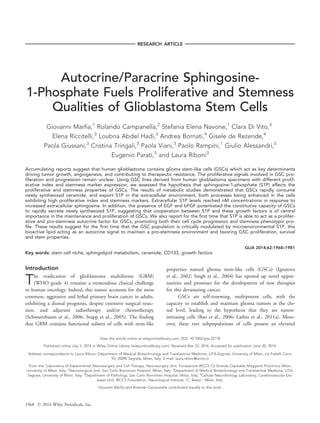
  • 5. number of MIB-1-stained nuclei in brown, reflecting active cellular proliferation, were evident in SC02 (Fig. 1A). The mean value of MIB-1 labeling index, determined by the Ki- 67/MIB-1 antibody, was 2063.6% in pt 1 and 6067.1% in pt 2. SC01 and SC02 Grow as Neurospheres and Exhibit In Vivo Tumorigenic Potential GSCs obtained from pt 1 and 2 GBM specimens were named SC01 and SC02, respectively. In appropriate culture conditions that favor GSC survival and proliferation, both GBM-derived cancer cell lines formed GSCs that grew as free floating clusters, namely neurospheres (Fig. 1B). GSCs were next validated in vivo for their tumorigenic potential in immunodeficient mice. After intracranial injec- tion, both GSC lines formed tumors in all injected mice, with histological appearance similar to human GBMs (Fig. 1C). The mean tumor volume 6 SD at sacrifice was 9.1 6 4.8 mm3 and 12.9 6 5.5 mm3 in mice injected with SC01 and SC02, respectively. The human origin of the tumor was demonstrated by the positive staining with an anti- human nuclei antibody, which identified cells positive to human marker in the slides (Salmaggi et al., 2006). These results, showing that both cell lines exhibit the capacity for sphere formation, and are tumorigenic in brains of immuno- compromised mice, demonstrated that SC01 and SC02 expressed functional features of GSCs (Singh et al., 2004). SC01 and SC02 Neurospheres Differ in the Expression of Stemness Markers In order to phenotype SC01 and SC02, we determined the expression of different putative stem/progenitor cell markers. As shown in Fig. 2, all the investigated markers were highly expressed in both GSCs, with CD90 as the highest (90%) expressed one. It should be noted that CD90 has been recently identified as a novel marker for GSCs in high-grade gliomas and as a more general marker than the recognized GSC marker CD133 (He et al., 2012). The expression of all other markers, including CD133 and CD15, was significantly higher in SC02 than SC01. SC01 and SC02 Differ in the Proliferation Rate and Cell Cycle Parameters In the used culture condition, the analyses of the cellular growth curves showed that, along with passages, the cell num- ber gradually increased, and this increase was much more pronounced in the SC02 line (Fig. 3A). Moreover, at both 2 and 7 days in culture, the proliferation index was significantly higher in SC02 than SC01 (Fig. 3B). The results of the cell cycle evaluation by flow cytome- try revealed differences in the number of cells undergoing mitosis between the two cell lines. In fact, when compared with SC01, the SC02 cell line exhibited a significant increase in the percentage of cells in the mitotic phases and a decrease in that of cells in G0/G1 phase (Fig. 3C). FIGURE 2: Slow- and fast-proliferating GSCs growing as neuro- sphere differ in stemness marker expression. The expression of different stemness and precursor markers in SC01 (grey) and SC02 (blue) cells, obtained by cytofluorimetric analyses is shown. Data are expressed as % of total cells and are the mean 6 SD of 3–5 independent experiments. *P 0.05; **P 0.01; ***P 0.001 SC02 versus SC01. Similar data were obtained at different cell passages. [Color figure can be viewed in the online issue, which is available at wileyonlinelibrary.com.] FIGURE 1: Ki-67 immunoreactivity of GBMs and tumor-derived neurospheres. A: Tumor tissue from pt 2 stained with anti-Ki-67 antibody. The image shows positive nuclear expression of Ki-67 in brown and negative nuclei counterstained in blue (magnification, 40x). B: Repre- sentative microscopic image of GSCs grown as neurosphere (magnification, 20x). C: Representative histological image (hematoxylin and eosin staining) of a brain tumor derived from GSCs after intracranial orthotopic xenograft of nude mice. [Color figure can be viewed in the online issue, which is available at wileyonlinelibrary.com.] 1972 Volume 62, No. 12
  • 6. SC01 and SC02 Differ in Sph Metabolism and Extracellular Release of S1P To follow S1P metabolism and its breakdown product, we used Sph labeled with tritium at the C3 position of the long chain base backbone. This label remains in the lipid through- out the phosphorylation and recycling steps of the sphingoid base, and thus allows tracking of all metabolites as long as the sphingoid backbone remains intact, as well as S1P irre- versible degradation as tritiated water. After administration, tritiated Sph was rapidly incorporated into the two GSC lines in a time-dependent fashion, and, after 120 min, total incor- porated radioactivity accounted for 197.9 6 20.7 in SC01, and 214.2 6 19.8 nCi/dish in SC02, that is approximately half of the administered radioactivity. At all investigated times, the level of intracellular 3 H-Sph was found low, repre- senting less than 10% of the total incorporated radioactivity in both cell lines, this level being 3-, 4-fold higher in SC01 than that of SC02 cells (Fig. 4). These data indicate that both GSC types are able to rapidly incorporate and metabo- lize exogenous Sph, and that the SC02 cells are particularly efficient in this metabolization. At different pulse times, the bulk of incorporated radio- activity was associated to the N-acylated derivatives of Sph, and, in less amounts, to the 1-phosphorylation products (Fig. 4). It is worth noting that the ratio between N-acylated and 1-phosphorylated metabolites was 21.3 and 15.5 in SC01, and 5.2 and 4.9 in SC02 at 15 and 120 min, respectively. The main N-acylated metabolites produced during pulse in both GSC lines were represented by Cer and complex sphingolipids, including sphingomyelin, and, in much lower amounts, GlcCer. All over pulse, Cer was by far the major 3 H-metabolite, its anabolic derivatives increasing with time (Fig. 5). A significant decrease in 3 H-Cer in SC02 compared with SC01 was observed after 120 min pulse (Fig. 5, upper panel). All over pulse, in SC02 cells a marked elevation of sphingomyelin and Glc-Cer was also evident; at 120 min, in SC02 these sphingolipids increased by about 110% and 250%, respectively, relative to SC01 (Fig. 5, lower panels). As reported above, the metabolites derived from the 1- phosphorylation process represented the second class of Sph derivatives (Fig. 4). Among them, intracellular S1P repre- sented only a minor fraction of 1-phosphorylated Sph metab- olites and, at 15 min pulse, its level was significantly lower in SC01 than SC02 (Fig. 6A). Thereafter, there was no relevant change in the cellular S1P level between SC01 and SC02. At all investigated times, tritiated water, represented the vast majority of Sph 1-phosphorylation products, its content being significantly higher (4-, 6-fold) in SC02 than in SC01 (Fig. 6B). After pulse with [C3-3 H]Sph, tritiated water derives from the oxidation of tritiated hexadecenal produced from 3 H-S1P by S1P lyase (Anelli et al., 2005). Our results FIGURE 3: Slow- and fast-proliferating GSCs differ in proliferation and cell cycle parameters. A: SC01 and SC02 were plated at the same initial density, and at each passage viable cells were counted; (B) GSCs at the 5th passage were plated, and after 2 and 7 days in culture, the proliferation index (PI) was calculated as described in “Materials and Methods”; (C) GSCs were plated as above, and cell cycle distribution was analyzed by flow cytometry. Similar data were obtained at different cell passages. *P 0.05; **P 0.01; ***P 0.001, SC02 versus SC01. [Color figure can be viewed in the online issue, which is available at wileyonlinelibrary.com.] Marfia et al.: S1P Prompts Growth and Stemness of GSCs December 2014 1973
  • 7. indicate that a rapid and efficient degradation of S1P, derived from the phosphorylation of exogenous Sph, occurs in GSCs. Besides tritiated water, the analyses of the culture media from both cell lines revealed the presence of labeled S1P (Fig. 6C,D), which increased in a time-dependent fashion in both cell lines. Remarkably, at all investigated times, the S1P levels in the extracellular milieu of SC02 were significantly higher than those of SC01 (Fig. 6C). At 120 min pulse extracellular S1P from SC02 accounted for about 4-fold that of intracellu- lar S1P, and about 9-fold that of SC01 (P 0.001). Extracellular Secretion of S1P by SC Cells is Highly Efficient and Stimulated by Growth Factors To better define the S1P export from GSCs, we next addressed the possibility that elevated levels of Sph might influence its metabolism and S1P export. After pulse of GSCs with 1 mM Sph, the percentage of phosphorylated metabo- lites, including both intracellular S1P and its catabolic prod- uct water, increased in respect with the N-acylated ones in both cell lines. In the presence of 1 mM Sph, the amount of FIGURE 4: Slow- and fast-proliferating GSCs differ in Sph metab- olism. SC01 (left panels) and SC02 (right panels) cells were pulsed with 20 nM 3 H-Sph for 15–120 min. At the end, cells and media were processed and analyzed as described in “Materials and Methods”. The radioactivity distribution (as % of total incor- porated radioactivity) among unmetabolized Sph (green), and its N-acylation (light yellow), and 1-phosphorylation (blue) products is shown. Data are the mean of three experiments, performed in duplicate. *P 0.05; ***P 0.001, SC02 versus SC01. [Color fig- ure can be viewed in the online issue, which is available at wileyonlinelibrary.com.] FIGURE 5: Cer consumption to SM and Glc-Cer is enhanced in fast-proliferating GSCs. Content of labeled Cer (A), sphingomy- elin (B), and Glc-Cer (C) in SC01 (light grey) and SC02 (blue) at 15–120 min pulse with 3 H-Sph. Data are expressed as percent of N-acylated metabolites, and are the mean 6 SD of 3 experi- ments. **P 0.01; ***P 0.001, SC02 versus SC01. [Color figure can be viewed in the online issue, which is available at wileyonlinelibrary.com.] 1974 Volume 62, No. 12
  • 8. released S1P was relevant, reaching on average the concentra- tion of 2 nM in both cell lines (data not shown). Since EGF and bFGF are recognized as autocrine signals in GSCs, we then evaluated the possible influence of these growth factors on S1P release. The results demonstrated that either in the absence or presence of EGF and bFGF, GSCs can release S1P, reflecting a constitutive S1P secretion by the cells. It is relevant to note that this secretion was significantly enhanced by the presence of EGF and bFGF (Fig. 7A,B), supporting an autocrine loop induced by these growth factors. Extracellular S1P Prompts GSC Proliferation and Stemness On the basis of the above results, it was of interest to investi- gate whether extracellular S1P can influence the GSC prolif- eration index and/or stemness. In this scenario, GSCs were cultured in S1P-enriched SCM medium for 48 h, and the number of viable cells was determined. As shown in Fig. 8A, the addition of S1P resulted in a significant increase in viable cells compared with the control group (P 0.01). These results suggest that extracellular S1P promotes GSC survival and/or proliferation. Trypan blue exclusion test revealed that few cells (less than 5%) were stained in control and S1P- treated cells (data not shown). Cytofluorimetric studies were performed. These studies, besides confirming the low level of dead cells in both control and S1P-treated cells, demonstrated that the number of cells in the early and late apoptosis, as well as the necrotic one was significantly reduced after S1P administration (Fig. 8B). However, due to the low levels of death cells, it is unlikely that the increase in cell number in S1P-treated cells results from reduced apoptosis/necrosis, and FIGURE 6: Slow- and fast-proliferating GSCs differ in S1P degradation and export. The radioactivity associated to (A) intracellular S1P (S1Pin), (B) water, and (C) extracellular S1P (S1Pout) in SC01 (dotted line) and SC02 (straight line) is shown at 15–120 min pulse with 3 H-Sph. Results are expressed as nCi/well, and are the mean 6 SD of three experiments in duplicate. **P 0.01; ***P 0.001, SC02 versus SC01. D: Representative autoradiographic image of the extracellular S1P containing fractions from SC01 and SC02, after 15–120 min pulse. [Color fig- ure can be viewed in the online issue, which is available at wileyonlinelibrary.com.] Marfia et al.: S1P Prompts Growth and Stemness of GSCs December 2014 1975
  • 9. it may indicate an effect on cell cycle progression. We then performed FACS analyses to determine the cell cycle profile in S1P-treated cells. After culturing in S1P-containing medium, quantitative analysis showed that cells in the G0/G1 phase decreased and those in the S and G2/M phases signifi- cantly increased (Fig. 8C), suggesting that GSCs cultured in S1P-containing medium transitioned from the G1 phase to the S and G2/M phases of the cell cycle. In agreement, we found that the proliferation index of S1P-treated cells was increased significantly in comparison with cells that did not receive the sphingoid molecule, and this effect was inhibited by FTY720 (Fig. 8D). Taken together, these results point out that extracellular S1P, after binding to S1PRs, promotes GSC survival and proliferation. In order to evaluate if the prolifer- ating effect of S1P was selective for GSCs or shared with non-stem GBM cells, we prepared primary cultures of GBM cells from tumor samples from pt 1 and 2, and evaluated the effect of S1P on their proliferation. These experiments dem- onstrated that 200 nM S1P significantly increased the prolif- eration of GBM cells from pt 1 and pt 2, by 26.8% and 38.2%, respectively (P 0.01 in both cases). Finally, we evaluated the effect of nanomolar concentra- tions of S1P on cell stemness. Remarkably, the results of the phenotypic characterization by FACS analyses demonstrated that S1P significantly increased the expression of different stemness and precursor markers in GSCs (Fig. 9, upper panel), including, among others, CD133 and CD15. This S1P effect could be due to either an induction of stem cell marker expression or a selective expansion of cells that are already expressing stem cell markers. In order to clarify this point, we evaluated the effect of S1P on stem marker expres- sion in primary cultures of GBM cells prepared from pt 1 and 2. We first found that primary GBM cells express very low levels of stemness markers. Indeed, as shown in Fig. 9 (lower panel), the percentages of CD1331 and CD151 cells in GBM cells from pt 2 were by far lower (15- and 25-fold) the corresponding value of SC02, their corresponding GSCs (Fig. 9, upper panel). We found similar differences in the stem marker expression by GBM cells from pt 1 and SC01. Of relevance, and differently from what we observed in GSCs, S1P exerted only modest, if any, effect on the stemness profile of primary GBM cells (Fig. 9, lower panel). Discussion A central unresolved issue with GSCs is their growth signal- ing and cell cycle control. This lack of progress is especially surprising given that the histopathologic assessment in the diagnosis of GBM includes the identification of a high tumor cell mitotic index (Persano et al., 2013; Pojo and Costa, 2011), as well as that a key determinant of GSCs is their capacity for extensive proliferation and self-renewing (Al-Hajj et al., 2004; Oliver and Wechsler-Reya, 2004). S1P is increasingly recognized as a critical growth factor for a number of cancer cell types and, among others, glioma cells (Yester et al., 2011). A role for S1P as a potential mito- genic factor in GSCs, however, remains uncharacterized. In this study we investigated the origin and role for S1P in GSC properties and specifically in GSC proliferation and stemness. To the best of our knowledge, this work is the first to address this issue. Among different GSC lines prepared from GBM speci- mens, on the bases of proliferation index and cell cycle parameters, we selected two lines, named SC01 and SC02, as representative of slow- and fast-proliferating cells, respectively. These GSC populations demonstrated heterogeneity not only FIGURE 7: Growth factors stimulate S1P export by GSCs. GSCs were pulsed with 20 nM 3 H-Sph in SCM without (- GFs) or with (1 GFs) EGF and bFGF for 120 min. A: Representative autoradio- graphic image of the extracellular S1P containing fractions from SC01 and SC02, separated by HPTLC. B: Radioactivity associated to extracellular S1P (S1Pout) is expressed as nCi/well and is the mean 6 SD of three experiments. **P 0.01; ***P 0.001, 1 GFs versus - GFs. [Color figure can be viewed in the online issue, which is available at wileyonlinelibrary.com.] 1976 Volume 62, No. 12
  • 10. in terms of proliferative potential, but also of expression of stem cell markers, the fast proliferative status of SC02 being paralleled by a higher expression of stemness and progenitor cell parameters. Metabolic studies revealed that both GSC lines were able to efficiently take up and metabolize Sph, but with sig- nificant differences between slow-proliferating and fast- proliferating cells. In particular, SC02 exhibited an extremely rapid Sph processing, and a significant higher ratio between 1-phosphorylated and N-acylated metabolites than SC01, indicating that in the two cell lines a different propensity exists in the use of Sph by the two different metabolic routes. Moreover, we show that the two cell types had different kinetics of Cer consumption. Both types of Cer consumption, which convert Cer to SM and GlcCer were potentiated in SC02, and appear functional to avoid Cer accumulation in these cells. A further result of the metabolic experiments was that GSCs constitutively exhibit the property to efficiently release S1P in the extracellular microenvironment, in line with our recent study (Riccitelli et al., 2013). The novel finding is that the proliferative properties of GSCs appeared as related to efficient and rapid release of newly produced S1P. Indeed extracellular S1P level was up to 10-fold higher in SC02 than SC01, suggesting that the high extent of S1P release by SC02 cells reflects, and most probably participates, in their FIGURE 8: Extracellular S1P promotes GSC survival and proliferation. SC02 cells were treated in the absence (Ct) or presence of 200 nM S1P alone (S1P) or with 100 nM FTY720 (S1P1FTY) for 48 hours. At the end, cell viability (A), apoptosis (B), cell cycle parameters (C), and proliferation index (PI) (D) were assessed as reported in “Materials and Methods”. *P 0.05; **P 0.01; ***P 0.001, S1P-treated versus Ct. Similar effects of S1P were obtained on SC01 cells. [Color figure can be viewed in the online issue, which is available at wileyonlinelibrary.com.] Marfia et al.: S1P Prompts Growth and Stemness of GSCs December 2014 1977
  • 11. proliferation and stemness features. In spite representative of different GSC lines, the limited number of GSC lines here used and the heterogeneity of GBMs warrant further studies to confirm these findings. Overall, these metabolic experiments demonstrate that fast proliferating cells exhibit an increased flux through the pathway converting Sph to S1P, paralleled by an increase of Cer to complex sphingolipids, the most relevant changes being gain of extracellular S1P and loss of intracellular Sph and Cer. Since the balance between S1P and Cer/Sph levels is believed to provide a rheostat mechanism determinant for cell proliferation and fate (Spiegel and Merrill, 1996), our results suggest that altering this rheostat in favor of S1P provides SC02 an advantage in their proliferative and stemness qual- ities. In agreement, a decrease in total ceramides and an increase in S1P content with increasing glioma grade were reported (Abuhusain et al., 2013; Riboni et al., 2002). It is becoming increasingly clear that GSC properties involve a complex interplay between GSCs and their micro- environment, and the GSC microenvironment provides essen- tial cues to their maintenance (Charles et al., 2012; Filatova et al., 2013; Heddleston et al., 2011). Further results here obtained underscore the relevance of the extracellular environ- ment in affecting the extent of S1P export. Indeed, as dis- cussed below, we found that the availability of Sph, as well as that of growth factors are critical factors influencing the extracellular levels of S1P in GSCs. When GSCs were fed with increased concentrations of Sph, released S1P was found in the nanomolar range, i.e. in the range of Kd for S1P binding to its receptors (Rosen et al., 2009). Free Sph originates from the intracellular degra- dation of complex sphingolipids, and high Sph may also result from its release from necrotic cells (Cavalli et al., 2002), particularly abundant in aggressive GBMs (Lacroix et al., 2001). The observed high capacity of S1P export sug- gests that, in circumstances of high sphingolipid degradation, or in perinecrotic regions where GSCs are enriched in (Seidel et al., 2010), S1P biosynthesis and release occur very rapidly and to a high extent in GSCs, providing these cells a favor- able environment. Extrinsically, GSCs are regulated by growth factors and, among them, EGF and bFGF, which play a crucial role as autocrine factors sustaining GSC self-renewal and prolifera- tion (Li et al., 2009; Soeda et al., 2008), and support the growth of GSC in culture (Lee et al., 2006). We provide evi- dence that the presence of bFGF and EGF significantly pro- moted the S1P export by GSCs into their extracellular milieu. Notably, in spite that S1P is a potent chemoattractant for neural stem cells, these cells are not able to export S1P in their external environment, even in the presence of the growth factors (Kimura et al., 2007). Our study supports that GSCs not only receive S1P signal from extracellular sources but, differently from neural stem cells, also efficiently trans- mit S1P as signal to manipulate their environment. Evolving data in GBMs suggest that multiple GSC pools exist within these tumors with different proliferative state or control mechanism, and that these pools may replenish each other (Piccirillo et al., 2009). Whether S1P originates from and/or participates to the dynamic heterogeneous nature of GSCs remains a future challenge. We continued by analyzing the response of GSCs to S1P, and found that GSCs switched to a faster growth mode when exposed to exogenous S1P, implying that S1P can act as an autocrine signal for GSCs not only to invade and survive, but also to grow and proliferate. Indeed, here we show for the first time that the extracellular S1P sends a proliferative FIGURE 9: Extracellular S1P enhances stemness in GSCs but not in GBM cells. SC02 cells (upper panel) and GBM cells from pt 2 (lower panel) were incubated in the absence (Ct) or presence of 200 nM S1P (S1P) for 48 h. At the end, the immunophenotypic profile was assessed by cytofluorimetric analyses. Results are expressed as percent of total cells and are the mean 6 SD of three independent experiments. **P 0.01; ***P 0.001, S1P- treated versus Ct. Similar effects of S1P were observed on SC01 and GBM cells from pt 1. [Color figure can be viewed in the online issue, which is available at wileyonlinelibrary.com.] 1978 Volume 62, No. 12
  • 12. signal to GSCs that is strong enough to promote cell cycle and progression into G2/M phase of GSCs. Further experi- ments showed that S1P was able to exert a proliferative effect also in primary GBM cells from both pt 1 and pt 2, in agree- ment with previous findings (Van Brocklyn et al., 2002; Yoshida et al., 2010). Thus, it appears that GSCs share with non-stem GBM cells the ability to respond to S1P with increased proliferation. We found that the proliferative action of S1P in GSCs was inhibited by FTY720, a Sph analogue that, after cellular internalization and phosphorylation, is released extracellularly and binds to S1P receptors. It has been demonstrated that phosphorylated FTY720 induces internalization and degradation of the S1P1 receptor, resulting in prolonged down-regulation of this receptor (Oo et al., 2007). Since S1P1 expression is increased in CD1331 cells, as well as in experimental and intracranial glioblastomas (Annabi et al., 2009), it appears reasonable that S1P1 might be involved in the proliferating effect of extracellular S1P in CD1331 GSCs. Of relevance, besides changing the growth pattern of GSCs, our study provides first evidence that S1P enhanced the stemness phenotype in GSCs, elevating the level of stem cell markers. In spite this evidence suggests that S1P may play a role to maintain GSCs undifferentiated, the pluripotency potential of S1P in GSCs remains to be characterized. The finding that S1P promotes stemness parameters in GSCs, might reside in its ability to promote either the expres- sion of different stem markers or the proliferation of GSCs that are already expressing stem cell markers. In non-stem pri- mary GBM cells, we observed that S1P did not induce stem- ness marker expression, thus suggesting that its effect resides in the selective expansion of GSCs. We observed that S1P treatment resulted in elevation of the CD1331 population of GSCs. It is worth noting that this subpopulation of GSCs was demonstrated to present a more malignant behavior, and to maintain the GSC pool in the tumor, also increasing its cellular heterogeneity (Lathia et al., 2011b; Zeppernick et al., 2008). Although at present the mechanisms of the stemness-promoting action of S1P are unknown, it appears reasonable that the S1P might induce the expansion of CD1331 GSCs, and/or their de- differentiation to CD1331 GSCs. Of interest, besides CD1331 cells, S1P increased the CD151 population too, and this population was reported to complement some part of CD133 function, as it survives better and proliferates faster than their negative counterpart (Zeppernick et al., 2008). We found that S1P also increased the number of cells expressing the hematopoietic/endothelial progenitor markers CD311 and CD341 . Intriguingly, CD311 and CD341 GSCs were found to co-express CD1331 , and to be more proliferating than the negative counterpart (Christensen et al., 2011). Overall, our study suggests that fast-proliferating GSCs possess a high propensity for secreting S1P, and that autocrine S1P acts as microenvironmental signal to increase the GSC population and to enhance their stem cell properties. Besides emphasizing the importance of S1P as a proliferative factor, our study underscores S1P export as a crucial step in connect- ing GSCs with their niche, and suggests that S1P secretion by GSCs may also have the effect of targeting functions of non- stem niche cells in a paracrine manner. In line with this, S1P has been reported to favor propagation of non-stem cell types present in the GSC niche, such as astrocytes (Bassi et al., 2006), endothelial cells (Rosen and Goetzl, 2005), and as confirmed in our study, in non-stem glioma cells, too (Van Brocklyn et al., 2002; Yoshida et al., 2010). In conclusion, this work implicates S1P to be an auto- crine/paracrine mediator acting as a mitogenic and stemness- favoring factor through direct effects in GSCs, and possibly through the induction of their niche. This prompts further studies to understand the mechanism of S1P secretion and action in GSC microenvironment, and suggests that the inhi- bition of S1P release from GSCs, not previously considered as a GBM therapeutic, could be a valuable strategy to curtail GBM progression. Acknowledgment Grant sponsor: Italian Ministry of University and Scientific and Technological Research PRIN and FIRST (to LR and PV) and by LR8 of IRCCS Foundation Neurological Insti- tute C. Besta. The authors would like to thank Dr. G. Razionale for proofreading the manuscript. References Abuhusain HJ, Matin A, Qiao Q, Shen H, Kain N, Day BW, Stringer BW, Daniels B, Laaksonen MA, Teo C, McDonald KL, Don AS. 2013. A metabolic shift favoring sphingosine 1-phosphate at the expense of ceramide controls glioblastoma angiogenesis. J Biol Chem 288:37355–37364. Al-Hajj M, Becker MW, Wicha M, Weissman I, Clarke MF. 2004. Therapeutic implications of cancer stem cells. Curr Opin Genet Dev 14:43–47. Anelli V, Bassi R, Tettamanti G, Viani P, Riboni L. 2005. Extracellular release of newly synthesized sphingosine-1-phosphate by cerebellar granule cells and astrocytes. J Neurochem 92:1204–1215. Annabi B, Lachambre MP, Plouffe K, Sartelet H, Beliveau R. 2009. Modulation of invasive properties of CD1331 glioblastoma stem cells: Arole for MT1-MMP in bioactive lysophospholipid signaling. Mol Carcinog 48: 910–919. Bao S, Wu Q, McLendon RE, Hao Y, Shi Q, Hjelmeland AB, Dewhirst MW, Bigner DD, Rich JN. 2006. Glioma stem cells promote radioresistance by preferential activation of the DNA damage response. Nature 444:756–760. Bassi R, Anelli V, Giussani P, Tettamanti G, Viani P, Riboni L. 2006. Sphingo- sine-1-phosphate is released by cerebellar astrocytes in response to bFGF Marfia et al.: S1P Prompts Growth and Stemness of GSCs December 2014 1979
  • 13. and induces astrocyte proliferation through Gi-protein-coupled receptors. Glia 53:621–630. Cavalli AL, Ligutti JA, Gellings NM, Castro E, Page MT, Klepper RE, Palade PT, McNutt WT, Sabbadini RA. 2002. The Role of TNFa and sphingolipid sig- naling in cardiac hypoxia: Evidence that cardiomyocytes release TNFa and sphingosine. Basic Appl Myol 12:167–175. Charles NA, Holland EC, Gilbertson R, Glass R, Kettenmann H. 2012. The brain tumor microenvironment. Glia 60:502–514. Christensen K, Schrïder HD, Kristensen BW. 2011. CD1331 niches and single cells in glioblastoma have different phenotypes. Neuro Oncol 104: 129–143. Estrada-Bernal A, Palanichamy K, Ray Chaudhury A, Van Brocklyn JR. 2012. Induction of brain tumor stem cell apoptosis by FTY720: A potential thera- peutic agent for glioblastoma. Neuro Oncol 14:405–415. Filatova A, Acker T, Garvalov BK. 2013. The cancer stem cell niche(s): The crosstalk between glioma stem cells and their microenvironment. Biochim Biophys Acta 1830:2496–2508. Furuya H, Shimizu Y, Kawamori T. 2011. Sphingolipids in cancer. Cancer Metastasis Rev 30:567–576. Hadjipanayis CG, Van Meir EG. 2009. Brain cancer propagating cells: Biol- ogy, genetics and targeted therapies. Trends Mol Med 15:519–530. He J, Liu Y, Zhu T, Zhu J, DiMeco F, Vescovi AL, Heth JA, Muraszko KM, Fan X, Lubman DM. 2012. CD90 is identified as a marker for cancer stem cells in primary high grade gliomas using tissue microarrays. Mol Cell Proteomics 11: 1–8. Heddleston JM, Hitomi M, Venere M, Flavahan WA, Yan K, Kim Y, Minhas S, Rich JN, Hjelmeland AB. 2011. Glioma stem cell maintenance: The role of the microenvironment. Curr Pharm Des 17:2386–2401. Ignatova TN, Kukekov VG, Laywell ED, Suslov ON, Vrionis FD, Steindler DA. 2002. Human cortical glial tumors contain neural stem-like cells expressing astroglial and neuronal markers in vitro. Glia 39:193–206. Kapitonov D, Allegood JC, Mitchell C, Hait NC, Almenara JA, Adams JK, Zipkin RE, Dent P, Kordula T, Milstien S, Spiegel S. 2009. Targeting sphingo- sine kinase 1 inhibits AKT signaling, induces apoptosis, and suppresses growth of human glioblastoma cells and xenografts. Cancer Res 69:6915– 6923. Kimura A, Ohmori T, Ohkawa R, Madoiwa S, Mimuro J, Murakami T, Kobayashi CE, Hoshino CY, Yatomi AY, Sakata DY. 2007. Essential roles of sphingosine 1-phosphate/S1P1 receptor axis in the migration of neural stem cells toward a site of spinal cord injury. Stem Cells 25:115–124. Krishnamoorthy S, Honn KV. 2011. Eicosanoids and other lipid mediators and the tumor hypoxic microenvironment. Cancer Metastasis Rev 30:613–618. Lacroix M, Abi-Said D, Fourney DR, Gokaslan ZL, Shi W, DeMonte F, Lang FF, McCutcheon IE, Hassenbusch SJ, Holland E, Hess K, Michael C, Miller D, Sawaya R. 2001. A multivariate analysis of 416 patients with glioblastoma multiforme: Prognosis, extent of resection, and survival. J Neurosurg 95:190– 198. Laks DR, Masterman-Smith M, Visnyei K, Angenieux B, Orozco NM, Foran I, Yong WH, Vinters HV, Liau LM, Lazareff JA, Mischel PS, Cloughesy TF, Horvath S, Kornblum HI. 2009. Neurosphere formation is an independent predictor of clinical outcome in malignant glioma. Stem Cells 27:980–987. Lathia JD, Heddleston JM, Venere M, Rich JN. 2011a. Deadly teamwork: Neural cancer stem cells and the tumor microenvironment. Cell Stem Cell 8: 482–485. Lathia JD, Hitomi M, Gallagher J, Gadani SP, Adkins J, Vasanji A, Liu L, Eyler CE, Heddleston JM, Wu Q, Minhas S, Soeda A, Hoeppner DJ, Ravin R, McKay RD, McLendon RE, Corbeil D, Chenn A, Hjelmeland AB, Park DM, Rich JN. 2011b. Distribution of CD133 reveals glioma stem cells self-renew through symmetric and asymmetric cell divisions. Cell Death Dis 2: e200. Lee J, Kotliarova S, Kotliarov Y, Li A, Su Q, Donin NM, Pastorino S, Purow BW, Christopher N, Zhang W, Park JK, Fine HA. 2006. Tumor stem cells derived from glioblastomas cultured in bFGF and EGF more closely mirror the phenotype and genotype of primary tumors than do serum-cultured cell lines. Cancer Cell 9:391–403. Le Stunff H, Milstien S, Spiegel S. 2004. Generation and metabolism of bio- active sphingosine-1-phosphate. J Cell Biochem 92:882–899. Li G, Chen Z, Hu YD, Wei H, Li D, Ji H, Wang DL. 2009. Autocrine factors sustain glioblastoma stem cell self-renewal. Oncol Rep 21:419–424. Liu X, Zhang QH, Yi GH. 2012. Regulation of metabolism and transport of sphingosine-1-phosphate in mammalian cells. Mol Cell Biochem 363:21–33. Louis DN, Ohgaki H, Wiestler OD, Cavenee WK, Burger PC, Jouvet A, Scheithauer BW, Kleihues P. 2007. The 2007 WHO classification of tumours of the central nervous system. Acta Neuropathol 114:97–109. Maceyka M, Harikumar KB, Milstien, S, Spiegel S. 2012. Sphingosine-1-phos- phate signaling and its role in disease. Trends Cell Biol 22:50–60. Mora R, Dokic I, Kees T, H€uber CM, Keitel D, Geibig R, Br€ugge B, Zentgraf H, Brady NR, Regnier-Vigouroux A. 2010. Sphingolipid rheostat alterations related to transformation can be exploited for specific induction of lysosomal cell death in murine and human glioma. Glia 58:1364–1383. Oliver TG, Wechsler-Reya RJ. 2004. Getting at the root and stem of brain tumors. Neuron 42:885–888. Oo ML, Thangada S, Wu MT, Liu CH, Macdonald TL, Lynch KR, Lin CY, Hla T. 2007. Immunosuppressive and anti-angiogenic sphingosine 1-phosphate receptor-1 agonists induce ubiquitinylation and proteasomal degradation of the receptor. J Biol Chem 282:9082–9089. Pallini R, Ricci-Vitiani L, Banna GL, Signore M, Lombardi D, Todaro M, Stassi G, Martini M, Maira G, Larocca LM, De Maria R. 2008. Cancer stem cell anal- ysis and clinical outcome in patients with glioblastoma multiforme. Clin Can- cer Res 14:8205–8212. Persano L, Rampazzo E, Basso G, Viola G. 2013. Glioblastoma cancer stem cells: Role of the microenvironment and therapeutic targeting. Biochem Phar- macol 85:612–622. Pettus BJ, Chalfant CE, Hannun YA. 2002. Ceramide in apoptosis: An over- view and current perspectives. Biochim Biophys Acta 1585:114–125. Piccirillo SG, Combi R, Cajola L, Patrizi A, Redaelli S, Bentivegna A, Baronchelli S, Maira G, Pollo B, Mangiola A, DiMeco F, Dalpra L, Vescovi AL. 2009. Distinct pools of cancer stem-like cells coexist within human glioblasto- mas and display different tumorigenicity and independent genomic evolu- tion. Oncogene 28:1807–1811. Pojo M, Costa BM. 2011. Molecular hallmarks of gliomas. In: Garami M, edi- tor. Molecular targets of CNS tumors. Rijeka: InTech. pp 177–200. Pyne NJ, Pyne S. 2010. Sphingosine 1-phosphate and cancer. Nat Rev Can- cer 10:489–503. Ralte AM, Sharma MC, Karak AK, Mehta VS, Sarkar C. 2001. Clinicopatholog- ical features, MIB-1 labelling index and apoptotic index in recurrent astrocytic tumors. Pathol Oncol Res 7:267–278. Riboni L, Campanella R, Bassi R, Villani R, Gaini SM, Martinelli-Boneschi F, Viani P, Tettamanti G. 2002. Ceramide levels are inversely associated with malignant progression of human glial tumors. Glia 39:105–113. Riboni L, Viani P, Tettamanti G. 2000. Estimating sphingolipid metabolism and trafficking in cultured cells using radiolabeled compounds. Methods Enzymol 311:656–682. Riccitelli E, Giussani P, Di Vito C, Condomitti G, Tringali C, Caroli M, Galli R, Viani P, Riboni L. 2013. Extracellular sphingosine-1-phosphate: A novel actor in human glioblastoma stem cell survival. PLoS ONE 8:e68229. Rosen H, Goetzl EJ. 2005. Sphingosine-1-phosphate and its receptors: An autocrine and paracrine network. Nat Rev Immunol 5:560–570. Rosen H, Gonzalez-Cabrera PJ, Sanna MG, Brown S. 2009. Sphingosine 1- phosphate receptor signaling. Annu Rev Biochem 78:743–768. Salmaggi A, Boiardi A, Gelati M, Russo A, Calatozzolo C, Ciusani E, Sciacca FL, Ottolina A, Parati EA, La Porta C, Alessandri G, Marras C, Croci D, De Rossi M. 2006. Glioblastoma-derived tumorospheres identify a population of 1980 Volume 62, No. 12
  • 14. tumor stem-like cells with angiogenic potential and enhanced multidrug resistance phenotype. Glia 54:850–860. Schmidt KF, Ziu M, Schmidt NO, Vaghasia P, Cargioli TG, Doshi S, Albert MS, Black PM, Carroll RS, Sun Y. 2004. Volume reconstruction techniques improve the correlation between histological and in vivo tumor volume meas- urements in mouse models of human gliomas. J Neurooncol 68:207–215. Schwartzbaum JA, Fisher JL, Aldape KD, Wrensch M. 2006. Epidemiology and molecular pathology of glioma. Nat Clin Pract Neurol 2:494–503. Seidel S, Garvalov BK, Wirta V, von Stechow L, Sch€anzer A, Meletis K, Wolter M, Sommerlad D, Henze AT, Nister M, Reifenberger G, Lundeberg J, Frisen J, Acker T. 2010. A hypoxic niche regulates glioblastoma stem cells through hypoxia inducible factor 2a. Brain 133:983–995. Singh SK, Hawkins C, Clarke ID, Squire JA, Bayani J, Hide T, Henkelman RM, Cusimano MD, Dirks PB. 2004. Identification of human brain tumour initiating cells. Nature 432:396–401. Soeda A, Inagaki A, Oka N, Ikegame Y, Aoki H, Yoshimura S, Nakashima S, Kunisada T, Iwama T. 2008. Epidermal growth factor plays a crucial role in mitogenic regulation of human brain tumor stem cells. J Biol Chem 283: 10958–1866. Spiegel S, Merrill AH Jr. 1996. Sphingolipid metabolism and cell growth reg- ulation. FASEB J 10:1388–1397. Stupp R, Mason WP, van den Bent MJ, Weller M, Fisher B, Taphoorn MJ, Belanger K, Brandes AA, Marosi C, Bogdahn U, Curschmann J, Janzer RC, Ludwin SK, Gorlia T, Allgeier A, Lacombe D, Cairncross JG, Eisenhauer E, Mirimanoff RO. 2005. Radiotherapy plus concomitant and adjuvant temozolo- mide for glioblastoma. N Engl J Med 352:987–996. Takuwa Y, Okamoto Y, Yoshioka K, Takuwa N. 2012. Sphingosine-1-phos- phate signaling in physiology and diseases. Biofactors 38:329–337. Van Brocklyn JR, Jackson CA, Pearl DK, Kotur MS, Snyder PJ, Prior TW. 2005. Sphingosine kinase-1 expression correlates with poor survival of patients with glioblastoma multiforme: Roles of sphingosine kinase isoforms in growth of glioblastoma cell lines. J Neuropathol Exp Neurol 64:695–705. Van Brocklyn J, Letterle C, Snyder P, Prior T. 2002. Sphingosine-1-phosphate stimulates human glioma cell proliferation through Gi-coupled receptors: Role of ERK/MAP kinase and phosphatidylinositol 3-kinase b. Cancer Lett 181:195–204. Yester JW, Tizazu E, Harikumar KB, Kordula T. 2011. Extracellular and intra- cellular sphingosine-1-phosphate in cancer. Cancer Metastasis Rev 30:577– 597. Yoshida Y, Nakada M, Harada T, Tanaka S, Furuta T, Hayashi Y, Kita D, Uchiyama N, Hayashi Y, Hamada J. 2010. The expression level of sphingosine-1-phosphate receptor type 1 is related to MIB-1 labelling index and predicts survival of glioblastoma patients. J Neuro Oncol 9:41–47. Zeppernick F, Ahmadi R, Campos B, Dictus C, Helmke BM, Becker N, Lichter P, Unterberg A, Radlwimmer B, Herold-Mende CC. 2008. Stem cell marker CD133 affects clinical outcome in glioma patients. Clin Cancer Res 14:123–129. Marfia et al.: S1P Prompts Growth and Stemness of GSCs December 2014 1981