Anatomy of spinal cord
•Download as DOC, PDF•
2 likes•786 views

Medicine PG poster presentation

Recommended
More Related Content
What's hot
What's hot (20)
Viewers also liked
Viewers also liked (20)
Similar to Anatomy of spinal cord
Similar to Anatomy of spinal cord (20)
More from Kurian Joseph
More from Kurian Joseph (20)
Recently uploaded
Recently uploaded (20)
Anatomy of spinal cord
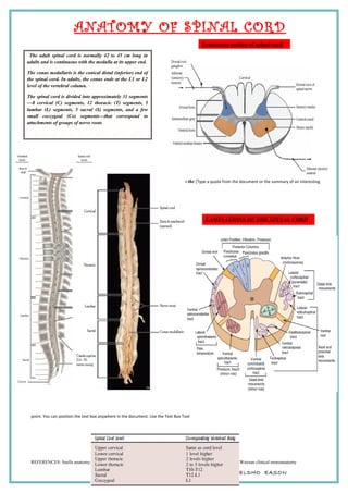
- 1. ANATOMY OF SPINAL CORD ood is supplied to the [Type a quote from the document or the summary of an interesting point. You can position the text box anywhere in the document. Use the Text Box Tool REFERENCES: Snells anatomy; Dejong’s neurologic examination, Hendelman atlas of functional neuroanatomy; Waxnan clinical neuroanatomy Dr YELDHO EASON Transverse section of spinal cord The adult spinal cord is normally 42 to 45 cm long in adults and is continuous with the medulla at its upper end. The conus medullaris is the conical distal (inferior) end of the spinal cord. In adults, the conus ends at the L1 or L2 level of the vertebral column. The spinal cord is divided into approximately 31 segments —8 cervical (C) segments, 12 thoracic (T) segments, 5 lumbar (L) segments, 5 sacral (S) segments, and a few small coccygeal (Co) segments—that correspond to attachments of groups of nerve roots LAMINATIONS OF THE SPINAL CORD
- 2. ANATOMY OF SPINAL CORD REFERENCES: Snells anatomy; Dejong’s neurologic examination, Hendelman atlas of functional neuroanatomy; Waxnan clinical neuroanatomy Dr YELDHO EASON VENOUS DRAINAGE OF SPINAL CORD. Venous drainage is by - six irregular plexiform channels - There is one each along the, anterior and posterior midlines and along the line of attachment of dorsal and ventral roots. - These are drained by radicular veins into epidural venous plexus - Batson’s plexus (valveless spinovertebral venous plexus) continues upwards into the intracranial cavity, which may be a means of transport of tumor cells. ARTERIAL SUPPLY OF SPINAL CORD. Arterial supply is via 1) One anterior spinal artery – a branch of vertebral artery – descends the entire length of spinal cord at the anterior median fissure – supplies anterior 2/3rd of spinal cord 2) Unpaired anterior medullary arteries - branch of lateral spinal artery, which in turn is a branch of intercostal artery - divides into anterior and posterior radicular branches to anastamose with anterior spinal and posterior spinal arteries respectively - supply the peripheral areas of the cord 3) A pair of posterior spinal arteries - branches of vertebral arteries - supply posterior horns and dorsal funiculi 4) Central arteries - branch of anterior spinal artery - supply central portions of the cord on both sides 5) Artery of Adamkiewicz - largest medullary artery - arises between T9 to L2 levels - reinforces the supply to lumbar segments Special points:: 1. Anastomosis is least efficient at the region of lateral columns 2. Cervical and lumbar segments have rich vascular supply 3. T1 to T4 segments are most vulnerable to ischemia 4. Anterior spinal artery thrombosis causes ischemia of anterior 2/3rd of the cord, sparing the posterior column, hence leads to dissociated sensory loss.