
Recommended
More Related Content
What's hot
What's hot (20)
Similar to Gastrointestinal_Disorder.pptx, GERD, Peptic Ulcer Diseases, Inflammatory Bowel Diseases, Alcoholic Liver Diseases
Similar to Gastrointestinal_Disorder.pptx, GERD, Peptic Ulcer Diseases, Inflammatory Bowel Diseases, Alcoholic Liver Diseases (20)
More from Dr. Kiran Dhamak
More from Dr. Kiran Dhamak (11)
Recently uploaded
Recently uploaded (20)
Gastrointestinal_Disorder.pptx, GERD, Peptic Ulcer Diseases, Inflammatory Bowel Diseases, Alcoholic Liver Diseases
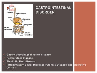
- 1. Gastro oesophageal reflux disease Peptic Ulcer Disease Alcoholic liver disease Inflammatory Bowel Diseases (Crohn’s Disease and Ulcerative Colitis) GASTROINTESTINAL DISORDER
- 2. The term gastrointestinal disorders refer to any condition or disease that occurs within the gastrointestinal tract. For example, gastro esophageal reflux disease (GERD), peptic ulcer disease (PUD), alcoholic liver disease (ALD), inflammatory bowel disease (IBD), etc. Gastrointestinal tract includes the mouth, esophagus, stomach, small intestine, large intestine, colon, rectum, biliary tract, gallbladder, liver, and pancreas. INTRODUCTION
- 3. Gastro esophageal reflux disease (GERD) is a chronic condition in which gastric content flow back into the esophagus, causing irritation to the esophageal mucosa with or without tissue damage. The GERD with tissue damage is known as esophagitis or erosion GERD and without tissue damage is known as non- erosive reflux disease (NERD) GASTRO ESOPHAGEAL REFLUX DISEASE (GERD)
- 4. Risk Factors Linked to GERD: Age and male sex are associated with a higher incidence of esophagitis. Obesity - Obese people 2.5 times more likely to have GERD than those with normal body mass index (BMI). Alcohol consumption Stress Smoking Coffee, chocolate, acidic and fatty foods (e.g. spicy foods, citrus, carbonated drinks Certain eating habits (e.g., heavy meals or lying down shortly after eating) ETIOPATHOGENESIS OF GERD
- 5. Hiatal hernia (It is a condition in which part of the stomach pushes up through the diaphragm muscle). Medication. A number of common drugs are associated with GERD e.g. Anticholinergic, benzodiazepines, calcium channel blockers, dopamine, nicotine nitrates, theophylline, estrogens, progesterone, glucagon, some prostaglandin, NSAIDs, bisphosphonates, antibiotics, etc. Pregnancy Gastrointestinal malformations and tumors latrogenic (e.g. after gastrectomy) Scleroderma Infectious esophagitis Asthma
- 6. The various mechanisms involved in the production of gastroesophageal reflux are inappropriate, transient relaxation of the lower esophageal sphincter (LES), pressure abnormalities in the LES (due to hormonal and neural mediators, food, drugs and patient lifestyle), poor esophageal clearance, delayed gastric emptying time and hiatal hernia. Untreated GERD may lead to Barrett esophagus, esophagitis, ulceration, stricture, and rarely adenocarcinoma (An esophageal stricture is an abnormal tightening of the esophagus)
- 7. Clinical manifestations of GERD can be classified as esophageal and extraesophageal manifestations of GERD. (A) Esophageal (Typical) Manifestations Retrosternal pain (Heart burn) Chest pain Epigastric pain Regurgitation mostly after meals Dysphagia (difficulty or discomfort in swallowing) Odynophagia (Painful swallowing) Nausea Belching Early satiety Bloating Water brash (Excessive salivation) Halitosis (Bad breath) CLINICAL MANIFESTATIONS OF GERD
- 8. (B) Extraesophageal (Atypical) Manifestations Chronic cough Asthma Laryngitis Bronchospasm Recurrent pulmonary infections Otitis media Dental erosion Discomfort in the nose Trouble sleeping
- 9. Diagnosis and Investigation of GERD A diagnosis of GERD based on the presence of typical symptoms is correct in only 70% of patients. Diagnostic studies, e.g, upper GI endoscopy, esophageal manometry test, 24-hours pH probe test and nuclear medicine gastric emptying study, may be indicated to confirm the diagnosis or to rule out other causes of symptoms.
- 10. The goals of GERD treatment are to control symptoms, to heal esophagitis, and prevent recurrent esophagitis or other complications. The treatment of GERD consists of lifestyle modification and initiating acid suppression therapy, preferably with proton pump inhibitors (PPIs). Sometimes surgical treatment with corrective anti reflux surgery is considered in select cases, e.g, patients who develop complications despite receiving optimal drug therapy. Patients with signs of GERD complications or other illness or do not respond to drug therapy should be considered for further diagnostic testing MANAGEMENT OF GERD
- 11. Non-pharmacological management of GERD includes patient education, lifestyle modification and surgical therapy. All patients should be educated regarding factors that may worsen the symptoms. Lifestyle modification is considered to be the first line of treatment of GERD. Lifestyle modification includes Avoid alcohol, chocolate, citrus juice, and tomato-based products. Weight loss (if overweight) Avoid peppermint, coffee, and possibly the onion family Avoid smoking Avoid eating at least 3 hours before bedtime, elevating the head of the bed Avoid skipping meals Avoid drugs that lower LES pressure [e.g., calcium channel blockers, beta alpha-adrenergic agonists, theophylline, nitrates, PDE-S inhibitors) or in esophagus (e.g, Ferrous sulfate, NSAIDs, bisphosphonates, etc.) NON-PHARMACOLOGICAL MANAGEMENT OF GERD
- 12. PPI drugs Cap. Omeprazole 20 mg once a day/BD OR Rabeprazole 20 mg or Pantoprazole 40 mg once a day OR Esomeprazole 40 mg once a Lansoprazole 30 mg once a day. All taken 30-60 minutes before meals for 2-8 weeks. Prokinetic agent Domperidone 30 mg sustained release. Other prokinetic agents that can be used are Metoclopramide, Itopride and Mosapride. PPIs irreversibly bind the proton pumps (H+/K+ ATPase), the final step in gastric acid secretion. Side effects with PPIs are generally well tolerated and minor such as diarrhea, abdominal pain, headache which resolves as treatment is discontinued. Other option include to suppress acid secretion is H₂ receptor antagonist e.g. Ranitidine, Cimetidine, Famotidine, Nizatidine, Roxatidine may be given intermittently to patients intolerant of PPIs. PHARMACOLOGICAL MANAGEMENT OF GERD
- 13. Antacids and alginates are effective in short- term or intermittent symptom antacid-alginate combination is recommended for episodic and postprandial symptoms. These preparations are usually taken after each meal and at bed time Further evaluation may be considered in patients non-responsive to this therapy include an upper Gl endoscopy with or without enhanced imaging and function testing.
- 15. Peptic ulcer disease is the presence of ulceration (an erosion or lesion) in the esophagus, stomach, and duodenum secondary due to pepsin and gastric acid secretion. The diameter of peptic ulcer is at least 0.5 cm and a depth that reaches the muscularis mucosae. Peptic ulcer is the main cause of upper gastrointestinal haemorrhage. INTRODUCTION
- 16. Types of peptic ulcer are (A) According to site of ulcer 1. Gastric ulcer (Stomach ulcer): It is a peptic ulcer of the gastric mucosa. 2 Duodenal ulcer: It is a peptic ulcer of the duodenal mucosa (B) According to cause 1. Helicobacter pylori associated peptic ulcers 2 NSAID induced peptic ulcers
- 17. Ulcer formation occurs when either the gastric protective mechanisms are disrupt and/or excessive acids or pepsin is secreted. The lining of the stomach and duodenum normally has a barrier of mucous to protect it from acidic digestive juices. If this protect barrier is damaged, the acid may cause inflammation and lesion of the lining. ETIOPATHOGENESIS OF PEPTIC ULCER
- 18. Peptic ulcer disease (PUD) may be due to any of the following Helicobacter pylori infection (most common) Prolonged nonselective NSAID use (possibly in combination with glucocorticoids) Medications such as steroids, SSRI, bisphosphonates, anticoagulants, potassium chloride or chemotherapeutic agents Lifestyle factors (e.g, alcohol use, smoking, caffeine, spicy food, etc.). Psychological factors (e.g, anxiety, stress, post-traumatic stress disorder (PTSD)
- 19. Stomach cancer Extensive burns (e.g, Curling's ulcer) Increased intracranial pressure (e.g, Cushing's ulcer) Conditions associated with an overproduction of stomach acid (hypersecretastates) (e.g. Zollinger-Ellison syndrome) Genetic factors Rarely systemic mastocytosis, hyperparathyroidism, radiation, systemic inflammation diseases (e.g, Crohn's disease, sarcoidosis)
- 20. Epigastric pain is the most common symptom of peptic ulcer but occurs only in minority of patients. It is characterized by a gnawing or burning sensation. Pain typically follows a daily pattern specific to the patient. In case of gastric ulcer. Pain increases shortly after eating, there is a possibility of weight loss due to fear of food intake and nightly pain is less common. In case of duodenal ulcer Pain increases 2-5 hours after eating, pain is relieved with food intake, there is a possibility of weight gain and nightly pain is more common. Other symptoms include belching, indigestion, gastrointestinal reflux, nausea and vomiting, loss of appetite, abdominal fullness, inability to tolerate fatty foods and heartburn CLINICAL MANIFESTATIONS OF PEPTIC ULCER
- 21. In advance cases upper abdominal pain, anorexia, cachexia. Hematemesis, melena, or anemia may suggest bleeding. Patient may show iron deficiency anaemia. Up to 70% of patients with peptic ulcers do not experience symptoms. Diagnosis and Investigation of Peptic Ulcer Stool Antigen Test Polymerase Chain Reaction (PCR) Urea breath test Biopsy CBC Liver Function Test GI Endoscopy
- 22. Drugs Dose (mg) Frequency Duration Proton Pump Inhibitors (PPI) 20 mg BD 14 Days Clarithromycin 500 mg BD Metronidazole 400 mg BD PI 20 mg BD 14 Days Amoxicillin 1 mg BD Metronidazole 400 mg TDS PPI 20 mg BD 14 Days Amoxicillin 1 mg BD Clarithromycin 500 mg BD PHARMACOLOGICAL MANAGEMENT OF PEPTIC ULCER H. Pylori Eradication Therapy
- 23. Proton Pump Inhibitor Oral Dose Frequency Omeprazole 20 mg Twice Daily Lansoprazole 30 mg Twice Daily Pantoprazole 40 mg Twice Daily Esomeprazole 20 mg Twice Daily Rabeprazole 20 mg Twice Daily Proton Pump Inhibitors
- 24. Oral Drug Regimens Used to Treat NSAID Induced Peptic Ulcers Drug Oral Dose Proton Pump Inhibitors Omeprazole 20-40 mg daily Lansoprazole 15-30 mg daily Pantoprazole 40 mg daily Esomeprazole 20-40 mg daily Rabeprazole 20 mg daily H2 Receptor Antagonists Ranitidine 150 mg twice daily/ 300 mg at bed time Famotidine 20 mg twice daily/ 40 mg at bed time Ulcer Protective (Cytoprotective Agent) Sucralfate 1 g four times daily/2 g twice daily
- 25. Proton Pump Inhibitors H2 Receptor Antagonists Ulcer Protective
- 26. ALCOHOLIC LIVER DISEASE (ALD)
- 27. Alcoholic liver disease (ALD) refers to a range of progressive liver damaging conditions caused by chronic and excessive alcohol consumption. Liver is the primary site for ethanol metabolism. Each time liver filters alcohol, some of its cells die. The liver can regenerate new cells, but chronic and excessive alcohol consumption an reduce its ability to regenerate. This can result in serious and permanent damage to the liver. INTRODUCTION
- 28. Chronic and excessive alcohol consumption produces a wide spectrum of hepatic lesions which are steatosis, hepatitis, fibrosis/cirrhosis and hepatocellular carcinoma. Steatosis is characterized by the deposition of fat in hepatocytes Steatosis can progress to steatohepatitis, which is a more severe, inflammatory type of liver injury. ETIOPATHOGENESIS OF ALCOHOLIC LIVER DISEASE (ALD) Steatohepatitis can lead to the development of fibrosis, during which there is excessive deposition of extracellular matrix proteins This stage may progress to cirrhosis, characterized by excessive liver scarring, vascular alterations, and eventual liver failure.
- 29. The three stages of ALD, which may or may not occur sequentially, are as follows: Stage I Alcoholic fatty liver: This is the stage of steatosis This stage occurs after acute alcohol ingestion, is typically asymptomatic and reversible with abstinence. Stage II Alcoholic hepatitis: This is the stage of steatohepatitis Alcoholic hepatitis can range in severity from asymptomatic to liver failure and death. Stage III Alcohol-related cirrhosis: This is the final stage of ALD This stage involves replacement of the normal hepatic parenchyma with extensive thick bands of fibrous tissue and regenerative nodules, which results in the clinical manifestations of portal hypertension and liver failure.
- 30. The mechanism of development of ALD involves hepatic degradation of ethanol to acetyl-CoA by alcohol dehydrogenase results in excess of NADH. The excess of NADH drives the formation of glycerol 3- phosphate (G3P) from dihydroxyacetone phosphate (DHAP Increase in both G3P and fatty acids cause increased triglyceride synthesis in the live accompanying inflammation (steatohepatitis). Chronic inflammation leads to hepatic fibrosis and sclerosis leading to portal hypertension and eventually cirrhosis. PATHOGENESIS OF ALD
- 31. Cofactors in the development of alcoholic liver disease (ALD) are Genetic factors: Rate of alcohol metabolism plays a role Gender: Women are more susceptible compared to men Diet and nutrition: Under nutrition and over nutrition Cigarette smoking Co-morbid conditions e.g. Non-alcoholic steatohepatitis (NASH), non- alcoholic fatty liver disease (NAFLD) viral hepatitis and hemochromatosis
- 32. 1. Alcoholic fatty liver It is reversible Mostly asymptomatic Feeling a sensation of pressure in the upper abdominal area Hepatomegaly soft in consistency Acute exacerbation with risk of hepatic failure is rare 2. Alcoholic hepatitis It is reversible in mild cases Nausea, loss of appetite, weight loss, low-grade fever with tachycardia Hepatomegaly with hepatic tenderness Jaundice, fatigue, and fever If portal hypertension ensues, splenomegaly, ascites, and/or variceal bleeding may develop CLINICAL MANIFESTATIONS OF ALCOHOLIC LIVER DISEASE (ALD)
- 33. 3. Alcohol-related cirrhosis It is irreversible and final stage of ALD Patients are often initially asymptomatic Nausea, vomiting, fatigue, malaise, anorexia, and weight loss Hepatomegaly Splenomegaly Pruritus Jaundice Ascites Telangiectasia (These are dilated small blood vessels on the skin or mucous membranes) Caput medusa (It is the cluster of swollen veins in abdomen). Palmar erythema (It is a skin condition that makes the palms of hands turn red) Petechiae and purpura Hormonal disorders Gynecomastia Hypogonadism Peripheral edema
- 34. Patient history of habitual alcohol intake of sufficient length and intensity, transaminase levels, and imaging studies are crucial for diagnosis of ALD. Laboratory tests Aspartate aminotransferase (AST) > Alanine aminotransferase (ALT) (both ↑ AST and ↑ ALT) ↑ Gamma-glutamyl transpeptidase (GGT) ↑ Serum ferritin ↑ CDT(carbohydrate-deficient transferring) ↑ Bilirubin ↑ Prothrombin time ↑ Glutamate dehydrogenase (GLDH) ↑ Ammonia ↓ Serum albumin ↓Cholinesterase DIAGNOSIS AND INVESTIGATION OF ALCOHOLIC LIVER DISEASE (ALD)
- 35. Treatment options include medication, lifestyle changes, and liver transplants. There are still no FDA approved pharmacological or nutritional therapies for treating patients with ALD. Treatment of ALD requires complete cessation of alcohol use. Also, patients should be encouraged to stop smoking. Disulfiram Naltrexone Acamprosate This drugs are approved abstinence and relapse prevention medications to treat alcohol addiction. MANAGEMENT OF ALCOHOLIC LIVER DISEASE (ALD)
- 36. INFLAMMATORY BOWEL DISEASES (CROHN’S DISEASE AND ULCERATIVE COLITIS)
- 37. There are two forms of idiopathic inflammatory bowel disease (IBD): Ulcerative Colitis Crohn's Disease
- 38. It is an inflammatory bowel disease (IBD) that causes inflammation and ulcers (sores) in your digestive tract. Ulcerative colitis affects the innermost lining of your large intestine, also called the colon, and rectum. In most people, symptoms usually develop over time, rather than suddenly. The Truelove-Witts Classification of UC
- 39. Montreal classification of severity of ulcerative colitis
- 40. Crohn's disease is a type of inflammatory bowel disease (IBD). It causes swelling of the tissues (inflammation) in your digestive tract, which can lead to abdominal pain, severe diarrhea, fatigue, weight loss and malnutrition. CROHN'S DISEASE Inflammation caused by Crohn's disease can involve different areas of the digestive tract in different people, most commonly the small intestine. This inflammation often spreads into the deeper layers of the bowel.
- 41. Infectious agents: Viruses L-Forms of bacteria Mycobacteria Chlamydia Genetics Metabolic defects Connective tissue Disorder Environmental Factors: Diet Smoking Use of NSAID’s Oral Contraceptive intake Immune Defects Altered host susceptibility Immune mediated mucosal damage ETIOPATHOGENESIS OF IBD
- 42. Psychological factors: Stress Emotional or physical trauma Occupation
- 43. Diarrhea, often with blood or pus Rectal bleeding — passing small amount of blood with stool Abdominal pain and cramping Rectal pain Urgency to defecate Inability to defecate despite urgency Weight loss Fatigue Fever In children, failure to grow CLINICAL MANIFESTATION OF UC
- 44. Diarrhea Fever Fatigue Abdominal pain and cramping Blood in your stool Mouth sores Reduced appetite and weight loss Pain or drainage near or around the anus due to inflammation from a tunnel into the skin (fistula) Inflammation of skin, eyes and joints Inflammation of the liver or bile ducts Kidney stones Iron deficiency (anemia) Delayed growth or sexual development, in children CLINICAL MANIFESTATION OF CD
- 45. Rehydration Supplementation of Nutritional Deficiencies Pharmacotherapy Supportive care: Antidiarrheal Agents Anticholinergic Drugs: Relieves Abdominal cramping Mild Disease: 5-aminosalicylic acid Derivatives: Mesalamine, Sulfasalazine, olsalazine Can be administered orally, as suppositories or as enemas If no improvement or 5-ASA agents are not tolerated Topical Corticosteroids: Budesonide Oral systemic Corticosteroids Pharmacological Management of UC
- 46. Pharmacological Management of CD Aminosalicylates: Sulfasalazine, Mesalazine, Olsalazine Antimicrobials: Ciprofloxacin, Metronidazole Corticosteroids: Prednisolone, Budesonide Thiopurines: Azathioprine or Mercaptopurine Calcineurin Antagonists: Cyclosporine