SlideShare a Scribd company logo
1 of 25
Saransh Khandelwal (BM23RESCH04003)
Technical Superintendent,
Department of Biomedical Engineering
IIT Hyderabad, 502284
PAGE 1
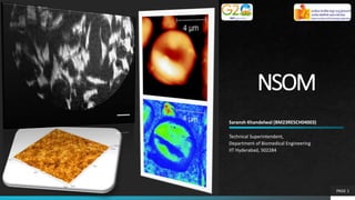
NSOM
Content
Outline
Introduction
Fundamentals of NSOM
Concept of Techniques involved
Application-based Research Paper
Conclusion and Future Aspects
PAGE 2
NSOM
Near-field scanning optical microscopy
(NSOM) a.k.a. scanning near-field
optical microscopy (SNOM)
It offers higher resolution around 50
nm (or even < 30 nm), depending on
tip aperture size.
Scanning probe technique developed
to surpass the spatial resolution
constraints that traditionally limit
conventional optical microscopy.
It provides simultaneous
measurements of the topography and
optical properties.
Introduction
PAGE 3
What is Resolution?
 Digital Imaging:
Image resolution is typically described in
PPI, which refers to how many pixels are
displayed per inch of an image. Higher
resolutions mean that there more pixels
per inch (PPI), resulting in more pixel
information and creating a high-quality,
crisp image.
PAGE 4
NSOM
Paper Review Conclusions Questions
Fig. 1: Similar images with lower and higher resolution.
What is Resolution?
 Microscopy:
The ability of a microscope to distinguish
details of a specimen or sample. In other
words, the minimum distance between
two distinct points of a specimen where
they can still be seen by the observer or
microscope camera as separate entities.
𝑅𝑒𝑠𝑜𝑙𝑢𝑡𝑖𝑜𝑛𝑑 =
𝜆
2 ∗ 𝑁𝐴
PAGE 5
NSOM
Paper Review Conclusions Questions
Fig. 2: Microscopic image of the sample taken with same magnification
and varying Numerical Aperture. [1]
Numerical Aperture (NA)
PAGE 6
NSOM
Paper Review Conclusions Questions
• NA is just a way of expressing the ability of an
objective lens to gather light information.
• ‘n’ represents refractive index
• ‘𝜃’ represent the maximum angle of acceptance
Fig. 3: Microscopic image of the sample taken with same magnification and
varying Numerical Aperture [2]
Abbe’s Limit
PAGE 7
NSOM
Paper Review Conclusions Questions
In general, the light propagates through space in an
unconfined manner and is the "normal" light utilized in
conventional microscopy
𝑅𝑒𝑠𝑜𝑙𝑢𝑡𝑖𝑜𝑛𝑑 =
.61𝝀
𝑁𝐴
In order to get better resolution, we need to trade-off
between Numerical Aperture (NA) or Wavelength (𝝀)….
How…?
Confining the wavelength i.e. by Aperture Manipulation
Near-Field Wavelength
PAGE 8
NSOM
Paper Review Conclusions Questions
• Traditionally, the lens collecting the scattering light is
placed several wavelengths of the illumination light far
away from the sample surface. This causes the
commonly known diffraction limit, that is far-field
optical techniques are limited to resolve features
approximately on the order of half of the wavelength of
the illuminating light due to the diffraction of light.
• The near-field (or evanescent) light consists of a non-
propagating field that exists near the surface of an
object at distances less than a single wavelength of
light. Light in the near-field has its greatest amplitude
in the region within the first few tens of nanometers of
the specimen surface. Because the near-field light
decays exponentially [3,5] within a distance less than the
wavelength of the light, it usually goes undetected.
Fig. 4: Diagram illustrating near field optics [3]
Near-Field Wavelength
PAGE 9
NSOM
Paper Review Conclusions Questions
The classical NSOM uses a tapered optical fibre probe and an aperture that is much smaller than the
wavelength of the light. After light passes through the cantilever tip with nano-aperture, a optical near-field (or
evanescent field) on the far side of this tip can be created that is not diffraction limited. Then the resolution in
near-field microscopy is directly affected by the size of the aperture and independent of the wavelength of the
light.
Fig. 5: Near-field technique [3]
NSOM Instrumentation
PAGE 10
NSOM
Paper Review Conclusions Questions
The classical NSOM uses a tapered optical fibre
probe and an aperture that is much smaller
than the wavelength of the light. After light
passes through the cantilever tip with nano-
aperture, a optical near-field (or evanescent
field) on the far side of this tip can be created
that is not diffraction limited. Then the
resolution in near-field microscopy is directly
affected by the size of the aperture and
independent of the wavelength of the light.
Fig. 6: General principles of NSOM instrumentation [3]
Fig. 7: SEM image of a coated NSOM tip. Grains in the Aluminium and the aperture at the end are visible [4]
NSOM Instrumentation
PAGE 11
NSOM
Paper Review Conclusions Questions
An illuminating probe aperture having a diameter
less than the wavelength of light is maintained in
the near field of the specimen surface. Because
close proximity or contact between the specimen
and probe (separation less than the wavelength)
is a general requirement for non-diffraction-
limited resolution.
An x-y-z scanner (usually piezoelectric) is utilized
to control the movement of the probe over the
specimen. The NSOM configuration positions the
objective in the far field, in the conventional
manner, for collection of the image-forming
optical signal.
Fig. 6: General principles of NSOM instrumentation [3]
NSOM Instrumentation
PAGE 12
NSOM
Paper Review Conclusions Questions
• Depending upon the design, the x-y-z scanner can either be
attached to the specimen or to the local probe. If the scanner and
specimen are coupled, then the specimen moves under the fixed
probe tip in a raster pattern to generate an image from the signal
produced by the tip-specimen interaction.
• The size of the area imaged is dependent only on the maximum
displacement that the scanner can produce.
• A computer simultaneously evaluates the probe position,
incorporating data obtained from the feedback system, and controls
the scanning of the tip and the separation of the tip and specimen
surface.
• The information is collected and recorded by the computer point-by-
point during the raster movement.
• Two-dimensional data sets gathered are subsequently compiled and
displayed as a three-dimensional reconstruction on a computer
monitor.
Fig. 8: NSOM imaging schematic [5]
X-Y-Z Scanner Mechanism
PAGE 13
NSOM
Paper Review Conclusions Questions
• The scanner must have low noise (small position fluctuations) and precision positioning capability.
• The required precision of the probe positioning usually necessitates that the entire instrument rest
on a vibration isolation table, or be suspended by some other means, to eliminate the transfer of
mechanical vibrations from the building to the instrument.
• it is necessary to maintain the probe in constant feedback above the specimen surface being
imaged.
• In case of unnoticed, damage to the probe tip or the specimen, is likely if the two come into
contact.
• The exponential variation of signal level with changing tip-to-specimen separation can produce
artifacts in the image that do not accurately represent optical information related to the specimen.
• Two most commonly employed mechanisms of tip positioning have been optical methods that
monitor the tip vibration amplitude (usually interferometric), and a non-optical tuning fork
technique.
Feedback Methods
PAGE 14
NSOM
Paper Review Conclusions Questions
Oscillatory Feedback Methods
• NSOM tip is almost always oscillated at the resonance frequency
of the probe. This allows lock-in detection techniques to be
utilized, which eliminates positional detection problems
associated with low-frequency noise and drift.
Shear-Force Feedback
• The shear-force feedback method laterally dithers the probe tip
at a mechanical resonance frequency in proximity to the
specimen surface.
Piezo-Electric Feedback
• Piezoelectric quartz tuning forks were first introduced into
scanning probe microscopy for use in scanning near-field
acoustic microscopy. Fig. 10: Feedback mechanism in NSOM [6]
NSOM Image
PAGE 15
NSOM
Paper Review Conclusions Questions
Fig. 11: NSOM image and topographic data of cleaned glass [6]
History of NSOM
PAGE 16
NSOM
Paper Review Conclusions Questions
• Edward H. Synge, beginning in 1928, published a series of articles that first conceptualized the idea
of an ultra-high resolution optical microscope. Synge's proposal suggested a new type of optical
microscope that would bypass the diffraction limit, but required fabrication of a 10-nanometer
aperture (much smaller than the light wavelength) in an opaque screen.
• In addition, Synge accurately outlined a number of the technical difficulties that building a near-
field microscope would present. Included in these were the challenges of fabricating the minute
aperture, achieving a sufficiently intense light source, specimen positioning at the nanometer scale,
and maintaining the aperture in close proximity to the specimen.
• In 1972, E. A. Ash and G. Nicholls demonstrated the near-field resolution of a sub-wavelength
aperture scanning microscope operating in the microwave region of the electromagnetic spectrum.
• In 1984, a research group at IBM Corporation's Zurich laboratory reported optical measurements at
a sub-diffraction resolution level.
• The IBM researchers employed a metal-coated quartz crystal probe on which an aperture was
fabricated at the tip, and designated the technique scanning near-field optical microscopy (SNOM).
• The Cornell group used electron-beam lithography to create apertures, smaller than 50
nanometers, in silicon and metal. The IBM team was able to claim the highest optical resolution of
25 nm, or one-twentieth of the 488-nanometer radiation wavelength.
Advantages
PAGE 17
NSOM
Paper Review Conclusions Questions
• Its ability to provide optical and spectroscopic data at high spatial resolution, in
combination with simultaneous topographic information.
• Typical resolutions for most NSOM instruments range around 50 nanometers,
which is only 5 or 6 times better than that achieved by scanning confocal
microscopy.
• For biological materials, specimen preparation is especially demanding, as
complete dehydration is generally required prior to carrying out sectioning or
coating.
• An additional limitation of other techniques such as AFM etc, is not able to take
advantage of the wide array of reporter dyes available to fluorescence
microscopy.
Applications
PAGE 18
NSOM
Paper Review Conclusions Questions
• Near-field optical spectroscopy: NSOM can be used to study the optical properties
of materials, such as plasmons and excitons, with high spatial and spectral
resolution.
• Nanolithography: NSOM can be used for high-resolution patterning of surfaces and
for direct writing of nanostructures with sub-10 nm resolution.
• Imaging of biological samples: NSOM can be used to image biological samples,
such as cells and tissues, with sub-50 nm resolution, providing detailed information
about the structure and dynamics of these samples.
• Semiconductor device characterization: NSOM can be used for imaging and
analyzing the properties of semiconductor devices, such as transistors and LEDs, at
the nanoscale.
• Quantum information processing: NSOM can be used for quantum information
processing, such as single-photon generation and detection, which is critical for
developing quantum technologies.
Limitations
PAGE 19
NSOM
Paper Review Conclusions Questions
Some of the limitations of near-field optical microscopy include:
• Practically zero working distance and an extremely small depth of field.
• Extremely long scan times for high resolution images or large specimen areas.
• Very low transmissivity of apertures smaller than the incident light wavelength.
• Only features at the surface of specimens can be studied.
• Fiber optic probes are cumbersome for imaging soft materials due to their high spring
constants, especially in shear-force mode.
PAGE 20
NSOM
Paper Review
Conclusions Questions
Paper Review
 Royal Society of Chemistry
 Indexed: PubMed, MEDLINE
and Science Citation Index
 Scattering-type Scanning Near-
field Optical Microscope
machine to an array of newly
available Quantum Cascade
laser” (QCL) diode lasers. It can
deliver SI datasets down to a
∼𝜆/1000 spatial resolution of
∼10 nm
PAGE 21
NSOM
Paper Review
Conclusions Questions
Results
 The resolution here is ∼140
times better than the
diffraction limit at this
wavelength and has
allowed to image chemical
contrast within the cell for
the first time.
Fig. 12: Left Image, AFM scan of the physical structure of a single red blood cell in a squamous epithelial
oesophageal biopsy. Right image, s-SNOM image [7]
PAGE 22
NSOM
Paper Review
Conclusions Questions
Results (Cont..)
 The s-SNOM phase
image, in this example,
approximately
corresponds to the
absorption in the
graphene layer, and
reveals regions of
differing thickness, and
the edge effects
responsible for the
device operation
Fig. 12: Graphene layer AFM topology, s-SNOM image and amplitude [7]
(a)
(b)
PAGE 23
NSOM Paper Review
Conclusions
Questions
Conclusion and Discussions
 For the s-SNOM it is the beginning, and the challenges are still mostly
technical. We believe it has a very bright future as a new scientific
research tool for a wide range of biomedical problems, but, already, we
are on the lookout for that elusive “unmet need”
 Although in its infancy, this instrument can already deliver ultra-detailed
chemical images whose spatial resolutions beat the normal diffraction
limit by a factor of ∼1000. This is easily enough to generate chemical
maps of the insides of single cells for the first time, and a range of new
possible scientific applications are explored
PAGE 24
NSOM Paper Review Conclusions
Questions
Questions…??
References
PAGE 25
1. https://microscopeclarity.com/what-is-microscope-resolution/
2. Live‐Cell Fluorescence Imaging, Jennifer C. Waters, Methods in Cell Biology, Elsevier, Volume 81, 2007, Pages 115-140
3. https://phys.libretexts.org/Courses/University_of_California_Davis/UCD%3A_Biophysics_241_Membrane_Biology/05%3A
_Experimental_Characterization_Spectroscopy_and_Microscopy/5.06%3A_Nearfield_Scanning_Optical_Microscopy_%28
NSOM%29
4. https://dunngroup.ku.edu/near-field-scanning-optical-microscopy
5. https://micro.magnet.fsu.edu/primer/techniques/nearfield/nearfieldintro.html
6. https://my.eng.utah.edu/~lzang/images/Lecture_16_NSOM.pdf
7. New IR imaging modalities for cancer detection and for intra-cell chemical mapping with a sub-diffraction mid-IR s-SNOM, H.
Amrania et. al, Royal Society of Chemistry, Faraday Discuss., 2016, 187, 539-553

More Related Content

What's hot

Nanoindentation
NanoindentationNanoindentation
NanoindentationSum K
 
Scanning Electron microscopy
Scanning Electron microscopy Scanning Electron microscopy
Scanning Electron microscopy sagar sen sen
 
Neutron scattering
Neutron scatteringNeutron scattering
Neutron scatteringIshfaq Ahmad
 
What is Cathodoluminescence? - DELMIC
What is Cathodoluminescence? - DELMICWhat is Cathodoluminescence? - DELMIC
What is Cathodoluminescence? - DELMICDelmic B.V.
 
Presentationon optical and electron microscopy by deepak kumar
Presentationon optical and electron  microscopy by deepak kumar Presentationon optical and electron  microscopy by deepak kumar
Presentationon optical and electron microscopy by deepak kumar Drx Kumar
 
Atomic force microscope
Atomic force microscopeAtomic force microscope
Atomic force microscopeSenthil Arasan
 
Metal matrix Nanocomposite
Metal matrix Nanocomposite Metal matrix Nanocomposite
Metal matrix Nanocomposite Abinaya536
 
X-Ray Diffraction Technique
X-Ray Diffraction TechniqueX-Ray Diffraction Technique
X-Ray Diffraction Techniquetabirsir
 
Raman spectroscopy for nanomaterials
Raman spectroscopy for nanomaterialsRaman spectroscopy for nanomaterials
Raman spectroscopy for nanomaterialskrishslide
 
X ray diffraction
X ray diffractionX ray diffraction
X ray diffractionFaraz Khan
 
1 basics of x ray powder diffraction
1 basics of x ray powder diffraction1 basics of x ray powder diffraction
1 basics of x ray powder diffractionGoenthan27
 

What's hot (20)

Photoluminescence
PhotoluminescencePhotoluminescence
Photoluminescence
 
Nanoindentation
NanoindentationNanoindentation
Nanoindentation
 
Scanning electron microscopy
Scanning electron microscopyScanning electron microscopy
Scanning electron microscopy
 
Scanning Electron microscopy
Scanning Electron microscopy Scanning Electron microscopy
Scanning Electron microscopy
 
Neutron scattering
Neutron scatteringNeutron scattering
Neutron scattering
 
What is Cathodoluminescence? - DELMIC
What is Cathodoluminescence? - DELMICWhat is Cathodoluminescence? - DELMIC
What is Cathodoluminescence? - DELMIC
 
Presentationon optical and electron microscopy by deepak kumar
Presentationon optical and electron  microscopy by deepak kumar Presentationon optical and electron  microscopy by deepak kumar
Presentationon optical and electron microscopy by deepak kumar
 
WDS
WDSWDS
WDS
 
Atomic force microscope
Atomic force microscopeAtomic force microscope
Atomic force microscope
 
Atomic Force Microscopy
Atomic Force MicroscopyAtomic Force Microscopy
Atomic Force Microscopy
 
AFMppt.ppt
AFMppt.pptAFMppt.ppt
AFMppt.ppt
 
Metal matrix Nanocomposite
Metal matrix Nanocomposite Metal matrix Nanocomposite
Metal matrix Nanocomposite
 
Neutron diffraction
Neutron diffractionNeutron diffraction
Neutron diffraction
 
X-Ray Diffraction Technique
X-Ray Diffraction TechniqueX-Ray Diffraction Technique
X-Ray Diffraction Technique
 
Raman spectroscopy for nanomaterials
Raman spectroscopy for nanomaterialsRaman spectroscopy for nanomaterials
Raman spectroscopy for nanomaterials
 
X ray diffraction
X ray diffractionX ray diffraction
X ray diffraction
 
Scanning probe microscope
Scanning probe microscopeScanning probe microscope
Scanning probe microscope
 
Powerpoint slides
Powerpoint slidesPowerpoint slides
Powerpoint slides
 
AFM.ppt
AFM.pptAFM.ppt
AFM.ppt
 
1 basics of x ray powder diffraction
1 basics of x ray powder diffraction1 basics of x ray powder diffraction
1 basics of x ray powder diffraction
 

Similar to Near Field Scattering Optical Microscopy (NSOM)

Confocal microscopy Likhith K
Confocal microscopy Likhith KConfocal microscopy Likhith K
Confocal microscopy Likhith KLIKHITHK1
 
OCT MACULA INTERPRETATION.
OCT MACULA  INTERPRETATION.OCT MACULA  INTERPRETATION.
OCT MACULA INTERPRETATION.ANUJA DHAKAL
 
Nanonics - Raman MultiProbe
Nanonics - Raman MultiProbeNanonics - Raman MultiProbe
Nanonics - Raman MultiProbeNanonics
 
Imaging in Glaucoma
Imaging in Glaucoma Imaging in Glaucoma
Imaging in Glaucoma Anujeet Paul
 
Alternatives to Point-Scan Confocal Microscopy
Alternatives to Point-Scan Confocal MicroscopyAlternatives to Point-Scan Confocal Microscopy
Alternatives to Point-Scan Confocal Microscopymchelen
 
OPTICAL COHERENCE DEMYSTIFIED
OPTICAL COHERENCE DEMYSTIFIED OPTICAL COHERENCE DEMYSTIFIED
OPTICAL COHERENCE DEMYSTIFIED DINESH and SONALEE
 
Optical coherence tomography(OCT) --macula
Optical coherence tomography(OCT) --maculaOptical coherence tomography(OCT) --macula
Optical coherence tomography(OCT) --maculaAkshay Nayak
 
Crack detection in railway track 2
Crack detection in  railway track 2Crack detection in  railway track 2
Crack detection in railway track 2Ankit Maurya
 
Pollen photos using a Scanning Electron Microscope
Pollen photos using a Scanning Electron MicroscopePollen photos using a Scanning Electron Microscope
Pollen photos using a Scanning Electron MicroscopeChris Cardew
 
Microscopy fin
Microscopy finMicroscopy fin
Microscopy finMUBOSScz
 
3 d imaging super resolution
3 d imaging super resolution3 d imaging super resolution
3 d imaging super resolutionredsea3h
 

Similar to Near Field Scattering Optical Microscopy (NSOM) (20)

Confocal microscopy Likhith K
Confocal microscopy Likhith KConfocal microscopy Likhith K
Confocal microscopy Likhith K
 
OCT MACULA INTERPRETATION.
OCT MACULA  INTERPRETATION.OCT MACULA  INTERPRETATION.
OCT MACULA INTERPRETATION.
 
Nanonics - Raman MultiProbe
Nanonics - Raman MultiProbeNanonics - Raman MultiProbe
Nanonics - Raman MultiProbe
 
OCT Machines
OCT Machines OCT Machines
OCT Machines
 
Optical coherence tomography
Optical coherence tomographyOptical coherence tomography
Optical coherence tomography
 
Imaging in Glaucoma
Imaging in Glaucoma Imaging in Glaucoma
Imaging in Glaucoma
 
Alternatives to Point-Scan Confocal Microscopy
Alternatives to Point-Scan Confocal MicroscopyAlternatives to Point-Scan Confocal Microscopy
Alternatives to Point-Scan Confocal Microscopy
 
Oct introduction
Oct  introductionOct  introduction
Oct introduction
 
Final oct
Final octFinal oct
Final oct
 
OPTICAL COHERENCE DEMYSTIFIED
OPTICAL COHERENCE DEMYSTIFIED OPTICAL COHERENCE DEMYSTIFIED
OPTICAL COHERENCE DEMYSTIFIED
 
Cbct
CbctCbct
Cbct
 
Optical coherence tomography(OCT) --macula
Optical coherence tomography(OCT) --maculaOptical coherence tomography(OCT) --macula
Optical coherence tomography(OCT) --macula
 
Microscopy - The Life of Bio science lab
Microscopy - The Life of Bio science labMicroscopy - The Life of Bio science lab
Microscopy - The Life of Bio science lab
 
Crack detection in railway track 2
Crack detection in  railway track 2Crack detection in  railway track 2
Crack detection in railway track 2
 
Pollen photos using a Scanning Electron Microscope
Pollen photos using a Scanning Electron MicroscopePollen photos using a Scanning Electron Microscope
Pollen photos using a Scanning Electron Microscope
 
OCT
OCTOCT
OCT
 
Microscopy fin
Microscopy finMicroscopy fin
Microscopy fin
 
Cone beam ct
Cone beam ctCone beam ct
Cone beam ct
 
3 d imaging super resolution
3 d imaging super resolution3 d imaging super resolution
3 d imaging super resolution
 
UNIT 4.pptx
UNIT 4.pptxUNIT 4.pptx
UNIT 4.pptx
 

Recently uploaded

Kisan Call Centre - To harness potential of ICT in Agriculture by answer farm...
Kisan Call Centre - To harness potential of ICT in Agriculture by answer farm...Kisan Call Centre - To harness potential of ICT in Agriculture by answer farm...
Kisan Call Centre - To harness potential of ICT in Agriculture by answer farm...Krashi Coaching
 
PSYCHIATRIC History collection FORMAT.pptx
PSYCHIATRIC   History collection FORMAT.pptxPSYCHIATRIC   History collection FORMAT.pptx
PSYCHIATRIC History collection FORMAT.pptxPoojaSen20
 
Solving Puzzles Benefits Everyone (English).pptx
Solving Puzzles Benefits Everyone (English).pptxSolving Puzzles Benefits Everyone (English).pptx
Solving Puzzles Benefits Everyone (English).pptxOH TEIK BIN
 
SOCIAL AND HISTORICAL CONTEXT - LFTVD.pptx
SOCIAL AND HISTORICAL CONTEXT - LFTVD.pptxSOCIAL AND HISTORICAL CONTEXT - LFTVD.pptx
SOCIAL AND HISTORICAL CONTEXT - LFTVD.pptxiammrhaywood
 
Arihant handbook biology for class 11 .pdf
Arihant handbook biology for class 11 .pdfArihant handbook biology for class 11 .pdf
Arihant handbook biology for class 11 .pdfchloefrazer622
 
A Critique of the Proposed National Education Policy Reform
A Critique of the Proposed National Education Policy ReformA Critique of the Proposed National Education Policy Reform
A Critique of the Proposed National Education Policy ReformChameera Dedduwage
 
Sanyam Choudhary Chemistry practical.pdf
Sanyam Choudhary Chemistry practical.pdfSanyam Choudhary Chemistry practical.pdf
Sanyam Choudhary Chemistry practical.pdfsanyamsingh5019
 
URLs and Routing in the Odoo 17 Website App
URLs and Routing in the Odoo 17 Website AppURLs and Routing in the Odoo 17 Website App
URLs and Routing in the Odoo 17 Website AppCeline George
 
Accessible design: Minimum effort, maximum impact
Accessible design: Minimum effort, maximum impactAccessible design: Minimum effort, maximum impact
Accessible design: Minimum effort, maximum impactdawncurless
 
BASLIQ CURRENT LOOKBOOK LOOKBOOK(1) (1).pdf
BASLIQ CURRENT LOOKBOOK  LOOKBOOK(1) (1).pdfBASLIQ CURRENT LOOKBOOK  LOOKBOOK(1) (1).pdf
BASLIQ CURRENT LOOKBOOK LOOKBOOK(1) (1).pdfSoniaTolstoy
 
The basics of sentences session 2pptx copy.pptx
The basics of sentences session 2pptx copy.pptxThe basics of sentences session 2pptx copy.pptx
The basics of sentences session 2pptx copy.pptxheathfieldcps1
 
Grant Readiness 101 TechSoup and Remy Consulting
Grant Readiness 101 TechSoup and Remy ConsultingGrant Readiness 101 TechSoup and Remy Consulting
Grant Readiness 101 TechSoup and Remy ConsultingTechSoup
 
Measures of Central Tendency: Mean, Median and Mode
Measures of Central Tendency: Mean, Median and ModeMeasures of Central Tendency: Mean, Median and Mode
Measures of Central Tendency: Mean, Median and ModeThiyagu K
 
microwave assisted reaction. General introduction
microwave assisted reaction. General introductionmicrowave assisted reaction. General introduction
microwave assisted reaction. General introductionMaksud Ahmed
 
MENTAL STATUS EXAMINATION format.docx
MENTAL     STATUS EXAMINATION format.docxMENTAL     STATUS EXAMINATION format.docx
MENTAL STATUS EXAMINATION format.docxPoojaSen20
 
How to Make a Pirate ship Primary Education.pptx
How to Make a Pirate ship Primary Education.pptxHow to Make a Pirate ship Primary Education.pptx
How to Make a Pirate ship Primary Education.pptxmanuelaromero2013
 
POINT- BIOCHEMISTRY SEM 2 ENZYMES UNIT 5.pptx
POINT- BIOCHEMISTRY SEM 2 ENZYMES UNIT 5.pptxPOINT- BIOCHEMISTRY SEM 2 ENZYMES UNIT 5.pptx
POINT- BIOCHEMISTRY SEM 2 ENZYMES UNIT 5.pptxSayali Powar
 

Recently uploaded (20)

Kisan Call Centre - To harness potential of ICT in Agriculture by answer farm...
Kisan Call Centre - To harness potential of ICT in Agriculture by answer farm...Kisan Call Centre - To harness potential of ICT in Agriculture by answer farm...
Kisan Call Centre - To harness potential of ICT in Agriculture by answer farm...
 
PSYCHIATRIC History collection FORMAT.pptx
PSYCHIATRIC   History collection FORMAT.pptxPSYCHIATRIC   History collection FORMAT.pptx
PSYCHIATRIC History collection FORMAT.pptx
 
Solving Puzzles Benefits Everyone (English).pptx
Solving Puzzles Benefits Everyone (English).pptxSolving Puzzles Benefits Everyone (English).pptx
Solving Puzzles Benefits Everyone (English).pptx
 
SOCIAL AND HISTORICAL CONTEXT - LFTVD.pptx
SOCIAL AND HISTORICAL CONTEXT - LFTVD.pptxSOCIAL AND HISTORICAL CONTEXT - LFTVD.pptx
SOCIAL AND HISTORICAL CONTEXT - LFTVD.pptx
 
Arihant handbook biology for class 11 .pdf
Arihant handbook biology for class 11 .pdfArihant handbook biology for class 11 .pdf
Arihant handbook biology for class 11 .pdf
 
A Critique of the Proposed National Education Policy Reform
A Critique of the Proposed National Education Policy ReformA Critique of the Proposed National Education Policy Reform
A Critique of the Proposed National Education Policy Reform
 
Sanyam Choudhary Chemistry practical.pdf
Sanyam Choudhary Chemistry practical.pdfSanyam Choudhary Chemistry practical.pdf
Sanyam Choudhary Chemistry practical.pdf
 
URLs and Routing in the Odoo 17 Website App
URLs and Routing in the Odoo 17 Website AppURLs and Routing in the Odoo 17 Website App
URLs and Routing in the Odoo 17 Website App
 
Accessible design: Minimum effort, maximum impact
Accessible design: Minimum effort, maximum impactAccessible design: Minimum effort, maximum impact
Accessible design: Minimum effort, maximum impact
 
BASLIQ CURRENT LOOKBOOK LOOKBOOK(1) (1).pdf
BASLIQ CURRENT LOOKBOOK  LOOKBOOK(1) (1).pdfBASLIQ CURRENT LOOKBOOK  LOOKBOOK(1) (1).pdf
BASLIQ CURRENT LOOKBOOK LOOKBOOK(1) (1).pdf
 
The basics of sentences session 2pptx copy.pptx
The basics of sentences session 2pptx copy.pptxThe basics of sentences session 2pptx copy.pptx
The basics of sentences session 2pptx copy.pptx
 
Grant Readiness 101 TechSoup and Remy Consulting
Grant Readiness 101 TechSoup and Remy ConsultingGrant Readiness 101 TechSoup and Remy Consulting
Grant Readiness 101 TechSoup and Remy Consulting
 
Measures of Central Tendency: Mean, Median and Mode
Measures of Central Tendency: Mean, Median and ModeMeasures of Central Tendency: Mean, Median and Mode
Measures of Central Tendency: Mean, Median and Mode
 
microwave assisted reaction. General introduction
microwave assisted reaction. General introductionmicrowave assisted reaction. General introduction
microwave assisted reaction. General introduction
 
Model Call Girl in Bikash Puri Delhi reach out to us at 🔝9953056974🔝
Model Call Girl in Bikash Puri  Delhi reach out to us at 🔝9953056974🔝Model Call Girl in Bikash Puri  Delhi reach out to us at 🔝9953056974🔝
Model Call Girl in Bikash Puri Delhi reach out to us at 🔝9953056974🔝
 
Código Creativo y Arte de Software | Unidad 1
Código Creativo y Arte de Software | Unidad 1Código Creativo y Arte de Software | Unidad 1
Código Creativo y Arte de Software | Unidad 1
 
MENTAL STATUS EXAMINATION format.docx
MENTAL     STATUS EXAMINATION format.docxMENTAL     STATUS EXAMINATION format.docx
MENTAL STATUS EXAMINATION format.docx
 
How to Make a Pirate ship Primary Education.pptx
How to Make a Pirate ship Primary Education.pptxHow to Make a Pirate ship Primary Education.pptx
How to Make a Pirate ship Primary Education.pptx
 
POINT- BIOCHEMISTRY SEM 2 ENZYMES UNIT 5.pptx
POINT- BIOCHEMISTRY SEM 2 ENZYMES UNIT 5.pptxPOINT- BIOCHEMISTRY SEM 2 ENZYMES UNIT 5.pptx
POINT- BIOCHEMISTRY SEM 2 ENZYMES UNIT 5.pptx
 
TataKelola dan KamSiber Kecerdasan Buatan v022.pdf
TataKelola dan KamSiber Kecerdasan Buatan v022.pdfTataKelola dan KamSiber Kecerdasan Buatan v022.pdf
TataKelola dan KamSiber Kecerdasan Buatan v022.pdf
 

Near Field Scattering Optical Microscopy (NSOM)

  • 1. Saransh Khandelwal (BM23RESCH04003) Technical Superintendent, Department of Biomedical Engineering IIT Hyderabad, 502284 PAGE 1 NSOM
  • 2. Content Outline Introduction Fundamentals of NSOM Concept of Techniques involved Application-based Research Paper Conclusion and Future Aspects PAGE 2
  • 3. NSOM Near-field scanning optical microscopy (NSOM) a.k.a. scanning near-field optical microscopy (SNOM) It offers higher resolution around 50 nm (or even < 30 nm), depending on tip aperture size. Scanning probe technique developed to surpass the spatial resolution constraints that traditionally limit conventional optical microscopy. It provides simultaneous measurements of the topography and optical properties. Introduction PAGE 3
  • 4. What is Resolution?  Digital Imaging: Image resolution is typically described in PPI, which refers to how many pixels are displayed per inch of an image. Higher resolutions mean that there more pixels per inch (PPI), resulting in more pixel information and creating a high-quality, crisp image. PAGE 4 NSOM Paper Review Conclusions Questions Fig. 1: Similar images with lower and higher resolution.
  • 5. What is Resolution?  Microscopy: The ability of a microscope to distinguish details of a specimen or sample. In other words, the minimum distance between two distinct points of a specimen where they can still be seen by the observer or microscope camera as separate entities. 𝑅𝑒𝑠𝑜𝑙𝑢𝑡𝑖𝑜𝑛𝑑 = 𝜆 2 ∗ 𝑁𝐴 PAGE 5 NSOM Paper Review Conclusions Questions Fig. 2: Microscopic image of the sample taken with same magnification and varying Numerical Aperture. [1]
  • 6. Numerical Aperture (NA) PAGE 6 NSOM Paper Review Conclusions Questions • NA is just a way of expressing the ability of an objective lens to gather light information. • ‘n’ represents refractive index • ‘𝜃’ represent the maximum angle of acceptance Fig. 3: Microscopic image of the sample taken with same magnification and varying Numerical Aperture [2]
  • 7. Abbe’s Limit PAGE 7 NSOM Paper Review Conclusions Questions In general, the light propagates through space in an unconfined manner and is the "normal" light utilized in conventional microscopy 𝑅𝑒𝑠𝑜𝑙𝑢𝑡𝑖𝑜𝑛𝑑 = .61𝝀 𝑁𝐴 In order to get better resolution, we need to trade-off between Numerical Aperture (NA) or Wavelength (𝝀)…. How…? Confining the wavelength i.e. by Aperture Manipulation
  • 8. Near-Field Wavelength PAGE 8 NSOM Paper Review Conclusions Questions • Traditionally, the lens collecting the scattering light is placed several wavelengths of the illumination light far away from the sample surface. This causes the commonly known diffraction limit, that is far-field optical techniques are limited to resolve features approximately on the order of half of the wavelength of the illuminating light due to the diffraction of light. • The near-field (or evanescent) light consists of a non- propagating field that exists near the surface of an object at distances less than a single wavelength of light. Light in the near-field has its greatest amplitude in the region within the first few tens of nanometers of the specimen surface. Because the near-field light decays exponentially [3,5] within a distance less than the wavelength of the light, it usually goes undetected. Fig. 4: Diagram illustrating near field optics [3]
  • 9. Near-Field Wavelength PAGE 9 NSOM Paper Review Conclusions Questions The classical NSOM uses a tapered optical fibre probe and an aperture that is much smaller than the wavelength of the light. After light passes through the cantilever tip with nano-aperture, a optical near-field (or evanescent field) on the far side of this tip can be created that is not diffraction limited. Then the resolution in near-field microscopy is directly affected by the size of the aperture and independent of the wavelength of the light. Fig. 5: Near-field technique [3]
  • 10. NSOM Instrumentation PAGE 10 NSOM Paper Review Conclusions Questions The classical NSOM uses a tapered optical fibre probe and an aperture that is much smaller than the wavelength of the light. After light passes through the cantilever tip with nano- aperture, a optical near-field (or evanescent field) on the far side of this tip can be created that is not diffraction limited. Then the resolution in near-field microscopy is directly affected by the size of the aperture and independent of the wavelength of the light. Fig. 6: General principles of NSOM instrumentation [3] Fig. 7: SEM image of a coated NSOM tip. Grains in the Aluminium and the aperture at the end are visible [4]
  • 11. NSOM Instrumentation PAGE 11 NSOM Paper Review Conclusions Questions An illuminating probe aperture having a diameter less than the wavelength of light is maintained in the near field of the specimen surface. Because close proximity or contact between the specimen and probe (separation less than the wavelength) is a general requirement for non-diffraction- limited resolution. An x-y-z scanner (usually piezoelectric) is utilized to control the movement of the probe over the specimen. The NSOM configuration positions the objective in the far field, in the conventional manner, for collection of the image-forming optical signal. Fig. 6: General principles of NSOM instrumentation [3]
  • 12. NSOM Instrumentation PAGE 12 NSOM Paper Review Conclusions Questions • Depending upon the design, the x-y-z scanner can either be attached to the specimen or to the local probe. If the scanner and specimen are coupled, then the specimen moves under the fixed probe tip in a raster pattern to generate an image from the signal produced by the tip-specimen interaction. • The size of the area imaged is dependent only on the maximum displacement that the scanner can produce. • A computer simultaneously evaluates the probe position, incorporating data obtained from the feedback system, and controls the scanning of the tip and the separation of the tip and specimen surface. • The information is collected and recorded by the computer point-by- point during the raster movement. • Two-dimensional data sets gathered are subsequently compiled and displayed as a three-dimensional reconstruction on a computer monitor. Fig. 8: NSOM imaging schematic [5]
  • 13. X-Y-Z Scanner Mechanism PAGE 13 NSOM Paper Review Conclusions Questions • The scanner must have low noise (small position fluctuations) and precision positioning capability. • The required precision of the probe positioning usually necessitates that the entire instrument rest on a vibration isolation table, or be suspended by some other means, to eliminate the transfer of mechanical vibrations from the building to the instrument. • it is necessary to maintain the probe in constant feedback above the specimen surface being imaged. • In case of unnoticed, damage to the probe tip or the specimen, is likely if the two come into contact. • The exponential variation of signal level with changing tip-to-specimen separation can produce artifacts in the image that do not accurately represent optical information related to the specimen. • Two most commonly employed mechanisms of tip positioning have been optical methods that monitor the tip vibration amplitude (usually interferometric), and a non-optical tuning fork technique.
  • 14. Feedback Methods PAGE 14 NSOM Paper Review Conclusions Questions Oscillatory Feedback Methods • NSOM tip is almost always oscillated at the resonance frequency of the probe. This allows lock-in detection techniques to be utilized, which eliminates positional detection problems associated with low-frequency noise and drift. Shear-Force Feedback • The shear-force feedback method laterally dithers the probe tip at a mechanical resonance frequency in proximity to the specimen surface. Piezo-Electric Feedback • Piezoelectric quartz tuning forks were first introduced into scanning probe microscopy for use in scanning near-field acoustic microscopy. Fig. 10: Feedback mechanism in NSOM [6]
  • 15. NSOM Image PAGE 15 NSOM Paper Review Conclusions Questions Fig. 11: NSOM image and topographic data of cleaned glass [6]
  • 16. History of NSOM PAGE 16 NSOM Paper Review Conclusions Questions • Edward H. Synge, beginning in 1928, published a series of articles that first conceptualized the idea of an ultra-high resolution optical microscope. Synge's proposal suggested a new type of optical microscope that would bypass the diffraction limit, but required fabrication of a 10-nanometer aperture (much smaller than the light wavelength) in an opaque screen. • In addition, Synge accurately outlined a number of the technical difficulties that building a near- field microscope would present. Included in these were the challenges of fabricating the minute aperture, achieving a sufficiently intense light source, specimen positioning at the nanometer scale, and maintaining the aperture in close proximity to the specimen. • In 1972, E. A. Ash and G. Nicholls demonstrated the near-field resolution of a sub-wavelength aperture scanning microscope operating in the microwave region of the electromagnetic spectrum. • In 1984, a research group at IBM Corporation's Zurich laboratory reported optical measurements at a sub-diffraction resolution level. • The IBM researchers employed a metal-coated quartz crystal probe on which an aperture was fabricated at the tip, and designated the technique scanning near-field optical microscopy (SNOM). • The Cornell group used electron-beam lithography to create apertures, smaller than 50 nanometers, in silicon and metal. The IBM team was able to claim the highest optical resolution of 25 nm, or one-twentieth of the 488-nanometer radiation wavelength.
  • 17. Advantages PAGE 17 NSOM Paper Review Conclusions Questions • Its ability to provide optical and spectroscopic data at high spatial resolution, in combination with simultaneous topographic information. • Typical resolutions for most NSOM instruments range around 50 nanometers, which is only 5 or 6 times better than that achieved by scanning confocal microscopy. • For biological materials, specimen preparation is especially demanding, as complete dehydration is generally required prior to carrying out sectioning or coating. • An additional limitation of other techniques such as AFM etc, is not able to take advantage of the wide array of reporter dyes available to fluorescence microscopy.
  • 18. Applications PAGE 18 NSOM Paper Review Conclusions Questions • Near-field optical spectroscopy: NSOM can be used to study the optical properties of materials, such as plasmons and excitons, with high spatial and spectral resolution. • Nanolithography: NSOM can be used for high-resolution patterning of surfaces and for direct writing of nanostructures with sub-10 nm resolution. • Imaging of biological samples: NSOM can be used to image biological samples, such as cells and tissues, with sub-50 nm resolution, providing detailed information about the structure and dynamics of these samples. • Semiconductor device characterization: NSOM can be used for imaging and analyzing the properties of semiconductor devices, such as transistors and LEDs, at the nanoscale. • Quantum information processing: NSOM can be used for quantum information processing, such as single-photon generation and detection, which is critical for developing quantum technologies.
  • 19. Limitations PAGE 19 NSOM Paper Review Conclusions Questions Some of the limitations of near-field optical microscopy include: • Practically zero working distance and an extremely small depth of field. • Extremely long scan times for high resolution images or large specimen areas. • Very low transmissivity of apertures smaller than the incident light wavelength. • Only features at the surface of specimens can be studied. • Fiber optic probes are cumbersome for imaging soft materials due to their high spring constants, especially in shear-force mode.
  • 20. PAGE 20 NSOM Paper Review Conclusions Questions Paper Review  Royal Society of Chemistry  Indexed: PubMed, MEDLINE and Science Citation Index  Scattering-type Scanning Near- field Optical Microscope machine to an array of newly available Quantum Cascade laser” (QCL) diode lasers. It can deliver SI datasets down to a ∼𝜆/1000 spatial resolution of ∼10 nm
  • 21. PAGE 21 NSOM Paper Review Conclusions Questions Results  The resolution here is ∼140 times better than the diffraction limit at this wavelength and has allowed to image chemical contrast within the cell for the first time. Fig. 12: Left Image, AFM scan of the physical structure of a single red blood cell in a squamous epithelial oesophageal biopsy. Right image, s-SNOM image [7]
  • 22. PAGE 22 NSOM Paper Review Conclusions Questions Results (Cont..)  The s-SNOM phase image, in this example, approximately corresponds to the absorption in the graphene layer, and reveals regions of differing thickness, and the edge effects responsible for the device operation Fig. 12: Graphene layer AFM topology, s-SNOM image and amplitude [7] (a) (b)
  • 23. PAGE 23 NSOM Paper Review Conclusions Questions Conclusion and Discussions  For the s-SNOM it is the beginning, and the challenges are still mostly technical. We believe it has a very bright future as a new scientific research tool for a wide range of biomedical problems, but, already, we are on the lookout for that elusive “unmet need”  Although in its infancy, this instrument can already deliver ultra-detailed chemical images whose spatial resolutions beat the normal diffraction limit by a factor of ∼1000. This is easily enough to generate chemical maps of the insides of single cells for the first time, and a range of new possible scientific applications are explored
  • 24. PAGE 24 NSOM Paper Review Conclusions Questions Questions…??
  • 25. References PAGE 25 1. https://microscopeclarity.com/what-is-microscope-resolution/ 2. Live‐Cell Fluorescence Imaging, Jennifer C. Waters, Methods in Cell Biology, Elsevier, Volume 81, 2007, Pages 115-140 3. https://phys.libretexts.org/Courses/University_of_California_Davis/UCD%3A_Biophysics_241_Membrane_Biology/05%3A _Experimental_Characterization_Spectroscopy_and_Microscopy/5.06%3A_Nearfield_Scanning_Optical_Microscopy_%28 NSOM%29 4. https://dunngroup.ku.edu/near-field-scanning-optical-microscopy 5. https://micro.magnet.fsu.edu/primer/techniques/nearfield/nearfieldintro.html 6. https://my.eng.utah.edu/~lzang/images/Lecture_16_NSOM.pdf 7. New IR imaging modalities for cancer detection and for intra-cell chemical mapping with a sub-diffraction mid-IR s-SNOM, H. Amrania et. al, Royal Society of Chemistry, Faraday Discuss., 2016, 187, 539-553

Editor's Notes

  1. https://www.biorxiv.org/content/10.1101/390948v1.full
  2. In addition to the optical information, NSOM can generate topographical or force data from the specimen in the same manner as the atomic force microscope. The two separate data sets (optical and topographical) can then be compared to determine the correlation between the physical structures and the optical contrast.
  3. Resolutionz = 2λ / [η • sin(α)]2 https://microscopeclarity.com/what-is-microscope-resolution/
  4. https://microscopeclarity.com/what-is-microscope-resolution/
  5. The far-field light propagates through space in an unconfined manner and is the "normal" light utilized in conventional microscopy. https://microscopeclarity.com/what-is-microscope-resolution/
  6. https://microscopeclarity.com/what-is-microscope-resolution/
  7. https://microscopeclarity.com/what-is-microscope-resolution/
  8. https://microscopeclarity.com/what-is-microscope-resolution/
  9. https://microscopeclarity.com/what-is-microscope-resolution/
  10. https://microscopeclarity.com/what-is-microscope-resolution/
  11. https://microscopeclarity.com/what-is-microscope-resolution/
  12. https://microscopeclarity.com/what-is-microscope-resolution/ agitate
  13. https://microscopeclarity.com/what-is-microscope-resolution/
  14. https://microscopeclarity.com/what-is-microscope-resolution/
  15. https://microscopeclarity.com/what-is-microscope-resolution/
  16. https://microscopeclarity.com/what-is-microscope-resolution/
  17. https://microscopeclarity.com/what-is-microscope-resolution/
  18. The whole process of analyzing ∼12,000 T cells and computing the capture statistics required ∼30 min Our conclusions are supported by the fact that the used CD4+ T cell and CD8+ T cell samples had manufacturer-reported purities of 94% and 93%, respectively