
Recommended
More Related Content
What's hot
What's hot (20)
Similar to Tertiary structure prediction- MODELLER, RASMOL
Similar to Tertiary structure prediction- MODELLER, RASMOL (20)
Recently uploaded
Recently uploaded (20)
Tertiary structure prediction- MODELLER, RASMOL
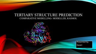
- 1. TERTIARY STRUCTURE PREDICTION COMPARATIVE MODELLING- MODELLER, RASMOL Presented by, Kaveri
- 2. WHAT IS A TERTIARY STRUCTURE OF PROTEIN? • Tertiary Structure is the folding of the total chain • Secondary structure linked by turns and loops. • Stability - non-bonding interactions & the disulfide bond. Tertiary structure
- 3. WHAT IS TERTIARY STRUCTURE PREDICTION? • Inference of the three-dimensional structure of a protein from its amino acid sequence. • Prediction of its folding and its secondary and tertiary structure from its primary structure. • Sites for calculating and displaying the 3-D structure of oligosaccharides and proteins. • Eg: ab initio, Homology modelling, protein threading etc
- 4. COMPARATIVE MODELLING • Predicts 3 dimensional structure of a given protein sequence (target) based on an alignment to one or more known protein structures (templates). • Uses experimentally determined protein structures as templates to predict the conformation of another protein that exhibits amino acid sequence homology.
- 5. • If similarity between the target sequence and the template sequence is detected, structural similarity can be assumed. • A similar structure can be predicted if sequence similarity is about or above 35%.
- 6. • The similarity of structures is very high in the so-called "core regions“. • core regions - comprised of secondary structure elements such as a-helices and b-sheets.
- 7. STEPS FOR COMPARATIVE MODELING 1. Find one or more proteins in the PDB that exhibit sequence similarity to your protein 2. Construct a sequence alignment 3. Decide which regions are structurally conserved (SCR): helices, strands 4. Construct loops either by analogy or from a database 5.Place side-chains: use existing conformational info and rotamer databases. 6. Refine the model: packing, surface accessibilities, energy calculations 7. Validate the model 8. Iterate all of the above steps to remove errors
- 8. MERITS AND DEMERITS MERITS • Enrichment of graphical methods • Formulation to incorporate domain knowledge • Effective for protein structure prediction • Solution for long range interaction DEMERITS • Manual feature extraction • Difficulty in verification • High complexity
- 9. MODELLER • Program for Comparative protein structure modelling by satisfaction of spatial restrains. • Used for comparative modeling of protein three-dimensional structures. • The user provides an alignment of a sequence to be modeled with known related structures MODELLER automatically calculates a model containing all non-hydrogen atoms. • Implements comparative protein structure modeling by satisfaction of spatial restraints.
- 10. • Additional tasks • de novo modeling of loops in protein structures • Optimization of various models of protein structure with respect to a flexibly defined objective function • Multiple alignment of protein sequences and/or structures • Clustering • Searching of sequence databases • Comparison of protein structures etc.
- 11. • MODELLER is written in Fortran-90 and is meant to run on a UNIX system. • Available free of charge to academic non-profit institutions. • https://salilab.org/modeller/ - website.
- 12. RASMOL • It is a molecular graphics bio-software used for the visualization of proteins, nucleic acids, and small molecules. • This molecular viewer is aimed at display, teaching and generation of publication quality images. • It allows user to display the molecule in a variety of structural representation. • Ras Mol can be downloaded from http://www.umass.edu/microbio/rasmol.
- 13. FEATURES • Ras Mol displays all or any part(s) of a molecule or molecular complex. • It can rotate the displayed image • Different parts of the structure can be coloured to convey structural information, and can be labeled • It can report information about atoms including name of the atom, bond distances, angle etc. • It can represent structures in different models (ball and stick models, ribbons, strands, cartoons etc.).
- 14. HOW TO USE RAS MOL ? 2 windows Graphics window command Line window Graphics window • The top of the graphics window has a menu bar of pull- down menus such as File, Display, Colours, Export, Options, and Help. • The molecules appears in initially as a wire –frame structure with a black back- ground. • With the help of the Display and Colours menus, we can change the display from wire-frame to back bone, sticks, space fill, or various ribbons, and we can also change the colour of the molecule.
- 15. Command Line window • It is used for selection of those portions of the molecules of interest. • The commands offer the possibility for changing the colour. • H-bonding involved in the secondary structure of the protein can be displayed. • Showing the ligands or coenzymes in space filling mode, all are colour coded to make the various features easily identifiable.
- 16. USES • It is a software to view, handle, and analyze macromolecular 3D structures. • It determines overall shape and size of a molecule. • To locate or predict possible binding sites of a ligand. • To study evolutionary process at molecular level.
- 17. THANK YOU