
Recommended
More Related Content
What's hot
What's hot (20)
Similar to Temporo mandibular joint
Similar to Temporo mandibular joint (20)
Recently uploaded
Recently uploaded (20)
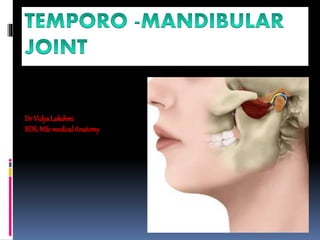
Temporo mandibular joint
- 2. It is the articulation of the base of the skull with the mandible It is a bicondylar variety of the synovial joint Articular surfaces of both bones are covered by white fibrocartilage Joint cavity is divided into upper menisco temporal & lower menisco mandibular compartments by articular disc
- 4. Bones forming the joint Above : Articular tubercle at the root of the zygoma & anterior part of the mandibular fossa of the temporal bone Below : Head of the mandible
- 6. Articular disc It is an oval plate of fibrocartilage Caps the head of the mandible Divides the joint completely into two compartments Attachment of the disc Attached to the inner aspect of fibrous capsule Blends in front with insertion of lateral pterygoid muscle Splits into two lamellae posteriorly- getting attached to squamotympanic fissure and posterior surface of neck of mandible
- 7. Ligaments of the T-M Joint Capsular ligament with synovial membrane Articular disc Lateral temporomandibular ligament Accessory ligaments- Stylomandibular ligament Sphenomandibular ligament
- 9. 1. Capsular ligament Envelops the joint Attachments Above : Articular tubercle –in front Squamotympanic fissure -behind Articular fossa –at the periphery Below : Attached around the neck of mandible In front : Blends with insertion of lateral pterygoid muscle Above the disc capsule is loose, below it is taut
- 11. 2. Lateral temporomandibular ligament Blends with lateral part of fibrous capsule Extends from the root of the zygoma to lateral surface and posterior margin of neck of mandible
- 12. 3. Sphenomandibular ligament Situated medial to the capsule Attachments Above Spine of the sphenoid Below Lingula of the mandible 4. Stylomandibular ligament Extends from the tip of the styloid process to the angle of the mandible
- 13. Relations of the joint In front- lateral ptertgoid ,temporalis muscle, masseteric nerves & vessels Behind – Parotid gland, auriculotemporal nerve, superficial temporal vesslels,external acoustic meatus Lateral –subcutaneous Medial – ,spine of sphenoid, middle meningeal artery, roots of auriculotemporal nerve Above- floor of the middle cranial fossa separated by thin plate of bone
- 14. Arterial supply Superficial temporal & maxillary artery Nerve supply Auriculotemporal nerve Masseteric nerve
- 17. Movements taking place in the joint Protrusion /Protraction Retraction Elevation Depression Side to side movements
- 19. Muscles producing movement Protrusion (protraction ) Both the pterygoid muscles Movement occurs in upper compatment Retraction Posterior fibres of temporalis Forceful retraction is assisted by middle and deep fibres of masseter, digastric , geniohyoid
- 22. Depression Lateral pterygoid muscle, geniohyoid, mylohyoid, digastric- Gravity Elevation Masseter,temporalis, medial pterygoid, Side to side movement Both pterygoids acting alternatively
- 26. Mechanism of depression At first there is forward rotation of mandibular condyle in lower compartment so that condyle come in contact with intermediate area of articular disc At second stage head of the mandible and articular disc glides forward in the upper compartment with the pull of lateral pterygoid muscle At the end ,head of mandible rotates forward till it comes to lie below articular tubercle with the pull of suprahyoid muscles Movement is initiated in the lower compartment by the rotation of mandibular head Forward gliding is further prevented in the upper compartment by posterior fibres of temporalis and articular disc, therefore the movement is completed by the lower compartment
- 27. Factors maintaining the stability of the joint Bones Articular tubercle – prevents forward dislocation Post glenoid tubercle-prevents backward dislocation Ligaments Muscles Protraction – Temporalis fibres Retraction – Lateral pterygoid Position of the mandible
- 28. Clinical anatomy Dislocation of the joint Forward dislocation is most common Mandible is displaced from articular tubercle to infratemporal fossa Cause is sudden spasm of lateral pterygoid while opening the jaw Reduction is done by traction on mandible to relieve spasm of masseter followed by elevation of mandible to throw back head into the articular fossa
- 32. Deatchment of articular disc from fibrous capsule results in derangement of joint Movement becomes painful & clicking sound appears during movement of the joint Degenerative changes can take place in the joint Injury to auriculotemporal nerve close to the joint result in laxity of the joint
- 33. TMJ Examination
- 34. THANKYOU
Editor's Notes
- Head of the mandible is elliptical and is flattened from side to side.Long axis of the head is oriented mediolaterally and at right angles to the plane of the ramus.
- The synovial membrane lines the inner aspect of capsule,but fails to cover the articular cartilage and disc.the articula disc consist of five parts-anterior extension, anterior thick band, intermediate zone,posterior thick band,bilamellar region.the articular disc is regarded as detached part of fibres of lateral pterygoid muscle
- Mensico temporal compartment allows for gliding movement and meniscomandibular compartment allows for rotation around both transverse axis as in elevation & depression ,vertical axis as in side to side movement
- Muscles attached to hyoid bone helps to fix the hyoid bone when mandible is depressed.