The Kidney: Structure and Functions in Homeostasis
•Download as PPTX, PDF•
0 likes•54 views

The document discusses the kidney and urine formation. It begins by describing the basic structures and functions of the kidney, including the renal cortex, medulla, nephrons, and blood supply. It then explains the three-step process of urine formation: filtration of blood in the glomerulus, reabsorption of water and solutes back into blood vessels, and secretion of wastes. Finally, it describes the composition of normal urine and how urine travels through the ureters, bladder and urethra to be excreted from the body.

Recommended
More Related Content
What's hot
What's hot (20)
Similar to The Kidney: Structure and Functions in Homeostasis
Similar to The Kidney: Structure and Functions in Homeostasis (20)
More from KALYANI SAUDAGAR
More from KALYANI SAUDAGAR (20)
Recently uploaded
Recently uploaded (20)
The Kidney: Structure and Functions in Homeostasis
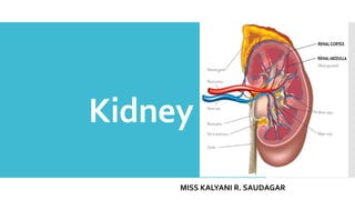
- 1. Kidney MISS KALYANI R. SAUDAGAR
- 2. CONTENTS INTRODUCTION SECTION 1 THE KIDNEYS SECTION 2 URINE FORMATION,TRANSPORTAND ELIMINATION SECTION 3 RENAL REGULATION OF BLOOD COMPOSITIONANDVOLUME SECTION 4 MINERAL HOMEOSTASIS APPENDIX APPENDIX : GLOSSARYOFTERMS APPENDIX : REFERENCES APPENDIX : CORRECT RESPONSES
- 3. SECTION1 THEKIDNEYS LEARNINGOBJECTIVES Aftercompletingthissection, youwillbeableto: •Describethefunctionsof thekidney •Identifythebasicstructures ofthekidney •Describethefunctionofthe Nephron •Describethestructures that transporturineoutofthe bodyafteritexitsthekidneys OVERVIEW OFTHE KIDNEYS The kidneys are a pair of kidney bean–shaped organs located just above the waist, between the peritoneum and the back of the abdomen.The two kidneys lie behind the liver and the intestines in the small of the back. They are partially protected by the 11th and 12th pair of ribs. As shown in Figure 1-1, the left kidney is slightly higher than the right, since the liver occupies the space above the kidney on the right side.2 The adrenal glands are a pair of flattened, pyramid-shaped glands that lie on top of the kidneys.They produce the hormones norepinephrine and epinephrine.These two hormones regulate the stress response, which increases heart rate and blood pressure.The adrenal glands also produce aldosterone, which will be discussed later in this module.1
- 4. CLINICAL CONNECTION The kidneys regulate bloodpressureby producingthe enzyme renin.Anincrease in the production ofrenin results in an increase in blood pressure.1-1 FUNCTIONS The kidneys are the hardest working organs of the urinary system.The other components of the urinary system serve mainly as passageways or for storage of urine. The functions of the kidneys include:1-1 • Excreting wastes and foreign substances in the urine • Regulating various properties of blood, including: – Ionic composition — by regulating the concentrations of several ions, such as sodium (Na+), potassium (K+), calcium (Ca2+), chloride (Cl-) and phosphate (HPO42-) – pH — by excreting hydrogen ions (H+) and conserving bicarbonate ions (HCO3-) – Osmolarity — by separately regulating the loss of water and solutes in the urine – Blood volume — by conserving or eliminating water in the urine, blood pressure is increased or decreased – Blood pressure — by secreting the enzyme renin, a component of the renin- angiotensin-aldosterone (RAA) system; renin causes an increase in blood pressure – Blood glucose levels — by synthesizing and releasing new glucose molecules Producing hormones: – Calcitriol — the active form of vitamin D, which helps regulate calcium levels – Erythropoietin (EPO) — which stimulates the production of red blood cells
- 5. FASTFACT Themainfunctionsofthe kidneysarethere- absorptionofsolutesand water,productionofurine, andexcretionofwastes.The kidneysalsoperform severalhomeostatic functions,including regulation ofelectrolytes,acid-base balance,andblood pressure.Theyalsoproduce thehormonesvitaminD anderythropoietin.1-1 Normal kidney functions collectively contribute to the maintenance of homeostasis, as summarized by Figure 1-2. Figure 1-2:The function of the kidneys in the maintenance of homeostasis.1-2 Normal kidney functions in homeostasis EXCRETARY REGULATION Nitrogenous waste products Drug metabolites and other waste Uric acid METABOLIC (ENDOCRINE) REGULATION Erythropoietin Renin-angiotensin-aldosteron Vitamine-d ELECTROLYTE BALANCE Sodium Phosphorus potassium Magnesium calcium ACID BASE BALANCE Metabolic compensation BODYWATER REGULATION Urine output Blood pressure
- 6. Figure1-3:Internalanatomyofthekidneys. 1-1 FASTFACT Thenephronsarethefunctionalunitsofthekidneys. Nephronsandcollectingductsperformthree basicfunctions:filtrationoftheblood,reabsorptionof waterandsolutes,andsecretionofwastes fromtheblood.Theseprocesseshelptomaintain homeostasiswithinthebody.1-1 STRUCTURE OFTHE KIDNEYS FAST FACT A typical adult kidney is 10–12 cm (4–5 in) long, 5–7 cm (2–3 in) wide, and 3 cm (1 in) thick, roughly the size of a bar of bath soap, and weighs 135-150 g (4.5–5 oz).1-1 If one was to slice a kidney in half (as illustrated by Figure 1-3), two distinct regions would be revealed: • A superficial, smooth-textured reddish exterior portion called the renal cortex • A deep, reddish-brown inner region called the renal medulla RENAL CORTEX The renal cortex refers to the smooth-textured area extending from the exterior (renal capsule) to the bases of striated, cone-shaped structures called renal pyramids and into the spaces between them.The renal capsule is the membrane that covers the surface of the kidney. RENAL MEDULLA The renal medulla consists of 8–18 renal pyramids.The base of each pyramid faces towards the renal cortex, and the apex of each pyramid, called the renal papilla, points to the renal hilum (the area of the kidney through which the ureter, blood vessels, lymphatic vessels, and nerves emerge).1-1The portions of renal cortex that extend between renal pyramids are called renal columns. A renal lobe consists of 1-1: • A renal pyramid •The overlying area of renal cortex • Half of each adjacent renal column NEPHRON Together the renal cortex and renal medulla constitute the functional portion of the kidney, called the parenchyma.The parenchyma contains approximately one million microscopic structures called nephron, which are the filtration units of the kidney.1-1
- 7. Figure1-4.Thefunctionalcomponentsof anephron.1-1 Each nephron consists of two parts (shown in Figure 1-4):1-1 • A renal corpuscle • A renal tubule A renal corpuscle is comprised of the glomerulus (a capillary network) plus the glomerular (Bowman’s) capsule, a double-walled epithelial cup that surrounds the glomerular capillaries.This configuration creates a thin, porous filtration membrane, which allows passage of water and small solutes, but prevents filtration of most plasma proteins, blood cells, and platelets.The filtered fluid passes into the renal tubule.1-1 The renal tubule is composed of three main sections, which are (in the order in which fluid passes):1-1 • Proximal convoluted tubule (attached to the glomerular capsule) • Loop of Henle (nephron loop) • Distal convoluted tubule PROXIMAL CONVOLUTED TUBULES The proximal convoluted tubule is attached to the glomerular capsule and is the first structure through which fluid passes.The largest amount of solute and water reabsorption from filtered fluid occurs in the proximal convoluted tubules. Reabsorbed substances include glucose, amino acids, lactic acid, water-soluble vitamins, and ions such as Na+, K+, Cl-, Ca2+, Mg2+, HCO3-and HPO42-. At the same time, waste products such as NH4+, urea and small amounts of creatinine are secreted into urine.1-1
- 8. CLINICALCONNECTION Acutekidneyinjury(AKI),an abruptandsustained declineinrenalfunction, leadstolossoftheaffected nephrons.In80%of patients,necrosis(death)of tubularcells,butnot glomeruliorsurrounding tissues,occurs.Causesof AKImayincludeoxygen deficiency,sepsisortoxic kidneyinjury.1-3 LOOP OF HENLE The loop of Henle connects the proximal and distal convoluted tubules.The descending part of this structure dips into the renal medulla. It next makes a hairpin turn and ascends to the renal cortex. Approximately 80–85% of nephrons reside in the outer portion of the renal cortex.The other 15–20% of nephrons exist deep in the cortex. Additional filtered ions and water are reabsorbed from the loop of Henle.1-1 DISTAL CONVOLUTED TUBULES Resorption of remaining Na+, Cl-, Ca2+ and water continue as fluid flows along the distal convoluted tubule. By the time the fluid reaches the end of the distal convoluted tubule, 90–95% of filtered solutes and water have returned to the bloodstream. Remaining substances that have not been reabsorbed are excreted in the urine.1-1 DUCTS, CALYCES ANDTHE RENAL PELVIS Distal convoluted tubules from several nephrons empty into a single collecting duct.The collecting ducts unite and later converge into large papillary ducts, which then drain into cuplike structures called calyces. Urine then moves into a single large cavity called the renal pelvis, and subsequently through the ureter into the urinary bladder.1-1 RENAL BLOOD SUPPLY The kidneys are supplied with numerous blood vessels due to their critical function in waste removal and the regulation of blood volume and composition. Despite their small size (less than 0.5% of total body mass), the kidneys receive 20–25% of the volume of blood pumped by the heart while at rest (resting cardiac output). In healthy adults, renal blood flows through both kidneys at a rate of approximately 1200 mL per minute.1-1
- 9. FASTFACT Whenthevolumeofthe bladderexceeds200–400mL, pressurewithinthebladder increases. Stretchreceptorsinthebladder transmitnerveimpulsesinto thespinalcord,triggeringa spinalreflexthatgivesthe sensationofafullbladder.This createsaconsciousdesireto urinate.Bycontrollingthe urethralsphincterandpelvic muscles,apersoncaninitiateor delayurination foralimitedperiod.1-1 LOWER URINARYTRACT Urine drains from the kidneys through the ureters into the bladder, and subsequently out through the urethra during urination (micturition).1-1 URETERS There are two ureters—one for each kidney.They transport urine from the renal pelvis to the urinary bladder. Along with gravity and hydrostatic pressure, muscular contractions push urine through the ureters.The ureters are long and thick-walled narrow tubes. They measure 25–30 cm (10–12 in) in length and vary in diameter from 1–10 mm between the renal pelvis and the urinary bladder. Like the kidneys, the ureters are positioned behind the peritoneum. They curve at their ends and pass through the wall of the bladder.1-1Although there is no anatomical valve between each ureter and the bladder, a physiological one exists. As the bladder fills with urine, it compresses the openings to the ureters, and prevents the backflow of urine. If this valve does not operate properly, microbes may travel up the ureters to cause a kidney infection.1-1 URINARY BLADDER The bladder stores urine. Situated in the pelvic cavity, the urinary bladder is a hollow, distensible muscular organ. In males it is directly in front of the rectum, whereas in females it is in front of the vagina and below the uterus. Held in place by folds of the peritoneum, the bladder becomes slightly distended and spherical as it fills with urine. When empty, the bladder collapses. Urinary bladder capacity ranges from 700–800 mL, and is slightly smaller in females due to the position of the uterus.1-1 URETHRA The urethra carries urine out of the body. It is a small tube that leads from the urinary bladder to the exterior of the body.1-1 • In females, the urethra has a length of 4 cm (1.5 in) and opens to the exterior through the external urethral orifice between the clitoris and the vaginal opening. • In males, the urethra is approximately 20 cm (8 in) long and extends from the urinary bladder to the exterior, but it first passes through the prostate, then through the deep muscles of the perineum, and lastly through the penis.
- 10. SECTION2 URINEFORMATION, TRANSPORTAND ELIMINATION LEARNINGOBJECTIVES Aftercompletingthissection, youwillbeableto: •Understandthenormal compositionofurine •Describetheprocessofurine formation •Describehowglomerular filtrationrate(GFR)isrelatedto kidneyfunction •Identifythedifferences betweenthereabsorptionand secretionprocesses COMPOSITION OF URINE Routine assessment of kidney function involves the evaluation of both the quantity and quality of urine, as well as the levels of waste in the blood.The analysis of the volume, physical, chemical, and microscopic properties of urine is called a urinalysis. It is one test physicians use to assist them in their routine assessment of kidney function and general physical health.1-1A normal adult will eliminate 1–2 liters of urine per day.The characteristics of normal urine are listed inTable 2-1. Table 2-1: Characteristics of Normal Urine.1-1 CHARACTERISTIC DESCRIPTION Volume 1–2 liters in 24 hours Can vary with fluid intake Color Yellow or amber Varies with concentration and diet Certain medications and certain diseases may also affect color Turbidity Transparent when freshly voided Becomes turbid (cloudy) upon standing Odor Mildly aromatic Becomes ammonia-like upon standing Can also vary with diet and certain diseases pH 4.6–8.0 (average of 6.0) Varies with diet Specific gravity (density) 1.001–1.035 Increases as concentration of solutes increases
- 11. CLINICALCONNECTION Indiabetesmellitus,thebodyis unabletoproduceoruse insulin.Becauseinsulinisnot availabletotransportglucose intothecells,excessglucoseis excretedintheurine,leadingto glucosuria.Becauseofthehigh levelsofglucoseinthefluid beingfilteredthroughtherenal tubules,thekidneys’abilityto reabsorbwaterisreduced.This leadstoexcessiveurine production,referredtoas polyuria.1 WATER AND SALT Water accounts for approximately 95% of the total volume of urine; the remaining 5% consists of electrolytes. Electrolytes are solutes derived from cellular metabolism and exogenous substances such as drugs. Specific hormones increase reabsorption of water and salts in the renal tubules. Factors such as fluid intake, diet and general health can also influence urine volume.1-1 INORGANIC SOLUTES Typical solutes normally present in urine include filtered and secreted electrolytes that are not reabsorbed. Inorganic solutes (ions), such as potassium, are secreted by cells in the collecting duct.1-1 ORGANIC SOLUTES A number of organic solutes are also found in urine, including1-1: • Urea from the breakdown of proteins • Creatinine from the breakdown of creatine phosphate in muscle fibers • Uric acid generated from the breakdown of nucleic acids • Urobilinogen from the breakdown of hemoglobin • Fatty acids, pigments, enzymes and hormones If body metabolism or kidney function is altered by disease, then substances not normally present may be found in the urine, or normal constituents may appear in abnormal amounts. For example, the presence of glucose in the urine, referred to as glucosuria, usually indicates diabetes mellitus.1-1 PROTEIN Normal urine is virtually free of protein.The presence of protein in the urine is called proteinuria.This condition can indicate improper functioning of the kidneys. For example, the plasma protein albumin usually appears in urine in only trace amounts due to its large size and inability to pass through the tiny openings (fenestrations) in the capillary walls that allow the kidneys to act as filters. If the permeability of filtration membranes increases due to injury, irritation or disease of the kidneys, or due to elevated blood pressure, albumin and other proteins can enter the urine.1-1 For example, microalbuminuria (small amounts of albumin in the urine) is an early manifestation of kidney disease related to diabetes, hypertension or glomerular damage.2-1
- 12. CLINICALCONNECTION Severaldifferenttestsareusedtoevaluatekidney disease.Thesetestsincludemeasurementof proteinoralbuminlevelsinurineandcalculation oftherateatwhichcreatinineisclearedinthe urine.Theurinaryalbumin-to-creatinineratio (UACR)isonemethodofquantifying proteinuria.2-1 Bloodtests,aswellasurinalysis,canbeusedto evaluatekidneyfunction.Bloodureanitrogen (BUN)isatestthatmeasuresthelevelsofblood nitrogenfromtheureathatisgeneratedbythe breakdownofaminoacids.BUNiselevatedwhen renaldiseaseorobstructionoftheurinarytract occursandglomerularfiltrationrate(GFR)is decreasedorwhentherenalbloodflowis reduced.1-1 EstimatesofGFR(eGFR)areconsideredthebest overallmeasureofthelevelofkidneyfunction. ToestimatetheGFR,predictionequationstake intoaccountserumcreatinineconcentrationand someorallofthefollowingvariables:age,gender, raceandbodysize.2-1Anemergingmethod ofestimatingGFRisbasedonbloodlevelsof cystatinC,asmallproteinfreelyfilteredbythe glomerularmembraneanddegradedinthe proximaltubules.2-2 Table 2-2 lists the components of urine, and the amount filtered, reabsorbed and excreted per day. Table 2-2: Substances filtered, reabsorbed and excreted by the healthy kidney.1-1 AssumingGFR is 180 liters per day. In addition to being filtered and reabsorbed, urea is secreted. After virtually all filtered K+ is reabsorbed in the convoluted tubules and loop of Henle, a variable amount of K+ is secreted in the collecting duct. Assuming normal plasma concentration of 0.96 mg/L and normal urine concentration of 0.095 mg/L.2-2 Multiplied by GFR of 180 L/day. SUBSTANCE FILTERED(ENTERD GLOMULAR CAPSUL/DAY) REABSORBED(RETURNED TO BLOOD/DAY) URINE(EXCRETED/DAY) Water 180 liters 178-179 liters 1-2 liters Proteins 2.0 g 1.9 g 0.1 g Sodium ions (Na+) 579 g 575 g 4 g Chloride ions (Cl-) 640 g 633.7 g 6.3 g Bicarbonate ions (HCO3-) 275 g 274.97 g 0.03 g Glucose 162 g 162 g 0 g Urea 54 g 24 g 30 g Potassium ions (K+) 29.6 g 29.6 g 2.0 g Uric acid 8.5 g 7.7 g 0.8 g Creatinine 1.6 g 0 g 1.6 g Cystatin C 0.17 g - 0.017 g
- 13. Figure2-1:Relationofthestructureofanephron toitsthreebasicfunctions:glomerularfiltration, tubularreabsorptionandtubularsecretion.1-2 GLOMERULAR FILTRATION As shown in Figure 2-1, glomerular filtration is the initial step towards urine production.Water and most of the solutes in blood plasma move across the walls of the glomerular capillaries (the smallest of the blood vessels) into the glomerular capsule, and into the renal tubule. Solutes that are in the fluid draining into the renal pelvis stay in the urine and are eventually excreted.1-1 FILTRATION PROCESS Filtration is the first basic function of the nephron.The endothelial cells of glomerular capillaries, plus a layer of epithelial cells that encircle the capillaries, form a leaky barrier known as the filtration membrane. It allows the filtration of most water and small solutes, but prevents the filtration of plasma proteins, blood cells and platelets.The filtration membrane is a sandwich-like assembly.A solute’s size and charge determine how easily it can pass through glomerular capillary membranes.1-1
- 14. FASTFACT Creatinineclearanceisatesttomeasurehow efficientlythekidneysremovecreatinineand otherwastesfromtheblood.Lowcreatinine clearanceindicatesimpairedkidneyfunction.2-3 CreatinineclearanceisrelatedtoGFR.Plasma levelsofcreatininearestableinhealthyindividuals becausecreatinineisproducedatasteadyrateby muscle.Creatinineinurinecomesfromexcretion ofplasmacreatinineinthekidneys.Thereisa steadydailyexcretionofurinecreatinin— however,overshortperiodsoftime(hours),the urinecreatinineconcentrationvarieswiththe stateofhydrationandconcentrationofurine.By measuringbloodandurinecreatininelevelsovera specificperiod,therateofcreatinineclearance (mL/min)canbecalculated.1-1 Thenormalrangeforserumcreatinineis approximately0.5–1.1mg/dLinadultfemales, andapproximately0.6–1.2mg/dLinadultmales. Thisrangemayvarybyage,genderandweight.2- 4 Pressure forces fluids and solutes through the capillary membrane.The same process occurs in glomerular capillaries as in other capillaries in the body. However, the volume of fluid filtered by the glomerular capillaries is much greater.Three factors permit this high throughput:1-2 • Glomerular capillaries have a large surface area •The filtration membrane is thin and porous • Glomerular capillary blood pressure is high, which creates resistance to the outflow of blood from the glomerulus and promotes filtration by pushing water and solutes through the capillary membrane Meanwhile, hydrostatic and osmotic pressures oppose filtration by pushing fluid back into the capillaries.1-1 GLOMERULAR FILTRATION RATE The glomerular filtration rate (GFR) refers to the amount of filtrate formed in all the renal corpuscles of both kidneys each minute. In adults, the average is 125 mL/min for men and 105 mL/min for women. Homeostasis of body fluids dictates that a relatively constant GFR is required.1-1 • If theGFR is too high, needed substances may pass through the renal tubules and be lost in the urine • If the GFR is too low, all of the filtrate may be reabsorbed and too few waste products excreted The kidneys, nervous system and hormones regulateGFR by1-1: •Adjusting blood flow into and out of the glomerulus •Altering the glomerular capillary surface available for filtration
- 15. CLINICALCONNECTION Acutekidneyinjury(AKI)isanabruptreductioninkidney function.Itisassociatedwithincreased serumcreatininelevels,decreasedGFR,andoliguria (reductioninurineoutput)formorethan sixhours.2-5LossofkidneyfunctioninAKImaybe reversible,requiringtemporarydialysis,ormay progresstoend-stagerenaldisease(ESRD),necessitating permanentrenalreplacementtherapy.2-6 Serumcreatininealoneisapoormarkerofearlyrenal dysfunction,asitisafunctionalmarkerthat isnotspecificforAKIandisinfluencedbynumerous nonrenalfactors.Consequently,development ofotherbiomarkersofearlykidneydamageisanintense areaofinvestigation.Severalbiomarkers havebeenidentifiedwithinthelastdecadethatcandetect AKIearlierthancreatinine.Themost promisingoftheseincludeneutrophilgelatinase-associated lipocalin(NGAL),cystatinC,kidney injurymolecule-1(KIM-1),liverfattyacidbindingprotein(L- FABP)andinterleukin-18(IL-18).1-3,2-7 Chronickidneydisease(CKD)referstoaprogressiveand usuallyirreversibledeclineinGFR. CKDoccursinstages,overaprolongedtimeframe.A patientwithCKDStage1hasanestimated GFR(eGFR)≥90mL/min/1.73m2.ByCKDStage5,theGFR isdiminishedto10–15%ofnormal (eGFRof<15mL/min/1.73m2).Thisfinalstagedenotes ESRD,necessitatingrenalreplacement therapybydialysisortransplant.1-1,2-1 TUBULAR REABSORPTIONAND SECRETION Approximately 99% of filtered water and many useful solutes are reabsorbed and returned to the blood as fluid flows along the renal tubule and through the collecting duct. As fluid flows through the renal tubule and collecting duct, the tubule and duct cells secrete materials such as wastes, drugs and excess ions into the fluid.Therefore, tubular reabsorption returns substances to the blood, and tubular secretion removes substances from the blood.1-1 REABSORPTION Reabsorption is the second basic function of the nephron and collecting duct. As fluid flows through the nephron, solutes such as glucose, amino acids, urea and ions, as well as small proteins and peptides, are returned to the blood. Most reabsorption occurs in the proximal convoluted tubule. Cells along the remaining length of the distal nephron further reabsorb water and selected ions in order to maintain homeostasis.1-1 FAST FACT The volume of blood that enters the proximal convoluted tubules every 30 minutes is greater than the total blood plasma volume of the body. This is possible because the normal rate of glomerular filtration is so high.1-1
- 16. CLINICALCONNECTION Severallowmolecularweightproteins andenzymesarefilteredthroughthe glomerulusandsubsequentlyreabsorbed intheproximaltubule.Tubularinjurymay increasetheconcentrationofthese proteinsintheurineeventhoughlessis beingfiltered.2-8SerumcystatinCisa markerofdecreasedGFR,whereasurine cystatinCisamarkeroftubularinjury.2-9 Acutekidneyinjury(AKI)inducesthe expressionofanumberofproteinsinthe renaltubules.Theseproteinsareexcreted inurineandarethusbeingevaluatedas specificbiomarkerstohelpdiagnoseAKI andpotentiallydifferentiatebetween differentsubtypesandpathologiesof kidneyinjury.Theyincludeinterleukin-18 (IL-18)andneutrophilgelatinase- associatedlipocalin(NGAL).1-3,2-7,2-10 SECRETION The third function of nephrons and collecting ducts is tubular secretion, the transfer of substances such as hydrogen ions (H+), potassium (K+), ammonium ions (NH4+), certain drugs from the blood and tubule cells into the tubular fluid.There are two critical outcomes to tubular secretion: the secretion of H+ helps to control blood pH, and the secretion of other waste products helps eliminate them from the body.1-1 In healthy individuals, creatinine is not secreted in significant amounts by the kidneys. Creatinine entering the urine comes from passive glomerular filtration. In severe renal disease, when plasma creatinine is high, active tubular secretion becomes important. HORMONAL REGULATION Tubular reabsorption and tubular secretion are regulated by hormones, which are described in Table 2-3.These hormones will be discussed in greater detail in the next section. Table 2-3: Hormonal regulation of tubular reabsorption and tubular secretion.1-1 HOEMONE MAJOR STIMULITHATTRIGGER RELEASE EFFECTS Angiotensin ll Low blood volume or low blood pressure stimulates renin-induced production of angiotensin Increased reabsorption of Na+, other solutes and water, which increases blood volume aldosterone Increased angiotensin ll level and increased level of plasma K+ promotes release of aldosteron by adrenal cortex Increased secretion of K+ and reabsorption of Na+,Cl- Increases reabsorption of water, which increases blood volume Antidiuretic hormone or vasopressin Increased osmolarity fluid or decreased blood volume promotes release of ADH from the posterior pituitary gland Iincreases facultative reabsorption of water, which decreases osmolarity of body fluids B-natriuretic peptide Stretching of the ventricles and atria of the heart stimulates secretion of BNP Increases excretion of Na+ in urine (natriuresis) Increases urine output (diuresis) Decreased blood volume
- 17. SECTION3 RENALREGULATIONOF BLOOD COMPOSITIONAND VOLUME LEARNINGOBJECTIVES Aftercompletingthis section,youwillbeableto: •Summarizetheroleofthe kidneysinregulatingblood composition •Understandhow hormonesactonthe kidneystoregulateblood volumeandpressure REGULATIONOF BLOOD COMPOSITION To perform correctly, the cells of the body require stable osmolarity, stable pH and glucose levels, and constant removal of wastes.These functions are provided, in part or in whole, by the kidneys.1-1 FAST FACT The osmolarity of a solution is a measure of the total number of dissolved particles (molecules and/or ions) per liter of solution.A similar term, osmolality, is the number of particles of solute per kilogram of water. Most body fluids and solutions used clinically are dilute, in which case there is less than a 1% difference between the two measures.1-1 IONIC COMPOSITIONAND OSMOLARITY The kidneys maintain a relatively constant blood osmolarity by regulating the loss of water and solutes. Reabsorption in the nephron collecting ducts helps to maintain this balance.1-1Most solute and water reabsorption from filtered fluid occurs in the proximal convoluted tubules. Sodium (Na+) ions are the major contributor to osmolarity and fluid volumes. Other ions, such as potassium (K+), calcium (Ca2+), and chloride (Cl-), must also be maintained within tight control to ensure proper cellular functioning.1-1 CLINICAL CONNECTION Parathyroid hormone (PTH) is a major regulator of the levels of calcium (Ca2+), magnesium (Mg2+) and phosphate (HPO42-) ions in the blood.The specific action of PTH is to increase the number and activity of cells called osteoclasts that break down mineralized bone.This releases calcium and phosphates from bone into the blood. PTH also acts on the kidneys. It slows the rate at which calcium and magnesium ions are lost from the blood into the urine, and it increases the loss of phosphate from the blood into the urine. PTH also stimulates the formation of calcitriol (the active form of vitamin D, often referred to as 1,25 dihydroxy Vitamin D).1 ACID-BASE BALANCE AND BICARBONATE To regulate acid-base balance (pH), the kidneys excrete variable amounts of hydrogen ions (H+) into the urine and conserve bicarbonate ions (HCO3-), which buffer H+ in the blood.The equilibrium between these two ions regulates blood pH. Slight changes in concentration can cause significant alterations in physiologic functions.1-1
- 18. CLINICAL CONNECTION Anemia(the condition ofhaving toofewred bloodcells) is common in patients with chronic kidney disease dueto a lackof erythropoietin.2-3 UREAWASTE Urea is generated from the breakdown of amino acids, and some is normally excreted in urine. After being filtered in the glomerulus, variable amounts of urea are reabsorbed or secreted in the tubules and collecting ducts. If the glomerular filtration rate decreases significantly, such as with renal disease, urea is not effectively eliminated in the urine and collects in the blood.This can be confirmed by the measurement of blood urea nitrogen (BUN).1-1 BLOODGLUCOSEAND GLUCONEOGENESIS The kidneys are able to synthesize and release new glucose molecules from substances other than glycogen or simple sugars in a process called gluconeogenesis.The kidneys convert the amino acid glutamine to glucose, thus helping to maintain normal blood glucose levels.1, 1-1 RED BLOODCELLS The kidneys regulate red blood cell formation through a hormone called erythropoietin (EPO).3-1 EPO is produced by the kidneys and increases the number of red blood cell precursors. In renal failure, EPO levels decrease and red blood cell production declines.3-1
- 19. REGULATIONOF BLOODVOLUMEAND PRESSURE Four different hormones affect the absorption of sodium ions, chloride ions and water, as well as the secretion of potassium ions by the renal tubules.The most important regulators of electrolyte reabsorption and secretion are angiotensin II and aldosterone.The major hormone for regulating water reabsorption is antidiuretic hormone (ADH). B-natriuretic peptide (BNP), and, to a lesser degree, atrial natriuretic peptide (ANP), also play a role in inhibiting both electrolyte and water reabsorption.1-1 RENIN-ANGIOTENSIN-ALDOSTERONE (RAA) SYSTEM The renin-angiotensin-aldosterone system controls the secretion of aldosterone hormone by the adrenal glands that lie on top of the kidneys. Aldosterone regulates the homeostasis of two mineral ions — sodium (Na+) and potassium (K+) — and helps to adjust blood pressure and blood volume.1 Figure 3-1 summarizes the effects of renin, angiotensin and aldosterone.1 • Dehydration, sodium deficiency or hemorrhage decrease blood volume and blood pressure • Low blood pressure stimulates the kidneys to secrete renin, an enzyme that converts angiotensinogen (a plasma protein produced in the liver) to angiotensin I • Angiotensin I travels through the bloodstream to the lungs, where angiotensin-converting enzyme (ACE) converts it to the active hormone, angiotensin II • Angiotensin II stimulates: – Contraction of smooth muscle in the walls of arterioles, which helps raise blood pressure towards normal – Secretion of aldosterone by the adrenal glands • Upon reaching the kidneys, aldosterone increases: – Reabsorption of sodium and water, so that less is lost in the urine – Secretion of potassium and hydrogen ions into the urine • Blood volume and blood pressure increase due to increased water reabsorption
- 21. Figure3-2:Negativefeedbackregulationofwater reabsorptionbyADH. ANTIDIURETIC HORMONE (VASOPRESSIN) Antidiuretic hormone (also known asADH or vasopressin) is released by the pituitary gland at the base of the brain. ADH regulates water reabsorption by increasing the water permeability of cells in the distal convoluted tubule and collecting duct. •When ADH concentrations are high, the kidneys produce as little as 400 to 500 mL of concentrated urine.This serves to retain fluid in the body and raise blood pressure. •When ADH levels are low, the kidneys produce a large amount of dilute urine. The regulation of ADH involves a negative feedback loop, shown in Figure 3-21-1: •The hypothalamus (a region of the brain that coordinates many hormonal and homeostatic processes) detects an increase in osmolarity (i.e., a decrease in water concentration and a concomitant increase in solutes). •The hypothalamus stimulates the pituitary gland to secrete ADH. • ADH makes the cells of the kidneys more permeable to water and increases water reabsorption. • Plasma osmolarity decreases and homeostasis is restored in the body, which subsequently stops the release of ADH A precipitous decrease in blood volume (e.g., due to hemorrhage or severe dehydration) is also a stimulus for ADH.1-1 B-NATRIURETIC PEPTIDE (BNP) A significant increase in blood volume stimulates the release of B-Natriuretic Peptide (BNP) from the heart.The importance of BNP for normal tubular function is unclear, but it can: • Inhibit reabsorption of sodium ions and water in the proximal convoluted tubule and collecting duct • Suppress the secretion of aldosterone andADH, thereby increasing the excretion of sodium in the urine and increasing urine output Both of these actions decrease blood volume and blood pressure.1-1 BNP (or its precursor, NT-pro-BNP) can be measured in many clinical laboratories to assess fluid overload.
- 22. SECTION4 MINERALHOMEOSTASIS LEARNINGOBJECTIVES Aftercompletingthis section,youwillbeableto: •Understandcalciumand phosphorushomeostasis •Relatetheparathyroidaxis totheendocrinefunctionof thekidney •Distinguishbetween calciumabsorption, resorptionandreabsorption OVERVIEW OF MINERAL HOMEOSTASIS Stable levels of inorganic minerals such as calcium (Ca2+), phosphate (HPO42-) and magnesium (Mg2+) are critical for normal physiological functioning. For example, calcium is essential for skeletal structure, proper functioning of nerve and muscle cells, blood clotting, and the function of numerous enzymes.4-1 Maintaining homeostasis of minerals is a finely tuned balancing act involving absorption from the gastrointestinal (GI) tract, reabsorption and secretion by the kidneys, and storage and resorption from bone.1 As illustrated in Figure 4-1, calcium homeostasis is maintained through a complex hormonal feedback loop that involves1, 4-1: • Calcitriol (also called 1,25-dihydroxyvitamin D3 or 1,25[OH]2D3), the active form of vitamin D, produced by the kidneys • Parathyroid hormone (PTH), produced by the parathyroid glands in the neck Figure 4-1:The parathyroid axis: a feedback loop involving parathyroid hormone (PTH) and calcitriol (1,25[OH]2D3) maintains calcium homeostasis.
- 23. FASTFACT Whatisthedifference between absorption,reabsorption and resorption? Absorptionistheintakeoffluidsor othersubstances bycellsofmucous membranes, suchastheabsorption ofcalciumbytheintestines.4-4 Reabsorption isthereturnofmost ofthefilteredwaterandmanyof thefiltered solutestothe bloodstream fromthekidneys.1-1 Resorptionistheremovalof minerals andcollagenfibers from bonebyosteoclasts.4-1All threeof theseprocessesarestimulated directlybyPTHorindirectly by activated vitaminD,whichthe kidneysproduceinresponseto PTH.1 INTESTINAL ABSORPTION As food is digested, dietary minerals are absorbed from the intestines. Calcitriol promotes the absorption of calcium and other minerals from the GI tract to the bloodstream.4-1 RENAL EXCRETION The kidneys filter and excrete minerals in the urine. PTH, the major regulator of mineral levels in the blood, acts on the kidneys in three ways:1 • Decreasing secretion of calcium and magnesium into the urine, thus raising their levels in the blood • Increasing excretion of phosphate in urine, thus lowering levels in the blood • Inducing conversion of inactive vitamin D to calcitriol by the kidneys BONE REMODELING AND RESORPTION Bone is a mineral reservoir that stores 99% of the body’s calcium, along with other minerals. Bone tissue is constantly remodeled and repaired, removing old bone and replacing it with new bone.4-1 • Cells called osteoclasts resorb (break down) old bone, releasing minerals such as calcium • Bone-building cells called osteoblasts form new bone, depositing minerals in the processTo maintain a constant level of calcium in the blood, the rate of bone remodeling is tightly controlled by hormones.4-1, 4-2, 4-3 • PTH stimulates resorption by osteoclasts, releasing calcium into the bloodstream • Calcitriol, produced by the kidneys, promotes normal bone mineralization and opposes PTH production • Calcitonin, produced by the thyroid gland, plays a lesser role by inhibiting osteoclasts and promoting calcium deposition by osteoblasts
- 24. IMPORTANCE OF VITAMIN D Vitamin D is an essential nutrient required for the optimal absorption of dietary calcium and phosphate and for normal bone formation.4-3 It is important to note that vitamin D itself is biologically inactive. A two-step process in the liver and then the kidney converts it to the active metabolite 1,25-dihydroxyvitamin D3 (1,25[OH]2D3), also known as calcitriol.4-3 The classical actions of vitamin D in kidneys, bone, parathyroid glands and the intestines pertain to maintaining calcium levels within a precise range to ensure normal cellular physiology and skeletal integrity.4-2, 4-3 Table 4-1 summarizes the classical calcemic effects of vitamin D. Table 4-1: Classical calcemic effects of vitamin D in various tissues. TISSUE EFFECTS Intestine Enhances absorption of dietery calcium and phosphate Kidney Self regulates vitamin D activation and degradation enhances renal calcium reabsorption Parathyroid Inhibits both PTH synthesis and secretion, as well as parathyroid cell growth bone Helps sustain normal bone mineralization and remoleding
- 25. REVIEW QUESTIONS: SECTION 1 Answers are provided at the end of this Learning Guide. 1.Which organs constitute the urinary system? 2.What are three functions of the kidneys? 3. At which location is the largest amount of water and solutes reabsorbed into the bloodstream? A) Distal tubule B) Glomerulus C) Proximal tubule D) Ureter 4.What is the path for fluid through a nephron? A -Bowman’s capsule → proximal convoluted tubule → loop of Henle → distal convoluted tubule B -Loop of Henle → Bowman’s capsule → proximal convoluted tubule → distal convoluted tubule C- Proximal convoluted tubule → Bowman’s capsule → distal convoluted tubule → loop of Henle 5. Complete the table by matching the organ (kidneys, ureters, urethra, urinary bladder) with its function: FUNCTION ORGAN Elimination of urine Production of urine Storage of urine Transport of urine
- 26. REVIEW QUESTIONS: SECTION 2 Answers are provided at the end of this LearningGuide. 1.Where does filtration take place? A) Collecting duct B ) Distal tubule C) Glomerulus D) Proximal tubule 2.Which of the following is not normally found in urine? A) Creatinine B) Glucose C ) Urea D) Water E) All of the above 3.What is currently considered the best way to assess kidney function? A) Blood urea nitrogen (BUN) B ) Creatinine clearance rate C) Estimates of glomerular filtration rate (GFR) D) Urinary albumin-to-creatinine ratio 4. During tubular reabsorption, _____% of filtered water is reabsorbed. A ) 50 B) 75 C ) 99
- 27. REVIEW QUESTIONS: SECTION 3 Answers are provided at the end of this Learning Guide. 1.Which of the following ions has the greatest influence on osmolarity? A ) Calcium B ) Phosphate C ) Potassium D ) Sodium 2.The kidneys respond to increases in blood pressure by: A) Excreting more water B) Filtering less sodium C ) Filtering more sodium D) Reabsorbing more water 3. If glomerular filtration rate declines, what is the effect on blood urea levels? A ) Increase B ) Decrease
- 28. REVIEW QUESTIONS: SECTION 4 Answers are provided at the end of this Learning Guide. 1.Where does activation of vitamin D to calcitriol take place? A ) Bone B) Gut C ) Kidneys D) Thyroid 2.Which is an effect of calcitriol? A) Decreases absorption of dietary calcium and phosphate B) Increases PTH synthesis and secretion C ) Promotes tubular secretion of calcium D) Sustains normal bone mineralization 3.Which is an effect of parathyroid hormone? A) Increases absorption of dietary calcium and phosphate B ) Promotes bone resorption C) Reduces serum calcium levels D ) Stimulates bone formation
- 29. APPENDIX : CORRECT RESPONSES SECTION 1 1. The kidneys, ureters, urinary bladder, and urethra 2.Any three of the following: • Regulation of blood ionic composition and pH • Regulation of blood volume and pressure • Maintenance of relatively constant blood osmolarity • Production of hormones calcitriol and erythropoietin • Regulation of blood glucose levels • Excretion of wastes and foreign substances 3. C 4. A 5. Functions Organ Elimination of urine urethra Production of urine kidneys Storage of urine Urinary bladder Transport of urine ureters
- 30. APPENDIX : CORRECT RESPONSES SECTION 1 SECTION 2 1. C 2. B 3. C 4. C SECTION 3 1. D 2. A 3. A SECTION 4 1. C 2. D 3. B
- 31. APPENDIX : GLOSSARY OF TERMS Absorption: Intake of fluids or other substances by cells of mucous membranes.4-4 Albumin: The major plasma protein, approximately 60% of the total, that is responsible for much of the osmotic pressure of the plasma. It acts as a transport protein for fatty acids, drugs and hormones among others. It is synthesized in the liver and its concentration is decreased in the context of renal disease.A-1 Aldosterone: A hormone produced by the adrenal cortex that promotes resorption of sodium and water by the kidneys and excretion of phosphorus in the urine.4-4 Anemia: Condition of the blood in which the number of functional red blood cells or their hemoglobin content is below normal.4-4 Antidiuretic hormone (ADH): Hormone produced by the hypothalamus, which stimulates reabsorption of water from the renal tubules and vasoconstriction of arterioles, raising blood pressure. Also called vasopressin.4-4 Calcitonin: A hormone produced by the thyroid gland that can lower the amount of blood calcium and phosphates by inhibiting bone resorption and by accelerating uptake of calcium and phosphate into bone.4-4 Calcitriol: The metabolically active form of the steroid hormone vitamin D, which is essential for the dietary absorption of calcium and phosphate. Also called 1,25-dihydroxyvitamin D3 (1,25[OH]2D3).A-2 Capillary: Microscopic blood vessel located between an arteriole and venule through which materials are exchanged between blood and interstitial fluid.4-4 Creatinine: A waste product from meat protein in the diet and from the muscles of the body.2-3 Cystatin C: A low-molecular-weight protein produced at a constant rate by all nucleated cells, freely filtered by the glomerulus, and almost completely degraded in the proximal renal tubules. Serves as a novel serum marker of GFR.2-2, A-3 Erythropoietin (EPO): A hormone made by the kidneys to help form red blood cells. Lack of this hormone may lead to anemia.2-3
- 32. GLOSSARYOF TERMS, CONTINUED Glomerular filtration rate (GFR):The amount of filtrate formed in all the renal corpuscles of both kidneys each minute (mL/min).1-1 Homeostasis:The condition in which the body’s internal environment remains relatively constant and within physiological limits.4-4 Hormone: A mediator released in one part of the body that regulates the activity of cells in another part of the body.1 Micturition:The act of expelling urine from the urinary bladder. Also called urination.1-1 Nephron:The working units of the kidneys, which remove wastes and extra fluids from the blood. Each kidney consists of about one million nephrons.2-3 Osmolality:The measure of the total number of dissolved particles per kilogram of water.1-1 Osmolarity:The measure of the total number of dissolved particles per liter of solution.1-1 Osteoblast: Cell that participates in bone formation by secreting some organic components and inorganic salts.4-4 Osteoclast: A large, multinucleated cell that resorbs (destroys) bone matrix.4-4 Parathyroid hormone (PTH): A hormone secreted by the parathyroid glands that increases blood calcium level and decreases blood phosphate level.4-4 Parenchyma:The functional part of the kidneys. Contains approximately one million nephrons — the filtration units of the kidneys.1-1
- 33. GLOSSARY OF TERMS, CONTINUED Peritoneum:The largest serous membrane of the body that lines the abdominal cavity and covers the viscera.4-4 Proteinuria: A condition in which the urine contains a large amount of protein, a sign that the kidneys are not functioning properly.2-3 Reabsorption, tubular:The return of most of the filtered water and filtered solutes to the bloodstream by the kidneys.1-1 Renin: Enzyme released by the kidneys in response to low blood pressure — the first step of the renin- angiotensin-aldosterone pathway that regulates aldosterone secretion.1 Renin-angiotensin-aldosterone (RAA) system: Pathway that regulates secretion of aldosterone to help control blood volume, blood pressure and levels of Na+, K+ and H+ in the blood.1 Resorption:The removal of minerals and collagen fibers from bone.4-1 Secretion, tubular: Movement of substances from the blood into the renal tubular fluid.4-4 Specific gravity:The ratio of the weight of a volume of a substance to the weight of an equal volume of distilled water.1-1 Urea: A waste product found in the blood and caused by the normal breakdown of protein in the liver.2- 3 Ureter: One of two tubules that connect the kidney with the urinary bladder.4-4 Urethra: Duct from the urinary bladder that conveys urine to the exterior.4-4 Urinalysis: Analysis of the volume and physical, chemical and microscopic properties of urine.4-4 Urinary bladder: Hollow muscular organ located in the pelvic cavity, behind the pubic bone; receives urine via the ureters and stores it until excreted via the urethra.4-4