
Recommended
More Related Content
What's hot
What's hot (20)
Similar to Physical Mapping.pptx
Similar to Physical Mapping.pptx (20)
Recently uploaded
Recently uploaded (20)
Physical Mapping.pptx
- 2. GENOME MAPPING Genome mapping is an important tool for locating a specific gene on a particular region of a chromosome and to determine relative distances between genes and molecular markers on the chromosomes. There are two types of Genome mapping: ● Genetic Mapping ● Physical Mapping
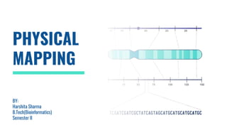
- 3. PHYSICAL MAPPING ● Physical mapping is a technique used in molecular biology to find the order and physical distance between DNA base pairs by DNA markers .It is one of the gene mapping techniques which can determine the sequence of DNA base pairs with high accuracy. ● A physical map, as related to genomics, is a graphical representation of physical locations of landmarks or markers (such as genes, variants and other DNA sequences of interest) within a chromosome or genome. ● Physical mapping uses DNA fragments and DNA markers to assemble larger DNA pieces. With the overlapping regions of the fragments,researchers can deduce the positions of the DNA bases. ● A physical map provides detail of the actual physical distance between genetic markers, as well as the number of nucleotides.
- 4. Types Of Physical Mapping Low Resolution Mapping Low-resolution physical mapping is typically capable of resolving DNA ranging from one base pair to several mega bases. In this category, most mapping methods involve generating a somatic cell hybrid panel, which is able to map any human DNA sequences, the gene of interest, to specific chromosomes of animal cells, such as those of mice and hamsters. High Resolution Mapping High-resolution physical mapping could resolve hundreds of kilobases to a single nucleotide of DNA. A major technique to map such large DNA regions is high resolution FISH Mapping which could be achieved by the hybridization of probes to extended interphase chromosomes or artificially extended chromatin. Since their hierarchic structure is less condensed comparing to prometaphase and metaphase chromosomes, the standard in- situ hybridization target, a high resolution of physical mapping could be produced.
- 5. Techniques Used To Create Physical Maps There are a several different techniques used for physical mapping. These include: ● Restriction mapping (fingerprint mapping and optical mapping) ● Fluorescent in situ hybridisation (FISH) mapping ● Sequence tagged site (STS) mapping.
- 6. Restriction Site Mapping ● Restriction mapping is a method used to map an unknown segment of DNA by breaking it into pieces and then identifying the locations of the breakpoints. ● This method relies upon the use of proteins called restriction enzymes, which can cut, or digest, DNA molecules at short, specific sequences called restriction sites. After a DNA segment has been digested using a restriction enzyme, the resulting fragments can be examined using a laboratory method called gel electrophoresis. ● One common method for constructing a restriction map involves digesting the unknown DNA sample in three ways. Here, two portions of the DNA sample are individually digested with different restriction enzymes, and a third portion of the DNA sample is double-digested with both restriction enzymes at the same time. ● Next, each digestion sample is separated using gel electrophoresis, and the sizes of the DNA fragments are recorded. The total length of the fragments in each digestion will be equal. However, because the length of each individual DNA fragment depends upon the positions of its restriction sites, each restriction site can be mapped according to the lengths of the fragments. ● The information from the double-digestion is particularly useful for correctly mapping the sites. The final drawing of the DNA segment that shows the positions of the restriction sites is called a restriction map.
- 8. ● Fluorescence in situ hybridization (abbreviated FISH) is a laboratory technique used to detect and locate a specific DNA sequence on a chromosome. ● In this technique, the full set of chromosomes from an individual is affixed to a glass slide and then exposed to a “probe”—a small piece of purified DNA tagged with a fluorescent dye. The fluorescently labeled probe finds and then binds to its matching sequence within the set of chromosomes. ● With the use of a special microscope, the chromosome and sub-chromosomal location where the fluorescent probe bound can be seen. ● Fluorescence in situ hybridization (FISH) is a molecular cytogenetic technique that allows the localization of a specific DNA sequence or an entire chromosome in a cell. It is utilized to diagnose genetic diseases, gene mapping, and identification of chromosomal abnormalities, and may also be used to study comparisons among the chromosomes' arrangements of genes of related species. ● FISH involves unwinding of the double helix structure and binding of the DNA of all probes attached to a fluorescent molecule with a specific sequence of sample DNA, which can be visualized under the fluorescent microscope. FISH fluorescence in situ hybridisation
- 9. FISH is useful, to help a researcher or clinician identify where a particular gene falls within an individual's chromosomes. The first step is to prepare short sequences of single-stranded DNA that match a portion of the gene the researcher is looking for. These are called probes. The next step is to label these probes by attaching one of a number of colors of fluorescent dye.DNA is composed of two strands of complementary molecules that bind to each other like chemical magnets. Since the researchers' probes are single- stranded, they are able to bind to the complementary strand of DNA, wherever it may reside on a person's chromosomes. When a probe binds to a chromosome, its fluorescent tag provides a way for researchers to see its location.
- 10. STS Sequenced Tagged Sites Mapping ● This technique maps the positions of short DNA sequences (between 200-500 base pairs in length) that are easily recognisable and only occur once in the genome. These short DNA sequences are called sequence-tagged sites (STSs). ● Sequence mapping resulted from DNA sequencing technology that allowed for the creation of detailed physical maps with distances measured in terms of the number of base pairs. The creation of genomic libraries and complementary DNA (cDNA) libraries (collections of cloned sequences or all DNA from a genome ) has speed up the process of physical mapping. ● A genetic site used to generate a physical map with sequencing technology (a sequence-tagged site, or STS) is a unique sequence in the genome with a known exact chromosomal location. ● An expressed sequence tag (EST) and a single sequence length polymorphism (SSLP) are common STSs. An EST is a short STS that is identified with cDNA libraries, while SSLPs are obtained from known genetic markers and provide a
- 11. ● To map a set of STS a collection of overlapping DNA fragments from a single chromosomes or the entire genome is required. ● To do this, genome is first broken into pieces. ● The fragments are then replicated up to 10 times in bacterial cells to create a library of DNA clones. ● The Polymerase Chain Reaction is then used to determine which fragments contain STS. ● Special primers are designed to bind to the either side of the STS to ensure that only a certain part of DNA is copied.
- 12. THANK YOU