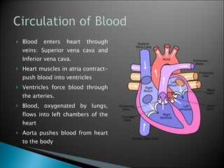
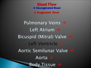
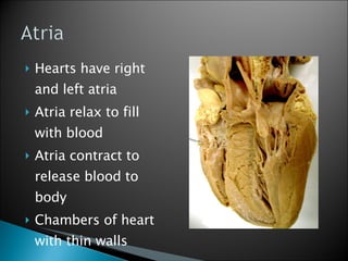
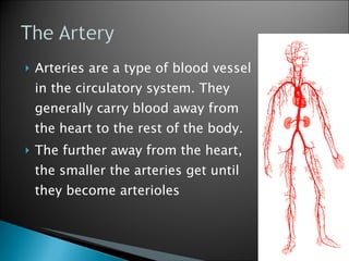
TJ Medical Society 2008 A Presentation brought to you by:  Alison Jarmas, Jerry Liu, Nikhil Prakash, Jimmy Wu, Judy Zeng, and the Officer Corps.
Blood Circulation and Atria
Blood enters heart through veins: Superior vena cava and Inferior vena cava.  Heart muscles in atria contract- push blood into ventricles Ventricles force blood through the arteries. Blood, oxygenated by lungs, flows into left chambers of the heart  Aorta pushes blood from heart to the body
Blood Flow   Deoxygenated Blood    Oxygenated  Blood  Blood from tissues throughout body     Superior and Inferior vena cava     Right Atrium     Tricuspid Valve   Right Ventricle     Pulmonary Semilunar Valve     Pulmonary Artery     Lungs   
Blood Flow   Deoxygenated Blood    Oxygenated  Blood  Pulmonary Veins     Left Atrium     Bicuspid (Mitral) Valve     Left Ventricle    Aortic Semilunar Valve   Aorta     Body Tissue  
 
Hearts have right and left atria Atria relax to fill with blood Atria contract to release blood to body Chambers of heart with thin walls
Diastole and Systole
Consists of cyclic periods of high or low blood pressure, measured in mmHg Systole – all four heart chambers simultaneously contract to propel blood through the cardiovascular system; causes increased pressure on arterial walls Diastole – the period of relaxation during which blood flows back into the atria and ventricles; causes decreased pressure on arterial walls Represented as systole/diastole (ie. 120/80)
RED – aortic pressure BLUE – left ventricular pressure YELLOW – left atrial pressure
Ventricles and the “Cardiums”
Ventricles Myocardium – muscle tissue Endocardium – tissue that lines the interior of heart Pericardium – sac that contains heart and roots of great blood vessels
Valves, Muscles, Tendinae
http://www.mitroflow.com/immagini/bassa/0_2005127191719-patients_chambers.jpg
Allows blood to enter ventricles Prevents backflow into atriums Connected to papillary muscles by chordae tendinae Tricuspid (right AV) 3 membrane flaps between right atrium and ventricle Bicuspid (mitral or left AV) 2 membrane flaps between left atrium and ventricle
Prevents backflow from arteries to ventricles 3 small membranous pockets; crescent-shaped flaps of endocardium Pulmonary (right) Connects pulmonary trunk to right ventricle Aortic (left) Connects aorta to left ventricle
Flaps of muscles connected to various valves on one end by chordae tendinae and to ventricular walls on the other Stabilize valves
“ heartstrings” Connective tissues connecting valves to papillary muscles Prevent valves from being turned inside out
www.byedr.com/medicine/1781-medicine-5.html
Aorta
The largest artery in the body: stems from LV and brings oxygenated blood to all parts of body in systemic circulation.   Diagram of Aorta in SHEEP
Pulmonary Circulation
Serves to oxygenate the depleted blood Circuit led in to by the pulmonary trunk (artery) Systemic circulation    vena cava    right atrium    right ventricle    pulmonary artery (right and left)     lungs (capillary beds)    pulmonary veins (right and left)    left atrium    left ventricle    aorta    systemic circulation
Arterioles intertwined with tubular bronchioles bring deoxygenated blood away from the heart Capillary beds in the saclike alveoli have increased surface area for gas exchange by diffusion (facilitated by a partial pressure gradient) Venules carry newly oxygenated blood back to the heart
 
A blood vessel that ALWAYS carries blood toward the heart Generally contains deoxygenated blood, except for the pulmonary and umbilical veins Path: Right Atrium    Right Ventricle    Pulmonary Arteries    Lungs    Becomes oxygenated Very low blood pressure- rely on skeletal-muscle pump Smallest veins are the venules
Three Layers Tunica Adventitia:  Outer covering of vessel; composed of connective tissue, collagen, and elastic fiber Tunica Media: Middle layer; composed of smooth muscle and elastic fiber Tunica Intima: Inner layer; composed of an elastic membrane lining and smooth endothelium
Types: Portal and Nonportal Superficial and Deep Pulmonary and Systemic Major Ones: Great Saphenous vein (GSV) – located in lower limbs Pulmonary veins: carry oxygenated air to the lungs Jugular Vein Inferior and Superior Vena Cava
Cranial/Caudal Vena Cava
Arteries
Arteries are a type of blood vessel in the circulatory system. They generally carry blood away from the heart to the rest of the body. The further away from the heart, the smaller the arteries get until they become arterioles
Arteries are tough on the outside, muscular in the middle to help force blood throughout, and smooth on the inside. Generally arteries are thicker than veins because they need to withstand greater pressure
Aorta Common carotids Common iliacs Pulmonary trunk
Arterioles, Venules, Capillaries
http://www.deconstructdesign.com/st_projects/mm_mania_2003/the_heart/functions.htm   Capillaries are the connectors between arterioles and venules.  Smallest blood vessels that are involved in regulation of osmotic pressure (internal body heat) Capillary wall consists of only the endothelium. Venules are small blood vessels that allow deoxygenated blood to flow from capillary beds to veins.  Has endothelial membrane, muscle/elastic tissue, and fibrous connective tissue.
Arterioles are the blood vessels from the arteries to the capillaries.  Blood pressure in arteries = result o work needed to pump cardiac output through the vascular resistance.
© TJHSST Medical Society 2007-2008

Sheep Heart Dissection

  • 1.
    TJ Medical Society2008 A Presentation brought to you by: Alison Jarmas, Jerry Liu, Nikhil Prakash, Jimmy Wu, Judy Zeng, and the Officer Corps.
  • 2.
  • 3.
    Blood enters heartthrough veins: Superior vena cava and Inferior vena cava. Heart muscles in atria contract- push blood into ventricles Ventricles force blood through the arteries. Blood, oxygenated by lungs, flows into left chambers of the heart Aorta pushes blood from heart to the body
  • 4.
    Blood Flow  Deoxygenated Blood  Oxygenated Blood Blood from tissues throughout body  Superior and Inferior vena cava  Right Atrium  Tricuspid Valve  Right Ventricle  Pulmonary Semilunar Valve  Pulmonary Artery  Lungs 
  • 5.
    Blood Flow  Deoxygenated Blood  Oxygenated Blood Pulmonary Veins  Left Atrium  Bicuspid (Mitral) Valve  Left Ventricle  Aortic Semilunar Valve  Aorta  Body Tissue 
  • 6.
  • 7.
    Hearts have rightand left atria Atria relax to fill with blood Atria contract to release blood to body Chambers of heart with thin walls
  • 8.
  • 9.
    Consists of cyclicperiods of high or low blood pressure, measured in mmHg Systole – all four heart chambers simultaneously contract to propel blood through the cardiovascular system; causes increased pressure on arterial walls Diastole – the period of relaxation during which blood flows back into the atria and ventricles; causes decreased pressure on arterial walls Represented as systole/diastole (ie. 120/80)
  • 10.
    RED – aorticpressure BLUE – left ventricular pressure YELLOW – left atrial pressure
  • 11.
    Ventricles and the“Cardiums”
  • 12.
    Ventricles Myocardium –muscle tissue Endocardium – tissue that lines the interior of heart Pericardium – sac that contains heart and roots of great blood vessels
  • 13.
  • 14.
  • 15.
    Allows blood toenter ventricles Prevents backflow into atriums Connected to papillary muscles by chordae tendinae Tricuspid (right AV) 3 membrane flaps between right atrium and ventricle Bicuspid (mitral or left AV) 2 membrane flaps between left atrium and ventricle
  • 16.
    Prevents backflow fromarteries to ventricles 3 small membranous pockets; crescent-shaped flaps of endocardium Pulmonary (right) Connects pulmonary trunk to right ventricle Aortic (left) Connects aorta to left ventricle
  • 17.
    Flaps of musclesconnected to various valves on one end by chordae tendinae and to ventricular walls on the other Stabilize valves
  • 18.
    “ heartstrings” Connectivetissues connecting valves to papillary muscles Prevent valves from being turned inside out
  • 19.
  • 20.
  • 21.
    The largest arteryin the body: stems from LV and brings oxygenated blood to all parts of body in systemic circulation. Diagram of Aorta in SHEEP
  • 22.
  • 23.
    Serves to oxygenatethe depleted blood Circuit led in to by the pulmonary trunk (artery) Systemic circulation  vena cava  right atrium  right ventricle  pulmonary artery (right and left)  lungs (capillary beds)  pulmonary veins (right and left)  left atrium  left ventricle  aorta  systemic circulation
  • 24.
    Arterioles intertwined withtubular bronchioles bring deoxygenated blood away from the heart Capillary beds in the saclike alveoli have increased surface area for gas exchange by diffusion (facilitated by a partial pressure gradient) Venules carry newly oxygenated blood back to the heart
  • 25.
  • 26.
    A blood vesselthat ALWAYS carries blood toward the heart Generally contains deoxygenated blood, except for the pulmonary and umbilical veins Path: Right Atrium  Right Ventricle  Pulmonary Arteries  Lungs  Becomes oxygenated Very low blood pressure- rely on skeletal-muscle pump Smallest veins are the venules
  • 27.
    Three Layers TunicaAdventitia: Outer covering of vessel; composed of connective tissue, collagen, and elastic fiber Tunica Media: Middle layer; composed of smooth muscle and elastic fiber Tunica Intima: Inner layer; composed of an elastic membrane lining and smooth endothelium
  • 28.
    Types: Portal andNonportal Superficial and Deep Pulmonary and Systemic Major Ones: Great Saphenous vein (GSV) – located in lower limbs Pulmonary veins: carry oxygenated air to the lungs Jugular Vein Inferior and Superior Vena Cava
  • 29.
  • 30.
  • 31.
  • 32.
    Arteries are atype of blood vessel in the circulatory system. They generally carry blood away from the heart to the rest of the body. The further away from the heart, the smaller the arteries get until they become arterioles
  • 33.
    Arteries are toughon the outside, muscular in the middle to help force blood throughout, and smooth on the inside. Generally arteries are thicker than veins because they need to withstand greater pressure
  • 34.
    Aorta Common carotidsCommon iliacs Pulmonary trunk
  • 35.
  • 36.
    http://www.deconstructdesign.com/st_projects/mm_mania_2003/the_heart/functions.htm Capillaries are the connectors between arterioles and venules. Smallest blood vessels that are involved in regulation of osmotic pressure (internal body heat) Capillary wall consists of only the endothelium. Venules are small blood vessels that allow deoxygenated blood to flow from capillary beds to veins. Has endothelial membrane, muscle/elastic tissue, and fibrous connective tissue.
  • 37.
    Arterioles are theblood vessels from the arteries to the capillaries. Blood pressure in arteries = result o work needed to pump cardiac output through the vascular resistance.
  • 38.
    © TJHSST MedicalSociety 2007-2008