SlideShare a Scribd company logo
1 of 15
Entry in to host cell:
Sendai and measles virus of family Paramyxoviridae(enveloped virus) directly fuced
with plasma membrane at neutral PH. These viruses bind with bind with cell surface
receptors via a viral intergral protein. Virus and cell membrane closely paired together by this
receptor ligand interaction.fusion induced by the viral glycoprotein Fusion protein (F) ,virla
nucleocapsid realeased in to the cell cytoplasm.
F protein is a type I integral membrane glycoprotein (theN terminus lies outside the
viral membrane) with similarities to influenza virus HA in its synthesis and structure. It is a
homotrimer that is synthesized as a precursor called F0 and cleaved during transit to the cell
surface by a host cell proteaseto produce two subunits, F1 and F2, held together by disulfi
debonds. Th e newly formed N-terminal 20 amino acids of the F1subunit, which are highly
hydrophobic, form a region called the fusion peptide because it inserts into target membranes
to initiate fusion. Viruses with the uncleaved F0 precursor can be produced in cells that lack
the protease responsible for its cleavage. Such virus particles are noninfectious; they bind
totarget cells but the viral genome does not enter. Cleavage of the F0 precursor is necessary
for fusion, not only because the fusion peptide is made available for insertion into the
plasmamembrane, but also to generate the metastable state of the protein that can undergo the
conformational rearrangements
needed for fusion. Because cleaved F-protein-mediated fusion can occur at neutral pH, it
must be controlled, both to ensure that virus particles fuse with only the appropriate cell and
to prevent aggregation of newly assembled virions. The fusion peptide of F1 is buried
between two subunits of the trimer in the pre-fusion protein. Conformational changes in F
protein lead to refolding of the protein, assembly of an _ -helical coiled coil, and movement
of the fusion peptide toward the cell membrane (Fig. 5.10). Such movement of the fusion
peptide has been described in atomic detail by comparing structures of the F protein before
and after fusion. The trigger that initiates conformational changes in the F protein is not
known. The results of experiments in which hemagglutinin-neuraminidase (HN) and F
glycoproteins are synthesized in cultured mammalian cells indicate that the fusion activity of
F protein is absent or inefficient if HN is not present. It has therefore been hypothesized that
an interaction between HN and F proteins is essential for fusion. It is thought that binding of
HN protein to its cellular receptor induces conformational changes, which in turn trigger
conformational change in the F protein, exposing the fusion peptide and making the protein
fusion competent (Fig. 5.10). The requirement for HN protein in F fusion activity has been
observed only with certain paramyxo viruses, including human parainfluenza virus type 3 and
mumps virus. As a result of fusion of the viral and plasma membranes, the viral nucleocapsid,
which is a ribonucleo protein (RNP) consisting of the strand viral RNA genome and the viral
proteins L, NP, and P, is released into the cytoplasm (Fig.5.10). Once in the cytoplasm, the L,
NP, and P proteins begin the synthesis of viral messenger RNAs (mRNAs), a process
discussed in Chapter 6. Because members of the Paramyxoviridae replicate in the cytoplasm,
fusion of the viral and plasma membranes achieves uncoating and delivery of the viral
genome to this cellular compartment in a single step. Fusion of human immunodeficiency
virus type 1 with the plasma membrane requires participation not only of the cell receptor
CD4 but also of an additional cellular protein. These proteins are cell surface receptors for
small molecules produced by many cells to attract and stimulate cells of the immune defense
system at sites of infection; hence, these small molecules are called chemotactic cytokines or
chemokines. Th e chemokine receptors on such cells comprise a large family of proteins with
seven membrane-spanning domains and are coupled to intracellular signal transduction
pathways. There are two major coreceptors for human immune deficiency virus type 1
infection. CXCr4 (a member of a family of chemokines characterized by having their first
two cysteines separated by a single amino acid) appears to be a specific coreceptor for virus
strains that infect T cells preferentially. The second is CCr5, a coreceptor for the
macrophage-tropicstrains of the virus. The chemokines that bind to this receptor activate both
T cells and macrophages, and the receptor is found on both types of cell. Individuals who are
homozygous for deletions in the CCr5 gene and produce nonfunctional coreceptors have no
discernible immune function abnormality, but they appear to be resistant to infection with
human immune deficiency virus type 1. Even heterozygous individuals seem to be somewhat
resistant to the virus. Other members of the CC chemokine receptor family (CCr2b and
CCr3)were subsequently found to serve as coreceptors for the virus.
Attachment to CD4 appears to create a high-affinity binding site on SU for CCr5. The atomic
structure of SU bound to CD4 revealed that binding of CD4 induces conformational changes
that expose binding sites for chemokine receptors(Fig. 5.10). Studies of CCr5 have shown
that the first N-terminal extracellular domain is crucial for coreceptor function, suggesting
that this sequence might interact with SU. An antibody molecule fused to both the CD4 and
CCr5binding sites is being explored as a therapeutic compound to block infection (Box 5.4).
Human immunodeficiency virus type 1 TM mediates envelope fusion with the cell
membrane. The high-affinitySU-CCr5 interaction may induce conformational changes in TM
to expose the fusion peptide, placing it near the cell membrane, where it can catalyze fusion
(Fig. 5.10). Such changes are similar to those that influenza virus HA undergoesupon
exposure to low pH. X-ray crystallographic analysis of fusion-active human immune
deficiency virus type 1 TM revealed that its structure is strikingly similar to that of the low-
pH fusogenic form of HA (see “Acid-Catalyzed Membrane Fusion” below.
Figure : Virus entry and movement in cells. Examples of genome uncoating at the plasma
membrane are shown on the left side of the cell. Fusion at the plasma membrane releases the
nucleocapsid into the cytoplasm. In some cases, the sub viral particle is transported on
microtubules toward the nucleus, where the nucleic acid is released. Uptake of virions by
clathrin-dependent endocytosis commences with binding to a specific cell surface receptor.
The ligand-receptor complex diffuses into an invagination of the plasma membrane coated
with the protein clathrin on the cytosolic side (clathrin-coated pits). The coated pit further
invaginates and pinches off, a process that is facilitated by the GTPase dynamin. The
resulting coated vesicle then fuses with an early endosome. Endosomes are acidic, as a result
ofthe activity of vacuolarproton ATPases. Particle uncoating usually occurs from early or late
endosomes. Late endosomes then fuse with lysosomes. Virus particles may enter cells by a
dynamin- and caveolin-dependent endocytic pathway (right side of the cell). Three types of
caveolar endocytosis have been identified. Dynamin 2-dependent endocytosis by caveolin 1-
containingcaveolae isobserved in cells infected with simian virus 40 and polyomavirus.
Dynamin 2-dependent, noncaveolar, lipid raft-mediated endocytosis occurs during echovirus
and rotavirus infection, while dynamin-independent, noncaveolar, raft-mediated endocytosis
is also observed during simian virus 40 and polyomavirus infection. This pathway brings
virions to the endoplasmic reticulum via the caveosome, a pH-neutral compartment. Clathrin-
and caveolin-independent endocytic pathways of viral entry have also been described (center
of cell). Movement of endocytic vesicles within cells occurs on microfilamentsor
microtubules, components of the cytoskeleton. Microfilaments are two-stranded helical
polymers of the ATPaseactin. They are dispersed throughout the cell but are most highly
concentrated beneath the plasma membrane, where they are connected via integrins and other
proteins to the extracellular matrix. Transport along microfilaments is accomplished by
myosin motors. Microtubules are 25-nm hollow cylinders made of the GTPase tubulin. They
radiate from the centrosome to the cell periphery. Movement on microtubules is carried out
by kinesin and dynein motors.
Figure : Penetration and uncoating at the plasma membrane.
(A) Overview. Entry of a member of the Paramyxoviridae, which bind to cell surface
receptors via the HN, H, or G glycoprotein. The fusion protein (F) then catalyzes membrane
fusion at the cell surface at neutral pH. The viral nucleocapsid, as RNP, is released into the
cytoplasm, where RNA synthesis begins.
(B) Model for F-protein-mediated membrane fusion. Binding of HN to the cell receptor
(red)induces conformational changes in HN that in turn induce conformational changes in the
F protein, moving the fusionpeptide from a buried position nearer to the cell membrane.
(C) Model of the role of chemokine receptors in human immunodeficiency virus type 1
fusion at the plasma membrane. For simplicity, the envelope glycoprotein is shown as a
monomer. Binding of SU to CD4 exposes a high-affinity chemokine receptor-binding site on
SU. The SU-chemokine receptor interaction leads to conformational changes in TM that
expose the fusion peptide and permit it to insert into the cell membrane, catalyzing fusion in a
manner similar to that proposed for influenza virus (cf. Fig. 5.12 and 5.13).
Blocking human immunodeficiency virus infection with two soluble cell receptors
Because viruses must bind to cell surface molecules to initiate replication, the use of soluble
receptors to block virus infection haslong been an attractive therapeutic option. Soluble CD4
receptors that block infection with human immunodeficiency virus type 1(HIV-1) have been
developed, but these have not been licensed because of their suboptimal potency. A newly
designed soluble receptor forHIV-1 overcomes this problem and provides broad and effective
protection against infection of cells and of nonhuman primates. A soluble form of CD4 fused
to an antibody molecule can block infection of mostHIV-1 isolates and has been shown to be
safe in humans, but its affinity for gp120 is low. Furthermore, human immunodeficiency
virus can also be spread from cell to cell by fusion, a process that is not blocked by
circulating, soluble CD4. Similarly, peptide mimics of theCCR5 coreceptor have been shown
to block infection, but their affinity for gp120 is also low. Combining the two gp120-binding
molecules solved the problem of low affinity and in addition provided protection against a
wide range of virus isolates. The entry inhibitor, calledeCD4-Ig, is a fusion of the first two
domains ofCD4 to the Fc domain of an antibody molecule, with the CCR5-mimicking
peptide at the carboxy terminus (illustrated). It binds strongly togp120 and blocks infection
with many different isolates of HIV-1, HIV-2, simian immune deficiency virus (SIV), and
HIV-1 resistant to broadly neutralizing monoclonal antibodies. The molecule blocks viral
infection at concentrations that might be achieved in humans (1.5 to 5.2 micrograms per
milliliter).When administered to mice, eCD4-Ig protected the animals from HIV-1. Rhesus
macaques inoculated with an adenovirus-associated virus (AAV) recombinant containing the
gene foreCD4-Ig were protected from infection with large quantities of virus for up to 34
weeks after immunization. Concentrations of eCD4-Ig in the sera of these animals ranged
from 17 to77 micrograms per milliliter. These results show that eCD4-Ig blocks HIV
infection with a wide range of isolates more effectively than previously studied broadly
neutralizing antibodies. Emergence of HIV variants resistant to neutralization with eCD4-Ig
would likely produce viruses that infect cells less efficiently, reducing their transmission.
eCD4-Ig is therefore an attractive candidate for therapy of HIV-1 infections. Whether
sustained production of the protein in humans will cause disease remains to be determined.
Because expression of the AAV genome persists for long periods, it might be advantageous
to include a kill-switch in the vector: a way of turning it off if something should go wrong.
Uncoating during Endocytosis :
Many viruses enter cells by the same pathways by which cellstake up
macromolecules. The plasma membrane, the limitingmembrane of the cell, permits nutrient
molecules to enterand waste molecules to leave, thereby ensuring an appropriateinternal
environment. Water, gases, and small hydrophobicmolecules such as ethanol can freely
traverse the lipid bilayer,but most metabolites and ions cannot. These essential
componentsenter the cell by specific transport processes. Integralmembrane proteins are
responsible for the transport of ions,sugars, and amino acids, while proteins and large
particles aretaken into the cell by phagocytosis or endocytosis. The formerprocess (Fig. 5.11)
is nonspecific, which means that any particleor molecule can be taken into the cell, and only
occurs inspecialized cell types such as dendritic cells and macrophages.
Clathrin-Mediated Endocytosis
A wide range of ligands, fluid, membrane proteins, and lipids are selectively taken
into cells from the extracellular milieuby clathrin-mediated endocytosis (Fig. 5.9 and 5.11),
also the mechanism of entry of many viruses. Ligands in the extra cellular medium bind to
cells via specific plasma membrane receptor proteins. The receptor-ligand assembly diff uses
along the membrane until it reaches an invagination that is coated on its cytoplasmic surface
by a cage-like lattice composed of the fibrous protein clathrin (Fig. 5.9). Such clathrin-
coatedpits can comprise as much as 2% of the surface area of a cell, and some receptors are
clustered over these areas even in the absence of their ligands. Following the accumulation of
receptor-ligand complexes, the clathrin-coated pit invaginates and then pinches off to form a
clathrin-coated vesicle. Within a few seconds, the clathrin coat is lost and the vesicles fuse
with small, smooth-walled vesicles located near the cell surface, called early endosomes . The
lumen of early endosomes is mildly acidic (pH 6.5 to 6.0), a result of energy-dependent
transport of protons into the interior of the vesicles by a membrane proton pump. The
contents of the early endosome are then transported via endosomal carrier vesicles to late
endosomes located close to the nucleus. The lumen of late endosomes is more acidic (pH 6.0
to 5.0). Late endosomes in turn fuse with lysosomes, which are vesicles containing a variety
of enzymes that degrade sugars, proteins, nucleic acids, and lipids. Viruses with a high pH
threshold for fusion, such as vesicular stomatitis virus, enter from early endosomes; most
enter the cytoplasm from late endosomes, and a few enter from lysosomes. Clathrin-mediated
endocytosis is a continuous but regulated process. For example, the uptake of vesicular
stomatitis virus into cells may be influenced by over 90 different cellular protein kinases.
Influenza virus, vesicular stomatitis virus and reovirus particles are taken into cells, not into
preexistingpits but mainly by clathrin-coated pits that form after virus binds to the cell
surface. It is not known how virus binding to the plasma membrane induces the formation of
the clathrin-coated pit.
Caveolar and Lipid Raft-Mediated Endocytosis
Although uptake of most viruses occurs by the clathrin mediated endocytic pathway,
some viruses enter by caveolinorraft -mediated endocytosis (Fig. 5.9). Th e caveolar pathway.
requires cholesterol (a major component of lipid rafts). Caveolae are distinguished from
clathrin-coated vesicles by their flask-like shape, their smaller size, the absence of a clathrin
coat, and the presence of a marker protein called caveolin. In the uninfected cell, caveolae
participate in transcytosis, signal transduction, and uptake of membrane components and
extracellular ligands. Binding of a virus particle to the cell surface activates signal
transduction pathways required for pinching off of the vesicle, which then moves within the
cytoplasm.
Disassembly of fi lamentous actin also occurs, presumably to facilitate movement of
the vesicle deeper into the cytoplasm. There it fuses with the caveosome, a larger
membranous organelle that contains caveolin (Fig. 5.9). In contrast to endosomes, the pH of
the caveosome lumen is neutral. Some viruses (e.g., echovirus type 1) penetrate the
cytoplasm from the caveosome. Others (simian virus 40, polyoma virus, coxsackievirusB3)
are sorted to the endoplasmic reticulum (ER) by a transport vesicle that lacks caveolin. These
viruses enter the cytoplasm by a process mediated by thiol oxidases present in the lumen of
the endoplasmic reticulum and by a component of the protein degradation pathway present in
the membrane. The study of virus entry by endocytosis can be confusing because some
viruses may enter cells by multiple routes, depending on cell type and multiplicity of
infection. For example, herpes simplex virus can enter cells by three different routes and
influenza A virus may enter cells by both clathrin-dependent and clathrin-independent
pathways.
Macropinocytosis
Macropinocytosis is a process by which extracellular fluid is taken into cells via large
vacuoles. It is triggered by ligands. and dependent on actin and a signaling pathway. It differs
from phagocytosis by the signaling pathways needed and can take place in many cell types.
Th is process serves as a pathway of entry for many viruses, including vaccinia virus,
herpesviruses, and ebola viruses. Upon receptor binding, viruses that enter cells via
macropinocytosis trigger a signaling cascade that leads to changes in cortical actin and
ruffling of the plasma membrane (Fig. 5.11). When these plasma membrane extensions
retract, the viruses are brought into macropinosomes and eventually leave these vesicles via
membrane fusion.
Membrane Fusion
The membranes of enveloped viruses fuse with those of the cell as a first step in
delivery of the viral nucleic acid. Membrane fusion takes place during many other cellular
processes, such as cell division, myoblast fusion, and exocytosis. Membrane fusion must be
regulated in order to maintain the integrity of the cell and its intracellular compartments.
Consequently, membrane fusion does not occur spontaneously but proceeds by specialized
mechanisms mediated by proteins. The two membranes must first come into closeproximity.
In cells, this reaction is mediated by interactions of integral membrane proteins that protrude
from the lipid bilayers, a targeting protein on one membrane and a docking protein on the
other. During entry of enveloped viruses, the virus and cell membranes are fi rst brought into
close contact by interaction of a viral glycoprotein with a cell receptor. The next step, fusion,
requires an even closer approach of the membranes, to within 1.5 nm of each other. This step
depends on the removal of water molecules from the membrane surfaces an energetically
unfavorable process. This step is hypothesized to occur when the viral glycoprotein
undergoes a structural rearrangement called “hairpinning” (Fig. 5.12).The precise mechanism
by which lipid bilayers fuse is not completely understood, but the action of fusion proteins is
thought to result in the formation of an opening called a fusion pore , allowing exchange of
material across the membranes (Box 5.5). The viral glycoprotein bound to a cell receptor, or a
different viral integral membrane protein, then catalyzes the fusion of the juxtaposed
membranes. Viral fusion proteins are integral membrane proteins, oft en glycoproteins, that
form homo- or hetero-oligomers. Virus-mediated fusion must be regulated to prevent viruses
from aggregating or to ensure that fusion does not occur in the incorrect cellular
compartment. In some cases, fusogenic potential is masked until the fusion protein interacts
with other integral membrane proteins. In others, low pH is required to expose fusion
domains. The activity of fusion proteins may also be regulated by cleavage of a precursor.
This requirement probably prevents premature activation of fusion potential during virus
assembly. Viral fusion proteins are often primed for fusion by proteolytic cleavage as they
move through the trans-Golgi network as described in Chapter 12. Proteases that catalyze
such cleavage are typically furin family convertases that either cleave the fusion proteins
directly (orthomyxoviruses, retroviruses, paramyxoviruses) orcleave a protein that masks the
fusion protein (alphaviruses, flaviviruses).
Figure : Mechanisms for the uptake of macromolecules from extracellular fluid. During
phagocytosis, large particles such as bacteria or cell fragments that come in contact with the
cell surface are engulfed by extensions of the plasma membrane. Phagosomes ultimately fuse
with lysosomes, resulting in degradation of the material within the vesicle. Macrophages use
phagocytosis to ingest bacteria and destroy them. Endocytosis comprises the invagination and
pinching off of small regions of the plasma membrane, resulting in the nonspecific
internalization of molecules (macropinocytosis) or the specific uptake of molecules bound to
cell surface receptors(receptor-mediated endocytosis). Macropinocytosis is a mechanism for
the uptake of extracellular fluid. It is triggered by ligand binding which initiates formation of
plasma membrane ruffling, which traps material in large vacuoles.
Figure : Influenza virus entry. The globular heads of native HA mediate binding of the virus
to sialicacid-containing cell receptors. The virus-receptor complex is endocytosed, and import
of H_ ions into the endosomeeacidifies the interior. Upon acidification, the viral HA
undergoes a conformational rearrangement that produces a fusogenic protein. The loop region
of native HA (yellow) becomes a coiled coil, moving the fusion peptides (red) to the top of
the molecule near the cell membrane. At the viral membrane, the long _-helix (purple) packs
against the trimercore, pulling the globular heads to the side. The long coiled coil bends, or
hairpins, bringing the fusion peptides and the trans membrane domains together. This
movement moves the cell and viral membranes close together so that fusion canoccur. To
allow release of vRNP into the cytoplasm, the H_ ions in the acidic endosome are pumped
into the particle interior by the M2 ion channel. As a result, vRNP is primed to dissociate
from M1 after fusion of the viral and endosomal membranes. The released vRNPs are
imported into the nucleus through the nuclear pore complex via a nuclear localization signal-
dependent mechanism (see “Import of Influenza Virus Ribonucleoprotein” below).
Fig. Membrane fusion proceeds through a hemifusion intermediate
Membrane fusion by flavivirus envelope glycoproteinE. Low pH causes
conformational changes in the viral glycoproteinsto produce the fusion-active forms. (A)
Ninety dimers of E tilethe surface of the virus particle. (B) The fusion loop is located at the
tipof domain II (yellow) where it is buried in dimers of the glycoprotein E.At low pH, the
dimers are disrupted, the proteins extend to form trimers,and the fusion peptide is directed
toward the cell membrane. The glycoproteinthen undergoes a rearrangement that brings the
fusion peptides, and transmembrane segments (purple) together. This movement brings
the viral and cell membranes together, allowing fusion.
Receptor Priming for Low-pH Fusion:
Two Entry Mechanisms Combined
During the entry of avian leukosis virus into cells, binding of the virus particle to the
cell receptor primes the viral fusion protein for low-pH-activated fusion. Avian leukosis
virus, like many other retroviruses with simple genomes, was believed to enter cells at the
plasma membrane in a pH-independent mechanism resembling that of members of the
Paramyxoviridae(Fig. 5.10). It is now known that binding of the viral membrane glycoprotein
(SU) to the cellular receptors of avian
leukosis viruses induces conformational rearrangements that convert the viral protein from a
native metastable state that is insensitive to low pH to a second metastable state. In this
state, exposure to low pH within the endosomal compartment leads to membrane fusion and
release of the viral capsid.
Figure .Entry of Ebolavirusintocells.
Virus particles bind cells via an unidentified attachment receptor and enter by
endocytosis. The mucin and glycan cap on the viral glycoprotein is removed by cellular
cysteine proteases, exposing binding sites for NPC1. The latter is required for fusion of the
viral and cell membranes, releasing the nucleocapsid into the cytoplasm.
Uncoating in the Cytoplasm by Ribosomes
Some enveloped RNA-containing viruses, such as Semliki Forest virus, contain
nucleocapsids that are disassembled in the cytoplasm by pH-independent mechanisms. The
icosahedral nucleocapsid of this virus is composed of a single viral protein, C protein, which
encloses the strand viral RNA. This structure is surrounded by an envelope containing viral
glycoproteins E1 and E2, which are arranged as heterodimers clustered into groups of three,
each cluster forming a spike on the virus surface. Fusion of the viral and endosomal
membranes exposes the nucleocapsid to the cytoplasm (Fig. 5.17). To begin translation of
strand viral RNA, the nucleocapsid must be disassembled, a process mediated by an abundant
cellular component the ribosome. Each ribosome binds three to six molecules of C protein,
disrupting the nucleocapsid. This process occurs while the nucleocapsid is attached to the
cytoplasmic side of the endosomal membrane (Fig. 5.17) and ultimately results in
disassembly. The uncoated viral RNA remains associated with cellular membranes, where
translation and replication begin.
Figure.Stepwise uncoating of adenovirus. (A) Adenoviruses bindthe cell receptor via the fiber
protein.Interaction of the penton base with anintegrin receptor leads to internalization
by endocytosis. Fibers are released fromthe capsid during uptake. Low pH in the
endosome causes destabilization of the capsid and release of protein VI (yellow
diamonds). The hydrophobic N terminus of protein VI disrupts the endosome
membrane, leading to release of a subviral particle into the cytoplasm. The capsid is
transported in the cytoplasm along microtubules and docks onto the nuclear pore
complex. (B) Electron micrograph of adenovirus type 2 particles bound to a microtubule
(top) and bound to the cytoplasmic face of the nuclear pore complex (bottom).
Bar in bottom panel, 200 nm.
Disrupting the Endosomal Membrane
Adenoviruses are composed of a double-stranded DNA genome packaged in an icosahedral
capsid. Internalization of most adenovirus serotypes by receptor-mediated endocytosis
requires attachment of fi ber to an integrin or Ig-like cell surface receptor and binding of the
penton base to a second cell receptor, the cellular vitronectin-binding integrins _ v _ 3 and _ v
_ 5 . Attachment is mediated by amino acid sequences in each of the fi ve subunits of the
adenovirus penton base that mimic the normal ligands of cell surface integrins. As the virus
particle is transported via the endosomes from the cell surface toward the nuclear membrane,
it undergoes multiple uncoating steps as structural proteins are removed sequentially (Fig.
5.18). As the endosome becomes acidifi ed, the viral capsid is destabilized, leading to release
of proteins from the capsid. Among these is protein VI, which causes disruption of the
endosomal membrane, thereby delivering the remainder of the particle into the cytoplasm. An
N-terminal amphipathic_ -helix of protein VI is probably responsible for its pH-dependent
membrane disruption activity. Th is region of the protein appears to be masked in the native
capsid by the hexon protein. The liberated subviral particle then docks onto the nuclear pore
complex (see “Import of DNA Genomes” below).
Forming a Pore in the Endosomal Membrane :
The genomes of nonenveloped picornaviruses are transferredacross the cell membrane by a
diff erent mechanism,as determined by structural information at the atomic level
and complementary genetic and biochemical data obtainedfrom studies of cell entry. The
interaction of poliovirus withits Ig-like cell receptor, CD155, leads to major conformational
rearrangements in the virus particle and the production of an expanded form called an altered
(A) particle (Fig. 5.19A). Portions of two capsid proteins, VP1 and VP4, move from
the inner surface of the capsid to the exterior. Th ese polypeptides are thought to form a pore
in the cell membrane that allows transport of viral RNA into the cytoplasm (Fig. 5.19B).
In support of this model, ion channel activity can be detected when A particles are added to
lipid bilayers.
Figure. Model for poliovirus entry into cells. (A) Overview. The native virion (160S) binds to
its cell receptor, CD155, and undergoes a receptor-mediated conformational transition
resulting in the formation of altered (A) particles. The viral RNA, shown as a curved green
line, leaves the capsid from within early endosomes close to the plasma
membrane. (B) Model of the formation of a pore in the cell membrane after poliovirus
binding. 1, poliovirus (shown in cross section, with capsid proteins purple) binds to CD155
(brown). 2, a conformational change leads to displacement of the pocket lipid (black). The
pocket may be occupied by sphingosine in the capsid of poliovirus type 1. The hydrophobic
N termini of VP1 (blue) are extruded and insert into the plasma membrane. 3, a pore is
formed in the membrane by VP4 and the VP1 N termini, through which the RNA is released
from the capsid into the cytosol.
Fig.Entry of Revovirus into the cells.
Movement of Viral and Subviral
Particles within Cells :
Viral and subviral particles move within the host cell duringentry and egress). However,
movement of molecules larger than 500 kDa does not occur by passivediffusion, because the
cytoplasm is crowded with organelles,high concentrations of proteins, and the cytoskeleton.
Rather,viral particles and their components are transported via theactin and microtubule
cytoskeletons. Such movement can be visualized in live cells by using fluorescently labeled
viral proteins. The cytoskeleton is a dynamic network of protein fi laments that extends
throughout the cytoplasm. It is composed of three types of fi lament—microtubules,
intermediate filaments, and microfi laments. Microtubules are organized in a polarized
manner, with minus ends situated at the microtubuleorganizing center near the nucleus, and
plus ends located at the cell periphery. This arrangement permits directed movement of
cellular and viral components over long distances. Actin filaments (microfilaments) typically
assist in virus movement close to the plasma membrane.
Virus-Induced Signaling via Cell Receptors
Binding of virus particles to cell receptors not only concentratesthe particles on the cell
surface, but also activates signalingpathways that facilitate virus entry and movement
within the cell or produce cellular responses that enhance virus propagation and/or affect
pathogenesis. Virus binding may lead to activation of protein kinases that trigger cascades
of responses at the plasma membrane, cytoplasm, and nucleus. Second messengers that
participate in signaling include phosphatidylinositides, diacylglycerides, and calcium;
regulators of membrane trafficking and actin dynamics also contribute to signaling. Virus-
receptor interactions also stimulate antiviral responses.
The reproduction of most DNA viruses, and some RNA viruses including retroviruses and
influenza viruses, begins in the cell nucleus. The genomes of these viruses must therefore be
imported from the cytoplasm. One way to accomplish thismovement is via the cellular
pathway for protein import into the nucleus. An alternative, observed in cells infected by
some retroviruses, is to enter the nucleus after the nuclear envelope breaks down during cell
division. When the nuclear envelope is reformed, the viral DNA is incorporated into the
nucleus together with cellular chromatin. This strategy restricts infection to cells that undergo
mitosis.

More Related Content

What's hot

adeno Virus assembly
adeno Virus assemblyadeno Virus assembly
adeno Virus assemblySh Naser
 
Organization and Regulation of Mitochondrial Protein Synthesis
Organization and Regulation of Mitochondrial Protein SynthesisOrganization and Regulation of Mitochondrial Protein Synthesis
Organization and Regulation of Mitochondrial Protein SynthesisHeena36363
 
Expression vectors
Expression vectorsExpression vectors
Expression vectorsKanchan Rawat
 
Adaptations of mycobacteria for survival in macrophages sameer tiwari
Adaptations of mycobacteria for survival in macrophages sameer tiwariAdaptations of mycobacteria for survival in macrophages sameer tiwari
Adaptations of mycobacteria for survival in macrophages sameer tiwariSameer Tiwari
 
notch signaling and acute leukemia
notch signaling and acute leukemianotch signaling and acute leukemia
notch signaling and acute leukemiaFereshteDorrazehi
 
TRISTETRAPROLIN - its role in inflammation and cancer
TRISTETRAPROLIN - its role in inflammation and cancerTRISTETRAPROLIN - its role in inflammation and cancer
TRISTETRAPROLIN - its role in inflammation and cancerHimadri Nath
 
Extrachromosomal replication of DNA
Extrachromosomal replication of DNAExtrachromosomal replication of DNA
Extrachromosomal replication of DNALubnaSSubair
 
Vector engineering and codon optimization
Vector engineering and codon optimizationVector engineering and codon optimization
Vector engineering and codon optimizationPiyush Jamwal
 
P 53 Tumour Biology
P 53 Tumour BiologyP 53 Tumour Biology
P 53 Tumour BiologyGaurav Dwivedi
 
Alternative splicing by kk sahu
Alternative splicing by kk sahuAlternative splicing by kk sahu
Alternative splicing by kk sahuKAUSHAL SAHU
 
Expression systems
Expression systemsExpression systems
Expression systemsBruno Mmassy
 
Pre trans splicing gene therapy
Pre trans splicing gene therapyPre trans splicing gene therapy
Pre trans splicing gene therapyfaraharooj
 

What's hot (20)

adeno Virus assembly
adeno Virus assemblyadeno Virus assembly
adeno Virus assembly
 
Organization and Regulation of Mitochondrial Protein Synthesis
Organization and Regulation of Mitochondrial Protein SynthesisOrganization and Regulation of Mitochondrial Protein Synthesis
Organization and Regulation of Mitochondrial Protein Synthesis
 
Expression vectors
Expression vectorsExpression vectors
Expression vectors
 
Adaptations of mycobacteria for survival in macrophages sameer tiwari
Adaptations of mycobacteria for survival in macrophages sameer tiwariAdaptations of mycobacteria for survival in macrophages sameer tiwari
Adaptations of mycobacteria for survival in macrophages sameer tiwari
 
Bharti
BhartiBharti
Bharti
 
notch signaling and acute leukemia
notch signaling and acute leukemianotch signaling and acute leukemia
notch signaling and acute leukemia
 
TRISTETRAPROLIN - its role in inflammation and cancer
TRISTETRAPROLIN - its role in inflammation and cancerTRISTETRAPROLIN - its role in inflammation and cancer
TRISTETRAPROLIN - its role in inflammation and cancer
 
Extrachromosomal replication of DNA
Extrachromosomal replication of DNAExtrachromosomal replication of DNA
Extrachromosomal replication of DNA
 
Notch signaling pathway
Notch signaling pathwayNotch signaling pathway
Notch signaling pathway
 
Cell signaling
Cell signalingCell signaling
Cell signaling
 
Vector engineering and codon optimization
Vector engineering and codon optimizationVector engineering and codon optimization
Vector engineering and codon optimization
 
P 53 Tumour Biology
P 53 Tumour BiologyP 53 Tumour Biology
P 53 Tumour Biology
 
Cell signaling
Cell signalingCell signaling
Cell signaling
 
Alternative splicing by kk sahu
Alternative splicing by kk sahuAlternative splicing by kk sahu
Alternative splicing by kk sahu
 
Cell signaling
Cell signalingCell signaling
Cell signaling
 
Expression systems
Expression systemsExpression systems
Expression systems
 
E.coli promoters
E.coli promotersE.coli promoters
E.coli promoters
 
Shuttle vector
Shuttle vectorShuttle vector
Shuttle vector
 
Pre trans splicing gene therapy
Pre trans splicing gene therapyPre trans splicing gene therapy
Pre trans splicing gene therapy
 
Cell Signaling
Cell SignalingCell Signaling
Cell Signaling
 

Similar to Entry in to host cell

Conjugation-Mechanism and significance
Conjugation-Mechanism and significanceConjugation-Mechanism and significance
Conjugation-Mechanism and significanceSukumar Sidtharth
 
Both influenza virus and (IFV), and Measles Virus (MV) are enveloped.pdf
Both influenza virus and (IFV), and Measles Virus (MV) are enveloped.pdfBoth influenza virus and (IFV), and Measles Virus (MV) are enveloped.pdf
Both influenza virus and (IFV), and Measles Virus (MV) are enveloped.pdfarchigallery1298
 
The Biology of HIV-AIDS Acquired immune deficiency syndrome (AIDS) is.pdf
The Biology of HIV-AIDS Acquired immune deficiency syndrome (AIDS) is.pdfThe Biology of HIV-AIDS Acquired immune deficiency syndrome (AIDS) is.pdf
The Biology of HIV-AIDS Acquired immune deficiency syndrome (AIDS) is.pdfaadyacouture
 
1a- What are three ways that bacteria can exchange genetic informati.pdf
1a- What are three ways that bacteria can exchange genetic informati.pdf1a- What are three ways that bacteria can exchange genetic informati.pdf
1a- What are three ways that bacteria can exchange genetic informati.pdfeyelineoptics
 
Microbiology antibiotic
Microbiology   antibioticMicrobiology   antibiotic
Microbiology antibioticMBBS IMS MSU
 
Host microbial interactions in periodontal diseases
Host microbial interactions in periodontal diseasesHost microbial interactions in periodontal diseases
Host microbial interactions in periodontal diseasesDr Heena Sharma
 
Antigen processing and presentation. Antigen – Antibody Interaction.
Antigen processing and presentation. Antigen – Antibody Interaction.Antigen processing and presentation. Antigen – Antibody Interaction.
Antigen processing and presentation. Antigen – Antibody Interaction.Md Azizul Haque
 
Conjugation, transposition and antibiotic resistance
Conjugation, transposition and antibiotic resistanceConjugation, transposition and antibiotic resistance
Conjugation, transposition and antibiotic resistanceAtika Noor
 
Differences between HIV-1 and MLV viruses
Differences between HIV-1 and MLV virusesDifferences between HIV-1 and MLV viruses
Differences between HIV-1 and MLV virusesdegarden
 
role of immune system in inflammation
role of immune system in inflammationrole of immune system in inflammation
role of immune system in inflammationAyesha Shaik
 
Insignt from nono medicine into chloroquine efficacy against COVID-19
Insignt from nono medicine into chloroquine efficacy against COVID-19Insignt from nono medicine into chloroquine efficacy against COVID-19
Insignt from nono medicine into chloroquine efficacy against COVID-19Valentina Corona
 
Pharmacology Science Research Project.docx
Pharmacology Science Research Project.docxPharmacology Science Research Project.docx
Pharmacology Science Research Project.docxwrite5
 
COMPLEMENTS system by dr naseeruddin .pptx
COMPLEMENTS system by dr naseeruddin .pptxCOMPLEMENTS system by dr naseeruddin .pptx
COMPLEMENTS system by dr naseeruddin .pptxdr shahida
 
Paxlovid and Molnupiravir What Are The Differences.pdf
Paxlovid and Molnupiravir What Are The Differences.pdfPaxlovid and Molnupiravir What Are The Differences.pdf
Paxlovid and Molnupiravir What Are The Differences.pdfDoriaFang
 

Similar to Entry in to host cell (20)

Conjugation-Mechanism and significance
Conjugation-Mechanism and significanceConjugation-Mechanism and significance
Conjugation-Mechanism and significance
 
Both influenza virus and (IFV), and Measles Virus (MV) are enveloped.pdf
Both influenza virus and (IFV), and Measles Virus (MV) are enveloped.pdfBoth influenza virus and (IFV), and Measles Virus (MV) are enveloped.pdf
Both influenza virus and (IFV), and Measles Virus (MV) are enveloped.pdf
 
The Biology of HIV-AIDS Acquired immune deficiency syndrome (AIDS) is.pdf
The Biology of HIV-AIDS Acquired immune deficiency syndrome (AIDS) is.pdfThe Biology of HIV-AIDS Acquired immune deficiency syndrome (AIDS) is.pdf
The Biology of HIV-AIDS Acquired immune deficiency syndrome (AIDS) is.pdf
 
Inflammation
InflammationInflammation
Inflammation
 
1a- What are three ways that bacteria can exchange genetic informati.pdf
1a- What are three ways that bacteria can exchange genetic informati.pdf1a- What are three ways that bacteria can exchange genetic informati.pdf
1a- What are three ways that bacteria can exchange genetic informati.pdf
 
Microbiology antibiotic
Microbiology   antibioticMicrobiology   antibiotic
Microbiology antibiotic
 
insertional mutagenesis
insertional mutagenesisinsertional mutagenesis
insertional mutagenesis
 
Host microbial interactions in periodontal diseases
Host microbial interactions in periodontal diseasesHost microbial interactions in periodontal diseases
Host microbial interactions in periodontal diseases
 
Antigen processing and presentation. Antigen – Antibody Interaction.
Antigen processing and presentation. Antigen – Antibody Interaction.Antigen processing and presentation. Antigen – Antibody Interaction.
Antigen processing and presentation. Antigen – Antibody Interaction.
 
Conjugation, transposition and antibiotic resistance
Conjugation, transposition and antibiotic resistanceConjugation, transposition and antibiotic resistance
Conjugation, transposition and antibiotic resistance
 
Differences between HIV-1 and MLV viruses
Differences between HIV-1 and MLV virusesDifferences between HIV-1 and MLV viruses
Differences between HIV-1 and MLV viruses
 
role of immune system in inflammation
role of immune system in inflammationrole of immune system in inflammation
role of immune system in inflammation
 
Pathogenesis of Mycoplasmosis
Pathogenesis of MycoplasmosisPathogenesis of Mycoplasmosis
Pathogenesis of Mycoplasmosis
 
HIV/AIDS
HIV/AIDSHIV/AIDS
HIV/AIDS
 
Hiv, aids
Hiv, aidsHiv, aids
Hiv, aids
 
Insignt from nono medicine into chloroquine efficacy against COVID-19
Insignt from nono medicine into chloroquine efficacy against COVID-19Insignt from nono medicine into chloroquine efficacy against COVID-19
Insignt from nono medicine into chloroquine efficacy against COVID-19
 
Pharmacology Science Research Project.docx
Pharmacology Science Research Project.docxPharmacology Science Research Project.docx
Pharmacology Science Research Project.docx
 
Immunology
ImmunologyImmunology
Immunology
 
COMPLEMENTS system by dr naseeruddin .pptx
COMPLEMENTS system by dr naseeruddin .pptxCOMPLEMENTS system by dr naseeruddin .pptx
COMPLEMENTS system by dr naseeruddin .pptx
 
Paxlovid and Molnupiravir What Are The Differences.pdf
Paxlovid and Molnupiravir What Are The Differences.pdfPaxlovid and Molnupiravir What Are The Differences.pdf
Paxlovid and Molnupiravir What Are The Differences.pdf
 

Recently uploaded

Vip profile Call Girls In Lonavala 9748763073 For Genuine Sex Service At Just...
Vip profile Call Girls In Lonavala 9748763073 For Genuine Sex Service At Just...Vip profile Call Girls In Lonavala 9748763073 For Genuine Sex Service At Just...
Vip profile Call Girls In Lonavala 9748763073 For Genuine Sex Service At Just...Monika Rani
 
Pests of mustard_Identification_Management_Dr.UPR.pdf
Pests of mustard_Identification_Management_Dr.UPR.pdfPests of mustard_Identification_Management_Dr.UPR.pdf
Pests of mustard_Identification_Management_Dr.UPR.pdfPirithiRaju
 
Kochi ❀CALL GIRL 84099*07087 ❀CALL GIRLS IN Kochi ESCORT SERVICE❀CALL GIRL
Kochi ❀CALL GIRL 84099*07087 ❀CALL GIRLS IN Kochi ESCORT SERVICE❀CALL GIRLKochi ❀CALL GIRL 84099*07087 ❀CALL GIRLS IN Kochi ESCORT SERVICE❀CALL GIRL
Kochi ❀CALL GIRL 84099*07087 ❀CALL GIRLS IN Kochi ESCORT SERVICE❀CALL GIRLkantirani197
 
Proteomics: types, protein profiling steps etc.
Proteomics: types, protein profiling steps etc.Proteomics: types, protein profiling steps etc.
Proteomics: types, protein profiling steps etc.Silpa
 
Feature-aligned N-BEATS with Sinkhorn divergence (ICLR '24)
Feature-aligned N-BEATS with Sinkhorn divergence (ICLR '24)Feature-aligned N-BEATS with Sinkhorn divergence (ICLR '24)
Feature-aligned N-BEATS with Sinkhorn divergence (ICLR '24)Joonhun Lee
 
Botany 4th semester series (krishna).pdf
Botany 4th semester series (krishna).pdfBotany 4th semester series (krishna).pdf
Botany 4th semester series (krishna).pdfSumit Kumar yadav
 
High Class Escorts in Hyderabad â‚č7.5k Pick Up & Drop With Cash Payment 969456...
High Class Escorts in Hyderabad â‚č7.5k Pick Up & Drop With Cash Payment 969456...High Class Escorts in Hyderabad â‚č7.5k Pick Up & Drop With Cash Payment 969456...
High Class Escorts in Hyderabad â‚č7.5k Pick Up & Drop With Cash Payment 969456...chandars293
 
Pests of cotton_Sucking_Pests_Dr.UPR.pdf
Pests of cotton_Sucking_Pests_Dr.UPR.pdfPests of cotton_Sucking_Pests_Dr.UPR.pdf
Pests of cotton_Sucking_Pests_Dr.UPR.pdfPirithiRaju
 
GBSN - Microbiology (Unit 1)
GBSN - Microbiology (Unit 1)GBSN - Microbiology (Unit 1)
GBSN - Microbiology (Unit 1)Areesha Ahmad
 
module for grade 9 for distance learning
module for grade 9 for distance learningmodule for grade 9 for distance learning
module for grade 9 for distance learninglevieagacer
 
TEST BANK For Radiologic Science for Technologists, 12th Edition by Stewart C...
TEST BANK For Radiologic Science for Technologists, 12th Edition by Stewart C...TEST BANK For Radiologic Science for Technologists, 12th Edition by Stewart C...
TEST BANK For Radiologic Science for Technologists, 12th Edition by Stewart C...ssifa0344
 
9999266834 Call Girls In Noida Sector 22 (Delhi) Call Girl Service
9999266834 Call Girls In Noida Sector 22 (Delhi) Call Girl Service9999266834 Call Girls In Noida Sector 22 (Delhi) Call Girl Service
9999266834 Call Girls In Noida Sector 22 (Delhi) Call Girl Servicenishacall1
 
Pests of cotton_Borer_Pests_Binomics_Dr.UPR.pdf
Pests of cotton_Borer_Pests_Binomics_Dr.UPR.pdfPests of cotton_Borer_Pests_Binomics_Dr.UPR.pdf
Pests of cotton_Borer_Pests_Binomics_Dr.UPR.pdfPirithiRaju
 
Nightside clouds and disequilibrium chemistry on the hot Jupiter WASP-43b
Nightside clouds and disequilibrium chemistry on the hot Jupiter WASP-43bNightside clouds and disequilibrium chemistry on the hot Jupiter WASP-43b
Nightside clouds and disequilibrium chemistry on the hot Jupiter WASP-43bSĂ©rgio Sacani
 
High Profile 🔝 8250077686 📞 Call Girls Service in GTB Nagar🍑
High Profile 🔝 8250077686 📞 Call Girls Service in GTB Nagar🍑High Profile 🔝 8250077686 📞 Call Girls Service in GTB Nagar🍑
High Profile 🔝 8250077686 📞 Call Girls Service in GTB Nagar🍑Damini Dixit
 
Factory Acceptance Test( FAT).pptx .
Factory Acceptance Test( FAT).pptx       .Factory Acceptance Test( FAT).pptx       .
Factory Acceptance Test( FAT).pptx .Poonam Aher Patil
 
SAMASTIPUR CALL GIRL 7857803690 LOW PRICE ESCORT SERVICE
SAMASTIPUR CALL GIRL 7857803690  LOW PRICE  ESCORT SERVICESAMASTIPUR CALL GIRL 7857803690  LOW PRICE  ESCORT SERVICE
SAMASTIPUR CALL GIRL 7857803690 LOW PRICE ESCORT SERVICEayushi9330
 
Forensic Biology & Its biological significance.pdf
Forensic Biology & Its biological significance.pdfForensic Biology & Its biological significance.pdf
Forensic Biology & Its biological significance.pdfrohankumarsinghrore1
 
Labelling Requirements and Label Claims for Dietary Supplements and Recommend...
Labelling Requirements and Label Claims for Dietary Supplements and Recommend...Labelling Requirements and Label Claims for Dietary Supplements and Recommend...
Labelling Requirements and Label Claims for Dietary Supplements and Recommend...Lokesh Kothari
 
GBSN - Microbiology (Unit 3)
GBSN - Microbiology (Unit 3)GBSN - Microbiology (Unit 3)
GBSN - Microbiology (Unit 3)Areesha Ahmad
 

Recently uploaded (20)

Vip profile Call Girls In Lonavala 9748763073 For Genuine Sex Service At Just...
Vip profile Call Girls In Lonavala 9748763073 For Genuine Sex Service At Just...Vip profile Call Girls In Lonavala 9748763073 For Genuine Sex Service At Just...
Vip profile Call Girls In Lonavala 9748763073 For Genuine Sex Service At Just...
 
Pests of mustard_Identification_Management_Dr.UPR.pdf
Pests of mustard_Identification_Management_Dr.UPR.pdfPests of mustard_Identification_Management_Dr.UPR.pdf
Pests of mustard_Identification_Management_Dr.UPR.pdf
 
Kochi ❀CALL GIRL 84099*07087 ❀CALL GIRLS IN Kochi ESCORT SERVICE❀CALL GIRL
Kochi ❀CALL GIRL 84099*07087 ❀CALL GIRLS IN Kochi ESCORT SERVICE❀CALL GIRLKochi ❀CALL GIRL 84099*07087 ❀CALL GIRLS IN Kochi ESCORT SERVICE❀CALL GIRL
Kochi ❀CALL GIRL 84099*07087 ❀CALL GIRLS IN Kochi ESCORT SERVICE❀CALL GIRL
 
Proteomics: types, protein profiling steps etc.
Proteomics: types, protein profiling steps etc.Proteomics: types, protein profiling steps etc.
Proteomics: types, protein profiling steps etc.
 
Feature-aligned N-BEATS with Sinkhorn divergence (ICLR '24)
Feature-aligned N-BEATS with Sinkhorn divergence (ICLR '24)Feature-aligned N-BEATS with Sinkhorn divergence (ICLR '24)
Feature-aligned N-BEATS with Sinkhorn divergence (ICLR '24)
 
Botany 4th semester series (krishna).pdf
Botany 4th semester series (krishna).pdfBotany 4th semester series (krishna).pdf
Botany 4th semester series (krishna).pdf
 
High Class Escorts in Hyderabad â‚č7.5k Pick Up & Drop With Cash Payment 969456...
High Class Escorts in Hyderabad â‚č7.5k Pick Up & Drop With Cash Payment 969456...High Class Escorts in Hyderabad â‚č7.5k Pick Up & Drop With Cash Payment 969456...
High Class Escorts in Hyderabad â‚č7.5k Pick Up & Drop With Cash Payment 969456...
 
Pests of cotton_Sucking_Pests_Dr.UPR.pdf
Pests of cotton_Sucking_Pests_Dr.UPR.pdfPests of cotton_Sucking_Pests_Dr.UPR.pdf
Pests of cotton_Sucking_Pests_Dr.UPR.pdf
 
GBSN - Microbiology (Unit 1)
GBSN - Microbiology (Unit 1)GBSN - Microbiology (Unit 1)
GBSN - Microbiology (Unit 1)
 
module for grade 9 for distance learning
module for grade 9 for distance learningmodule for grade 9 for distance learning
module for grade 9 for distance learning
 
TEST BANK For Radiologic Science for Technologists, 12th Edition by Stewart C...
TEST BANK For Radiologic Science for Technologists, 12th Edition by Stewart C...TEST BANK For Radiologic Science for Technologists, 12th Edition by Stewart C...
TEST BANK For Radiologic Science for Technologists, 12th Edition by Stewart C...
 
9999266834 Call Girls In Noida Sector 22 (Delhi) Call Girl Service
9999266834 Call Girls In Noida Sector 22 (Delhi) Call Girl Service9999266834 Call Girls In Noida Sector 22 (Delhi) Call Girl Service
9999266834 Call Girls In Noida Sector 22 (Delhi) Call Girl Service
 
Pests of cotton_Borer_Pests_Binomics_Dr.UPR.pdf
Pests of cotton_Borer_Pests_Binomics_Dr.UPR.pdfPests of cotton_Borer_Pests_Binomics_Dr.UPR.pdf
Pests of cotton_Borer_Pests_Binomics_Dr.UPR.pdf
 
Nightside clouds and disequilibrium chemistry on the hot Jupiter WASP-43b
Nightside clouds and disequilibrium chemistry on the hot Jupiter WASP-43bNightside clouds and disequilibrium chemistry on the hot Jupiter WASP-43b
Nightside clouds and disequilibrium chemistry on the hot Jupiter WASP-43b
 
High Profile 🔝 8250077686 📞 Call Girls Service in GTB Nagar🍑
High Profile 🔝 8250077686 📞 Call Girls Service in GTB Nagar🍑High Profile 🔝 8250077686 📞 Call Girls Service in GTB Nagar🍑
High Profile 🔝 8250077686 📞 Call Girls Service in GTB Nagar🍑
 
Factory Acceptance Test( FAT).pptx .
Factory Acceptance Test( FAT).pptx       .Factory Acceptance Test( FAT).pptx       .
Factory Acceptance Test( FAT).pptx .
 
SAMASTIPUR CALL GIRL 7857803690 LOW PRICE ESCORT SERVICE
SAMASTIPUR CALL GIRL 7857803690  LOW PRICE  ESCORT SERVICESAMASTIPUR CALL GIRL 7857803690  LOW PRICE  ESCORT SERVICE
SAMASTIPUR CALL GIRL 7857803690 LOW PRICE ESCORT SERVICE
 
Forensic Biology & Its biological significance.pdf
Forensic Biology & Its biological significance.pdfForensic Biology & Its biological significance.pdf
Forensic Biology & Its biological significance.pdf
 
Labelling Requirements and Label Claims for Dietary Supplements and Recommend...
Labelling Requirements and Label Claims for Dietary Supplements and Recommend...Labelling Requirements and Label Claims for Dietary Supplements and Recommend...
Labelling Requirements and Label Claims for Dietary Supplements and Recommend...
 
GBSN - Microbiology (Unit 3)
GBSN - Microbiology (Unit 3)GBSN - Microbiology (Unit 3)
GBSN - Microbiology (Unit 3)
 

Entry in to host cell

  • 1. Entry in to host cell: Sendai and measles virus of family Paramyxoviridae(enveloped virus) directly fuced with plasma membrane at neutral PH. These viruses bind with bind with cell surface receptors via a viral intergral protein. Virus and cell membrane closely paired together by this receptor ligand interaction.fusion induced by the viral glycoprotein Fusion protein (F) ,virla nucleocapsid realeased in to the cell cytoplasm. F protein is a type I integral membrane glycoprotein (theN terminus lies outside the viral membrane) with similarities to influenza virus HA in its synthesis and structure. It is a homotrimer that is synthesized as a precursor called F0 and cleaved during transit to the cell surface by a host cell proteaseto produce two subunits, F1 and F2, held together by disulfi debonds. Th e newly formed N-terminal 20 amino acids of the F1subunit, which are highly hydrophobic, form a region called the fusion peptide because it inserts into target membranes to initiate fusion. Viruses with the uncleaved F0 precursor can be produced in cells that lack the protease responsible for its cleavage. Such virus particles are noninfectious; they bind totarget cells but the viral genome does not enter. Cleavage of the F0 precursor is necessary for fusion, not only because the fusion peptide is made available for insertion into the plasmamembrane, but also to generate the metastable state of the protein that can undergo the conformational rearrangements needed for fusion. Because cleaved F-protein-mediated fusion can occur at neutral pH, it must be controlled, both to ensure that virus particles fuse with only the appropriate cell and to prevent aggregation of newly assembled virions. The fusion peptide of F1 is buried between two subunits of the trimer in the pre-fusion protein. Conformational changes in F protein lead to refolding of the protein, assembly of an _ -helical coiled coil, and movement of the fusion peptide toward the cell membrane (Fig. 5.10). Such movement of the fusion peptide has been described in atomic detail by comparing structures of the F protein before and after fusion. The trigger that initiates conformational changes in the F protein is not known. The results of experiments in which hemagglutinin-neuraminidase (HN) and F glycoproteins are synthesized in cultured mammalian cells indicate that the fusion activity of F protein is absent or inefficient if HN is not present. It has therefore been hypothesized that an interaction between HN and F proteins is essential for fusion. It is thought that binding of HN protein to its cellular receptor induces conformational changes, which in turn trigger conformational change in the F protein, exposing the fusion peptide and making the protein fusion competent (Fig. 5.10). The requirement for HN protein in F fusion activity has been observed only with certain paramyxo viruses, including human parainfluenza virus type 3 and mumps virus. As a result of fusion of the viral and plasma membranes, the viral nucleocapsid, which is a ribonucleo protein (RNP) consisting of the strand viral RNA genome and the viral proteins L, NP, and P, is released into the cytoplasm (Fig.5.10). Once in the cytoplasm, the L, NP, and P proteins begin the synthesis of viral messenger RNAs (mRNAs), a process discussed in Chapter 6. Because members of the Paramyxoviridae replicate in the cytoplasm, fusion of the viral and plasma membranes achieves uncoating and delivery of the viral genome to this cellular compartment in a single step. Fusion of human immunodeficiency virus type 1 with the plasma membrane requires participation not only of the cell receptor CD4 but also of an additional cellular protein. These proteins are cell surface receptors for small molecules produced by many cells to attract and stimulate cells of the immune defense system at sites of infection; hence, these small molecules are called chemotactic cytokines or chemokines. Th e chemokine receptors on such cells comprise a large family of proteins with
  • 2. seven membrane-spanning domains and are coupled to intracellular signal transduction pathways. There are two major coreceptors for human immune deficiency virus type 1 infection. CXCr4 (a member of a family of chemokines characterized by having their first two cysteines separated by a single amino acid) appears to be a specific coreceptor for virus strains that infect T cells preferentially. The second is CCr5, a coreceptor for the macrophage-tropicstrains of the virus. The chemokines that bind to this receptor activate both T cells and macrophages, and the receptor is found on both types of cell. Individuals who are homozygous for deletions in the CCr5 gene and produce nonfunctional coreceptors have no discernible immune function abnormality, but they appear to be resistant to infection with human immune deficiency virus type 1. Even heterozygous individuals seem to be somewhat resistant to the virus. Other members of the CC chemokine receptor family (CCr2b and CCr3)were subsequently found to serve as coreceptors for the virus. Attachment to CD4 appears to create a high-affinity binding site on SU for CCr5. The atomic structure of SU bound to CD4 revealed that binding of CD4 induces conformational changes that expose binding sites for chemokine receptors(Fig. 5.10). Studies of CCr5 have shown that the first N-terminal extracellular domain is crucial for coreceptor function, suggesting that this sequence might interact with SU. An antibody molecule fused to both the CD4 and CCr5binding sites is being explored as a therapeutic compound to block infection (Box 5.4). Human immunodeficiency virus type 1 TM mediates envelope fusion with the cell membrane. The high-affinitySU-CCr5 interaction may induce conformational changes in TM to expose the fusion peptide, placing it near the cell membrane, where it can catalyze fusion (Fig. 5.10). Such changes are similar to those that influenza virus HA undergoesupon exposure to low pH. X-ray crystallographic analysis of fusion-active human immune deficiency virus type 1 TM revealed that its structure is strikingly similar to that of the low- pH fusogenic form of HA (see “Acid-Catalyzed Membrane Fusion” below.
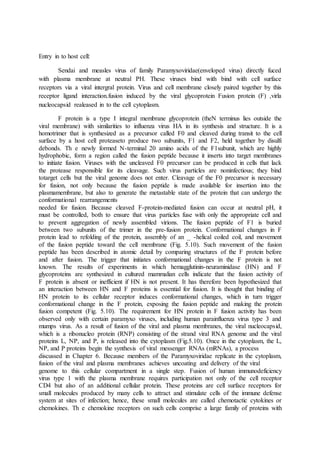
  • 3. Figure : Virus entry and movement in cells. Examples of genome uncoating at the plasma membrane are shown on the left side of the cell. Fusion at the plasma membrane releases the nucleocapsid into the cytoplasm. In some cases, the sub viral particle is transported on microtubules toward the nucleus, where the nucleic acid is released. Uptake of virions by clathrin-dependent endocytosis commences with binding to a specific cell surface receptor. The ligand-receptor complex diffuses into an invagination of the plasma membrane coated with the protein clathrin on the cytosolic side (clathrin-coated pits). The coated pit further invaginates and pinches off, a process that is facilitated by the GTPase dynamin. The resulting coated vesicle then fuses with an early endosome. Endosomes are acidic, as a result ofthe activity of vacuolarproton ATPases. Particle uncoating usually occurs from early or late endosomes. Late endosomes then fuse with lysosomes. Virus particles may enter cells by a dynamin- and caveolin-dependent endocytic pathway (right side of the cell). Three types of caveolar endocytosis have been identified. Dynamin 2-dependent endocytosis by caveolin 1- containingcaveolae isobserved in cells infected with simian virus 40 and polyomavirus. Dynamin 2-dependent, noncaveolar, lipid raft-mediated endocytosis occurs during echovirus and rotavirus infection, while dynamin-independent, noncaveolar, raft-mediated endocytosis is also observed during simian virus 40 and polyomavirus infection. This pathway brings virions to the endoplasmic reticulum via the caveosome, a pH-neutral compartment. Clathrin- and caveolin-independent endocytic pathways of viral entry have also been described (center of cell). Movement of endocytic vesicles within cells occurs on microfilamentsor microtubules, components of the cytoskeleton. Microfilaments are two-stranded helical polymers of the ATPaseactin. They are dispersed throughout the cell but are most highly concentrated beneath the plasma membrane, where they are connected via integrins and other proteins to the extracellular matrix. Transport along microfilaments is accomplished by
  • 4. myosin motors. Microtubules are 25-nm hollow cylinders made of the GTPase tubulin. They radiate from the centrosome to the cell periphery. Movement on microtubules is carried out by kinesin and dynein motors. Figure : Penetration and uncoating at the plasma membrane. (A) Overview. Entry of a member of the Paramyxoviridae, which bind to cell surface receptors via the HN, H, or G glycoprotein. The fusion protein (F) then catalyzes membrane fusion at the cell surface at neutral pH. The viral nucleocapsid, as RNP, is released into the cytoplasm, where RNA synthesis begins. (B) Model for F-protein-mediated membrane fusion. Binding of HN to the cell receptor (red)induces conformational changes in HN that in turn induce conformational changes in the F protein, moving the fusionpeptide from a buried position nearer to the cell membrane. (C) Model of the role of chemokine receptors in human immunodeficiency virus type 1 fusion at the plasma membrane. For simplicity, the envelope glycoprotein is shown as a monomer. Binding of SU to CD4 exposes a high-affinity chemokine receptor-binding site on SU. The SU-chemokine receptor interaction leads to conformational changes in TM that
  • 5. expose the fusion peptide and permit it to insert into the cell membrane, catalyzing fusion in a manner similar to that proposed for influenza virus (cf. Fig. 5.12 and 5.13). Blocking human immunodeficiency virus infection with two soluble cell receptors Because viruses must bind to cell surface molecules to initiate replication, the use of soluble receptors to block virus infection haslong been an attractive therapeutic option. Soluble CD4 receptors that block infection with human immunodeficiency virus type 1(HIV-1) have been developed, but these have not been licensed because of their suboptimal potency. A newly designed soluble receptor forHIV-1 overcomes this problem and provides broad and effective protection against infection of cells and of nonhuman primates. A soluble form of CD4 fused to an antibody molecule can block infection of mostHIV-1 isolates and has been shown to be safe in humans, but its affinity for gp120 is low. Furthermore, human immunodeficiency virus can also be spread from cell to cell by fusion, a process that is not blocked by circulating, soluble CD4. Similarly, peptide mimics of theCCR5 coreceptor have been shown to block infection, but their affinity for gp120 is also low. Combining the two gp120-binding molecules solved the problem of low affinity and in addition provided protection against a wide range of virus isolates. The entry inhibitor, calledeCD4-Ig, is a fusion of the first two domains ofCD4 to the Fc domain of an antibody molecule, with the CCR5-mimicking peptide at the carboxy terminus (illustrated). It binds strongly togp120 and blocks infection with many different isolates of HIV-1, HIV-2, simian immune deficiency virus (SIV), and HIV-1 resistant to broadly neutralizing monoclonal antibodies. The molecule blocks viral infection at concentrations that might be achieved in humans (1.5 to 5.2 micrograms per milliliter).When administered to mice, eCD4-Ig protected the animals from HIV-1. Rhesus macaques inoculated with an adenovirus-associated virus (AAV) recombinant containing the gene foreCD4-Ig were protected from infection with large quantities of virus for up to 34 weeks after immunization. Concentrations of eCD4-Ig in the sera of these animals ranged from 17 to77 micrograms per milliliter. These results show that eCD4-Ig blocks HIV infection with a wide range of isolates more effectively than previously studied broadly neutralizing antibodies. Emergence of HIV variants resistant to neutralization with eCD4-Ig would likely produce viruses that infect cells less efficiently, reducing their transmission. eCD4-Ig is therefore an attractive candidate for therapy of HIV-1 infections. Whether sustained production of the protein in humans will cause disease remains to be determined. Because expression of the AAV genome persists for long periods, it might be advantageous to include a kill-switch in the vector: a way of turning it off if something should go wrong. Uncoating during Endocytosis : Many viruses enter cells by the same pathways by which cellstake up macromolecules. The plasma membrane, the limitingmembrane of the cell, permits nutrient molecules to enterand waste molecules to leave, thereby ensuring an appropriateinternal environment. Water, gases, and small hydrophobicmolecules such as ethanol can freely traverse the lipid bilayer,but most metabolites and ions cannot. These essential componentsenter the cell by specific transport processes. Integralmembrane proteins are responsible for the transport of ions,sugars, and amino acids, while proteins and large particles aretaken into the cell by phagocytosis or endocytosis. The formerprocess (Fig. 5.11) is nonspecific, which means that any particleor molecule can be taken into the cell, and only occurs inspecialized cell types such as dendritic cells and macrophages. Clathrin-Mediated Endocytosis
  • 6. A wide range of ligands, fluid, membrane proteins, and lipids are selectively taken into cells from the extracellular milieuby clathrin-mediated endocytosis (Fig. 5.9 and 5.11), also the mechanism of entry of many viruses. Ligands in the extra cellular medium bind to cells via specific plasma membrane receptor proteins. The receptor-ligand assembly diff uses along the membrane until it reaches an invagination that is coated on its cytoplasmic surface by a cage-like lattice composed of the fibrous protein clathrin (Fig. 5.9). Such clathrin- coatedpits can comprise as much as 2% of the surface area of a cell, and some receptors are clustered over these areas even in the absence of their ligands. Following the accumulation of receptor-ligand complexes, the clathrin-coated pit invaginates and then pinches off to form a clathrin-coated vesicle. Within a few seconds, the clathrin coat is lost and the vesicles fuse with small, smooth-walled vesicles located near the cell surface, called early endosomes . The lumen of early endosomes is mildly acidic (pH 6.5 to 6.0), a result of energy-dependent transport of protons into the interior of the vesicles by a membrane proton pump. The contents of the early endosome are then transported via endosomal carrier vesicles to late endosomes located close to the nucleus. The lumen of late endosomes is more acidic (pH 6.0 to 5.0). Late endosomes in turn fuse with lysosomes, which are vesicles containing a variety of enzymes that degrade sugars, proteins, nucleic acids, and lipids. Viruses with a high pH threshold for fusion, such as vesicular stomatitis virus, enter from early endosomes; most enter the cytoplasm from late endosomes, and a few enter from lysosomes. Clathrin-mediated endocytosis is a continuous but regulated process. For example, the uptake of vesicular stomatitis virus into cells may be influenced by over 90 different cellular protein kinases. Influenza virus, vesicular stomatitis virus and reovirus particles are taken into cells, not into preexistingpits but mainly by clathrin-coated pits that form after virus binds to the cell surface. It is not known how virus binding to the plasma membrane induces the formation of the clathrin-coated pit. Caveolar and Lipid Raft-Mediated Endocytosis Although uptake of most viruses occurs by the clathrin mediated endocytic pathway, some viruses enter by caveolinorraft -mediated endocytosis (Fig. 5.9). Th e caveolar pathway. requires cholesterol (a major component of lipid rafts). Caveolae are distinguished from clathrin-coated vesicles by their flask-like shape, their smaller size, the absence of a clathrin coat, and the presence of a marker protein called caveolin. In the uninfected cell, caveolae participate in transcytosis, signal transduction, and uptake of membrane components and extracellular ligands. Binding of a virus particle to the cell surface activates signal transduction pathways required for pinching off of the vesicle, which then moves within the cytoplasm. Disassembly of fi lamentous actin also occurs, presumably to facilitate movement of the vesicle deeper into the cytoplasm. There it fuses with the caveosome, a larger membranous organelle that contains caveolin (Fig. 5.9). In contrast to endosomes, the pH of the caveosome lumen is neutral. Some viruses (e.g., echovirus type 1) penetrate the cytoplasm from the caveosome. Others (simian virus 40, polyoma virus, coxsackievirusB3) are sorted to the endoplasmic reticulum (ER) by a transport vesicle that lacks caveolin. These viruses enter the cytoplasm by a process mediated by thiol oxidases present in the lumen of the endoplasmic reticulum and by a component of the protein degradation pathway present in the membrane. The study of virus entry by endocytosis can be confusing because some viruses may enter cells by multiple routes, depending on cell type and multiplicity of infection. For example, herpes simplex virus can enter cells by three different routes and influenza A virus may enter cells by both clathrin-dependent and clathrin-independent pathways.
  • 7. Macropinocytosis Macropinocytosis is a process by which extracellular fluid is taken into cells via large vacuoles. It is triggered by ligands. and dependent on actin and a signaling pathway. It differs from phagocytosis by the signaling pathways needed and can take place in many cell types. Th is process serves as a pathway of entry for many viruses, including vaccinia virus, herpesviruses, and ebola viruses. Upon receptor binding, viruses that enter cells via macropinocytosis trigger a signaling cascade that leads to changes in cortical actin and ruffling of the plasma membrane (Fig. 5.11). When these plasma membrane extensions retract, the viruses are brought into macropinosomes and eventually leave these vesicles via membrane fusion. Membrane Fusion The membranes of enveloped viruses fuse with those of the cell as a first step in delivery of the viral nucleic acid. Membrane fusion takes place during many other cellular processes, such as cell division, myoblast fusion, and exocytosis. Membrane fusion must be regulated in order to maintain the integrity of the cell and its intracellular compartments. Consequently, membrane fusion does not occur spontaneously but proceeds by specialized mechanisms mediated by proteins. The two membranes must first come into closeproximity. In cells, this reaction is mediated by interactions of integral membrane proteins that protrude from the lipid bilayers, a targeting protein on one membrane and a docking protein on the other. During entry of enveloped viruses, the virus and cell membranes are fi rst brought into close contact by interaction of a viral glycoprotein with a cell receptor. The next step, fusion, requires an even closer approach of the membranes, to within 1.5 nm of each other. This step depends on the removal of water molecules from the membrane surfaces an energetically unfavorable process. This step is hypothesized to occur when the viral glycoprotein undergoes a structural rearrangement called “hairpinning” (Fig. 5.12).The precise mechanism by which lipid bilayers fuse is not completely understood, but the action of fusion proteins is thought to result in the formation of an opening called a fusion pore , allowing exchange of material across the membranes (Box 5.5). The viral glycoprotein bound to a cell receptor, or a different viral integral membrane protein, then catalyzes the fusion of the juxtaposed membranes. Viral fusion proteins are integral membrane proteins, oft en glycoproteins, that form homo- or hetero-oligomers. Virus-mediated fusion must be regulated to prevent viruses from aggregating or to ensure that fusion does not occur in the incorrect cellular compartment. In some cases, fusogenic potential is masked until the fusion protein interacts with other integral membrane proteins. In others, low pH is required to expose fusion domains. The activity of fusion proteins may also be regulated by cleavage of a precursor. This requirement probably prevents premature activation of fusion potential during virus assembly. Viral fusion proteins are often primed for fusion by proteolytic cleavage as they move through the trans-Golgi network as described in Chapter 12. Proteases that catalyze such cleavage are typically furin family convertases that either cleave the fusion proteins directly (orthomyxoviruses, retroviruses, paramyxoviruses) orcleave a protein that masks the fusion protein (alphaviruses, flaviviruses).
  • 8. Figure : Mechanisms for the uptake of macromolecules from extracellular fluid. During phagocytosis, large particles such as bacteria or cell fragments that come in contact with the cell surface are engulfed by extensions of the plasma membrane. Phagosomes ultimately fuse with lysosomes, resulting in degradation of the material within the vesicle. Macrophages use phagocytosis to ingest bacteria and destroy them. Endocytosis comprises the invagination and pinching off of small regions of the plasma membrane, resulting in the nonspecific internalization of molecules (macropinocytosis) or the specific uptake of molecules bound to cell surface receptors(receptor-mediated endocytosis). Macropinocytosis is a mechanism for the uptake of extracellular fluid. It is triggered by ligand binding which initiates formation of plasma membrane ruffling, which traps material in large vacuoles. Figure : Influenza virus entry. The globular heads of native HA mediate binding of the virus to sialicacid-containing cell receptors. The virus-receptor complex is endocytosed, and import of H_ ions into the endosomeeacidifies the interior. Upon acidification, the viral HA undergoes a conformational rearrangement that produces a fusogenic protein. The loop region of native HA (yellow) becomes a coiled coil, moving the fusion peptides (red) to the top of the molecule near the cell membrane. At the viral membrane, the long _-helix (purple) packs against the trimercore, pulling the globular heads to the side. The long coiled coil bends, or
  • 9. hairpins, bringing the fusion peptides and the trans membrane domains together. This movement moves the cell and viral membranes close together so that fusion canoccur. To allow release of vRNP into the cytoplasm, the H_ ions in the acidic endosome are pumped into the particle interior by the M2 ion channel. As a result, vRNP is primed to dissociate from M1 after fusion of the viral and endosomal membranes. The released vRNPs are imported into the nucleus through the nuclear pore complex via a nuclear localization signal- dependent mechanism (see “Import of Influenza Virus Ribonucleoprotein” below). Fig. Membrane fusion proceeds through a hemifusion intermediate
  • 10. Membrane fusion by flavivirus envelope glycoproteinE. Low pH causes conformational changes in the viral glycoproteinsto produce the fusion-active forms. (A) Ninety dimers of E tilethe surface of the virus particle. (B) The fusion loop is located at the tipof domain II (yellow) where it is buried in dimers of the glycoprotein E.At low pH, the dimers are disrupted, the proteins extend to form trimers,and the fusion peptide is directed toward the cell membrane. The glycoproteinthen undergoes a rearrangement that brings the fusion peptides, and transmembrane segments (purple) together. This movement brings the viral and cell membranes together, allowing fusion. Receptor Priming for Low-pH Fusion: Two Entry Mechanisms Combined During the entry of avian leukosis virus into cells, binding of the virus particle to the cell receptor primes the viral fusion protein for low-pH-activated fusion. Avian leukosis virus, like many other retroviruses with simple genomes, was believed to enter cells at the plasma membrane in a pH-independent mechanism resembling that of members of the Paramyxoviridae(Fig. 5.10). It is now known that binding of the viral membrane glycoprotein (SU) to the cellular receptors of avian leukosis viruses induces conformational rearrangements that convert the viral protein from a native metastable state that is insensitive to low pH to a second metastable state. In this state, exposure to low pH within the endosomal compartment leads to membrane fusion and release of the viral capsid. Figure .Entry of Ebolavirusintocells.
  • 11. Virus particles bind cells via an unidentified attachment receptor and enter by endocytosis. The mucin and glycan cap on the viral glycoprotein is removed by cellular cysteine proteases, exposing binding sites for NPC1. The latter is required for fusion of the viral and cell membranes, releasing the nucleocapsid into the cytoplasm. Uncoating in the Cytoplasm by Ribosomes Some enveloped RNA-containing viruses, such as Semliki Forest virus, contain nucleocapsids that are disassembled in the cytoplasm by pH-independent mechanisms. The icosahedral nucleocapsid of this virus is composed of a single viral protein, C protein, which encloses the strand viral RNA. This structure is surrounded by an envelope containing viral glycoproteins E1 and E2, which are arranged as heterodimers clustered into groups of three, each cluster forming a spike on the virus surface. Fusion of the viral and endosomal membranes exposes the nucleocapsid to the cytoplasm (Fig. 5.17). To begin translation of strand viral RNA, the nucleocapsid must be disassembled, a process mediated by an abundant cellular component the ribosome. Each ribosome binds three to six molecules of C protein, disrupting the nucleocapsid. This process occurs while the nucleocapsid is attached to the cytoplasmic side of the endosomal membrane (Fig. 5.17) and ultimately results in disassembly. The uncoated viral RNA remains associated with cellular membranes, where translation and replication begin. Figure.Stepwise uncoating of adenovirus. (A) Adenoviruses bindthe cell receptor via the fiber protein.Interaction of the penton base with anintegrin receptor leads to internalization by endocytosis. Fibers are released fromthe capsid during uptake. Low pH in the endosome causes destabilization of the capsid and release of protein VI (yellow diamonds). The hydrophobic N terminus of protein VI disrupts the endosome membrane, leading to release of a subviral particle into the cytoplasm. The capsid is transported in the cytoplasm along microtubules and docks onto the nuclear pore complex. (B) Electron micrograph of adenovirus type 2 particles bound to a microtubule (top) and bound to the cytoplasmic face of the nuclear pore complex (bottom). Bar in bottom panel, 200 nm. Disrupting the Endosomal Membrane Adenoviruses are composed of a double-stranded DNA genome packaged in an icosahedral capsid. Internalization of most adenovirus serotypes by receptor-mediated endocytosis requires attachment of fi ber to an integrin or Ig-like cell surface receptor and binding of the penton base to a second cell receptor, the cellular vitronectin-binding integrins _ v _ 3 and _ v _ 5 . Attachment is mediated by amino acid sequences in each of the fi ve subunits of the adenovirus penton base that mimic the normal ligands of cell surface integrins. As the virus particle is transported via the endosomes from the cell surface toward the nuclear membrane, it undergoes multiple uncoating steps as structural proteins are removed sequentially (Fig. 5.18). As the endosome becomes acidifi ed, the viral capsid is destabilized, leading to release of proteins from the capsid. Among these is protein VI, which causes disruption of the endosomal membrane, thereby delivering the remainder of the particle into the cytoplasm. An N-terminal amphipathic_ -helix of protein VI is probably responsible for its pH-dependent membrane disruption activity. Th is region of the protein appears to be masked in the native capsid by the hexon protein. The liberated subviral particle then docks onto the nuclear pore complex (see “Import of DNA Genomes” below).
  • 12. Forming a Pore in the Endosomal Membrane : The genomes of nonenveloped picornaviruses are transferredacross the cell membrane by a diff erent mechanism,as determined by structural information at the atomic level and complementary genetic and biochemical data obtainedfrom studies of cell entry. The interaction of poliovirus withits Ig-like cell receptor, CD155, leads to major conformational rearrangements in the virus particle and the production of an expanded form called an altered (A) particle (Fig. 5.19A). Portions of two capsid proteins, VP1 and VP4, move from the inner surface of the capsid to the exterior. Th ese polypeptides are thought to form a pore in the cell membrane that allows transport of viral RNA into the cytoplasm (Fig. 5.19B). In support of this model, ion channel activity can be detected when A particles are added to lipid bilayers.
  • 13. Figure. Model for poliovirus entry into cells. (A) Overview. The native virion (160S) binds to its cell receptor, CD155, and undergoes a receptor-mediated conformational transition resulting in the formation of altered (A) particles. The viral RNA, shown as a curved green line, leaves the capsid from within early endosomes close to the plasma membrane. (B) Model of the formation of a pore in the cell membrane after poliovirus binding. 1, poliovirus (shown in cross section, with capsid proteins purple) binds to CD155 (brown). 2, a conformational change leads to displacement of the pocket lipid (black). The pocket may be occupied by sphingosine in the capsid of poliovirus type 1. The hydrophobic N termini of VP1 (blue) are extruded and insert into the plasma membrane. 3, a pore is formed in the membrane by VP4 and the VP1 N termini, through which the RNA is released from the capsid into the cytosol.
  • 14. Fig.Entry of Revovirus into the cells. Movement of Viral and Subviral Particles within Cells : Viral and subviral particles move within the host cell duringentry and egress). However, movement of molecules larger than 500 kDa does not occur by passivediffusion, because the cytoplasm is crowded with organelles,high concentrations of proteins, and the cytoskeleton. Rather,viral particles and their components are transported via theactin and microtubule cytoskeletons. Such movement can be visualized in live cells by using fluorescently labeled viral proteins. The cytoskeleton is a dynamic network of protein fi laments that extends throughout the cytoplasm. It is composed of three types of fi lament—microtubules, intermediate filaments, and microfi laments. Microtubules are organized in a polarized manner, with minus ends situated at the microtubuleorganizing center near the nucleus, and plus ends located at the cell periphery. This arrangement permits directed movement of cellular and viral components over long distances. Actin filaments (microfilaments) typically assist in virus movement close to the plasma membrane.
  • 15. Virus-Induced Signaling via Cell Receptors Binding of virus particles to cell receptors not only concentratesthe particles on the cell surface, but also activates signalingpathways that facilitate virus entry and movement within the cell or produce cellular responses that enhance virus propagation and/or affect pathogenesis. Virus binding may lead to activation of protein kinases that trigger cascades of responses at the plasma membrane, cytoplasm, and nucleus. Second messengers that participate in signaling include phosphatidylinositides, diacylglycerides, and calcium; regulators of membrane trafficking and actin dynamics also contribute to signaling. Virus- receptor interactions also stimulate antiviral responses. The reproduction of most DNA viruses, and some RNA viruses including retroviruses and influenza viruses, begins in the cell nucleus. The genomes of these viruses must therefore be imported from the cytoplasm. One way to accomplish thismovement is via the cellular pathway for protein import into the nucleus. An alternative, observed in cells infected by some retroviruses, is to enter the nucleus after the nuclear envelope breaks down during cell division. When the nuclear envelope is reformed, the viral DNA is incorporated into the nucleus together with cellular chromatin. This strategy restricts infection to cells that undergo mitosis.