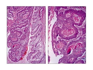
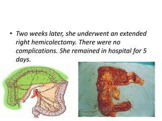
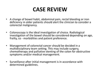
2. A 79 - year - old woman with altered
bowel habit and weight loss
3. Main complains
Mrs MS is a 79 - year - old woman who attends
your outpatient clinic with her daughter.
-constipation
- vague abdominal discomfort.
-lost her appetite
-her clothes have become looser over the last
few months.
4. • What are your main concerns?
• What other information do you need to
know?
6. Weight loss
• Establish the amount of weight lost over what
time period. Has it been purposeful or non -
intentional?
– Malignancy ?
– Malabsorptive?
– Inflammatory processes?
9. History of the disease
She explains that she has felt constipated for the
last year. Her bowel habit is regular, once per
day, with hard, pellet - like stool. There is no
excessive straining at stool and no rectal
bleeding.
Her abdominal discomfort is deep and constant.
It is located around the epigastrum and does
not radiate elsewhere.
It has no relieving or aggravating factors
10. History of the disease
• She feels bloated most of the day. She does
not have any nausea and never vomits. Her
appetite has declined and she has probably
lost about 6 kg in weight over the last 2
months (almost two dress sizes for her). She is
happy with this.
• She feels tired and lethargic but puts this
down to her age.
12. Family history
• There is a family history of diabetes and
ischaemic heart disease but no colorectal
cancer.
13. Past medical history
2016-non - insulin dependent type 2 diabetes mellitus.
2020-ischaemic heart disease (recently diagnosed 6 months ago by her
general practitioner),
- 2020- claudication on walking 200 m and She considers herself to be
well.
Her medications include aspirin, lisinopril, atorvastatin and metformin.
She was recently started on iron supplements for anaemia. She has
used lactulose as needed for constipated stool, usually once a month
at most.
She is an ex - smoker of 20 years with a 30 pack – year history.
16. On examination she has conjunctival pallor.
There is no lymphadenopathy.
Her abdomen is soft and tender in the
epigastrium with no obvious masses or
organomegaly.
There is nothing to find on rectal exam.
17. RED FLAG
Change of bowel habit, abdominal pain, rectal
bleeding or iron deficiency in older patients
are alarm features to suggest malignancy.
18. What investigations do you choose?
Blood tests
-Full blood count
-Iron, feritine
-urea, creatinine, electrolytes
-liver function
-calcium
-thyroid function
20. What are the other options instead
of colonoscopy?
• CT colonography with oral gastrografin to
opacify the stool.
21. When do you choose
radiological investigation?
If the patient was not fit for sedation or bowel
preparation for colonoscopy
If the patient refused an invasive procedure
22. What if a lesion is found?
• If a mass lesion is detected on imaging, an
endoscopic examination would be needed to
confirm radiological findings and obtain
histology.
• A staging CT scan is also appropriate to look
for the extent of disease and guide
management.
24. She proceeded to CT colonography given her age
and co - morbidities.
This demonstrated thickening of a region in the
ascending colon and scattered diverticula to
the splenic flexure
25.
26. • She proceeded to have a colonoscopy, aware
of the findings on the CT scan.
• She was admitted as an inpatient and given
intravenous rehydration pre – and
postoperatively.
• The colonoscopy revealed a polypoid mass
lesion causing partial obstruction in the
ascending colon
27.
28.
29. What are the management options?
• Surgery
• Chemotherapy
• Palliation of symptoms
30. The case was re - discussed at the MDT meeting and
surgical resection was deemed appropriate .
She underwent an exercise tolerance test and managed 3
min 25 s of the Bruce protocol, limited by dyspnoea.
There was evidence of ST depression consistent with
coronary artery disease. She was reviewed by the
cardiologists and commenced on anti - anginal
medication.
• This improved her symptoms. Echocardiogram
confirmed good left ventricular function and no
valvular disease.
31. • Two weeks later, she underwent an extended
right hemicolectomy. There were no
complications. She remained in hospital for 5
days.
32. • Histology revealed a T3,N0,M0 moderately
differentiated mucinous adenocarcinoma of
the ascending colon
33. • She was offered, but declined, adjuvant
chemotherapy due to the potential risks and
complications. She remains well 1 year later.
34. What about follow - up?
• She requires a surveillance colonoscopy 1 year
postoperatively.
35. CASE REVIEW
• A change of bowel habit, abdominal pain, rectal bleeding or iron
deficiency in older patients should alert the clinician to consider a
colorectal malignancy.
• Colonoscopy is the ideal investigation of choice. Radiological
investigation of the bowel should be considered depending on age,
frailty, co - morbidities and patient preference.
• Management of colorectal cancer should be decided in a
multidisciplinary team setting. This may include surgery,
chemotherapy and palliative stenting of the colon for obstructive
symptoms and/or medical management.
• Surveillance after initial management is in accordance with
determined guidelines.