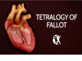
Tetralogy of Fallot Heart Defect Overview
•Download as PPTX, PDF•
5 likes•332 views

Tetralogy of Fallot is a congenital heart defect caused by four abnormalities present at birth: pulmonary valve stenosis, ventricular septal defect, overriding aorta, and right ventricular hypertrophy. This causes oxygen-poor blood to circulate through the body, resulting in blue-tinged skin. Symptoms vary in severity and include cyanosis, fainting, and fatigue. While the cause is often unknown, risk factors include certain infections or conditions in the mother during pregnancy. Diagnosis is via echocardiogram, catheterization, or chest x-ray, and treatment involves prostaglandin infusion, corrective surgery, or managing spells through positioning and medication.

Recommended
More Related Content
What's hot
What's hot (20)
Similar to Tetralogy of Fallot Heart Defect Overview
Similar to Tetralogy of Fallot Heart Defect Overview (20)
More from garvsuthar
More from garvsuthar (20)
Recently uploaded
Recently uploaded (20)
Tetralogy of Fallot Heart Defect Overview
- 2. Overview Tetralogy of Fallot is a rare condition caused by a combination of four heart defects that are present at birth (congenital). Pulmonary valve stenosis Ventricular septal defect Overriding aorta Right ventricular hypertrophy
- 3. . These defects, which affect the structure of the heart, cause oxygen-poor blood to flow out of the heart and to the rest of the body. Infants and children with tetralogy of Fallot usually have blue- tinged skin because their blood doesn't carry enough oxygen. Tetralogy of Fallot is often diagnosed during infancy or soon after. However, tetralogy of Fallot might not be detected until later in life in some adults, depending on the severity of the defects and symptoms.
- 5. Etiology Tetralogy of Fallot occurs during fetal growth, when the baby's heart is developing. While factors such as poor maternal nutrition, viral illness or genetic disorders might increase the risk of this condition, in most cases the cause of tetralogy of Fallot is unknown.
- 6. Tet spells Sometimes, babies who have tetralogy of Fallot will suddenly develop deep blue skin, nails and lips after crying or feeding, or when agitated. These episodes are called tet spells and are caused by a rapid drop in the amount of oxygen in the blood. Tet spells are most common in young infants, around 2 to 4 months old. Toddlers or older children might instinctively squat when they're short of breath. Squatting increases blood flow to the lungs.
- 8. Pulmonary valve stenosis - Narrowing (constriction) of the pulmonary valve reduces blood flow to the lungs. The narrowing might also affect the muscle beneath the pulmonary valve. In some severe cases, the pulmonary valve doesn't form properly (pulmonary atresia) and causes reduced blood flow to the lungs.
- 9. Ventricular septal defect. - Blood from the left ventricle also flows back to the right ventricle in an inefficient manner. This ability for blood to flow through the ventricular septal defect reduces the supply of oxygenated blood to the body and eventually can weaken the heart.
- 10. Overriding aorta - Normally the aorta — the main artery leading out to the body — branches off the left ventricle. In tetralogy of Fallot, the aorta is shifted slightly to the right and lies directly above the ventricular septal defect. In this position the aorta receives blood from both the right and left ventricles, mixing the oxygen-poor blood from the right ventricle with the oxygen-rich blood from the left ventricle.
- 11. Right ventricular hypertrophy - When the heart's pumping action is overworked, it causes the muscular wall of the right ventricle to thicken. Over time this might cause the heart to stiffen, become weak and eventually fail.
- 12. Sign And Symptoms - Tetralogy of Fallot symptoms vary, depending on the extent of obstruction of blood flow out of the right ventricle and into the lungs. A bluish coloration of the skin caused by blood low in oxygen (cyanosis) Loss of consciousness (fainting) Clubbing of fingers and toes — an abnormal, rounded shape of the nail bed Poor weight gain Dyspnea Tiring easily during play or exercise A heart murmur
- 13. Risk factors - While the exact cause of tetralogy of Fallot is unknown, various factors might increase the risk of a baby being born with this condition. These risk factors include: A viral illness during pregnancy, such as rubella (German measles) Alcoholism during pregnancy Poor nutrition during pregnancy A mother older than age 40 A parent who has tetralogy of Fallot The presence of Down syndrome or DiGeorge syndrome
- 14. Diagnosis Echocardiography. Cardiac catheterization Chest X-ray
- 15. Treatment For symptomatic neonates, prostaglandin E1 infusion For hypercyanotic spells, knee-chest positioning, calming, oxygen, IV fluids, and sometimes drugs Surgical repair Neonates with severe cyanosis may be palliated with an infusion of prostaglandin E1 to open the ductus arteriosus and thereby increase pulmonary blood flow.
- 16. Hyper- cyanotic spells - Hypercyanotic spells require immediate intervention. The first steps are to Place infants in a knee-chest position (older children usually squat spontaneously and do not develop hypercyanotic spells) Establish a calm environment Give supplemental oxygen Give IV fluids for volume expansion If the spell persists, standard medical therapy includes morphine, phenylephrine, and beta-blockers (propranolol or esmolol)