Pulmonary stenosis
•
22 likes•8,660 views

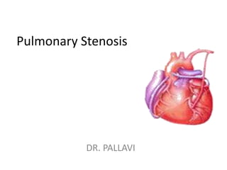
Pulmonary stenosis (also called pulmonic stenosis) is when the pulmonary valve (the valve between the right ventricle and the pulmonary artery) is too small, narrow, or stiff. Symptoms of pulmonary stenosis depend on how small the narrowing of the pulmonary valve is

Recommended
More Related Content
What's hot
What's hot (20)
Similar to Pulmonary stenosis
Similar to Pulmonary stenosis (20)
More from DR .PALLAVI PATHANIA
More from DR .PALLAVI PATHANIA (20)
Recently uploaded
Recently uploaded (20)
Pulmonary stenosis
- 2. Pulmonary valve: • a valve that has leaflets that are partially fused together. • a valve that has thick leaflets that do not open all the way. • the area above or below the pulmonary valve is narrowed.
- 3. Pulmonary Stenosis Rare, usually congenital in origin Common as PDA, flow of blood to the pulmonary artery due to narrowing blood flows back to right ventricle and right atrium right ventricle hypertrophy to compensate for blood volume and force blood to the pulmonary artery
- 4. Pathophysiology • The right ventricle - work harder • It fails to pump forward efficiently. • Pressure builds up in the right atrium • Fluid retention and swelling may occur. • There is a higher than average chance of developing an infection in the lining of the heart known as bacterial endocarditis.
- 5. Symptoms of pulmonary stenosis. • heavy or rapid breathing • shortness of breath • Fatigue, fainting • rapid heart rate • swelling in the feet, face, eyelids, and/or abdomen • Cyanosis in terminal stages • Severe cases: Distended jugular veins, hepatomegaly, ascites
- 6. Diagnostic Evaluation • Auscultation: systolic ejection murmur heard best at the left upper sternal border; ejection click. • ECG: varies; normal in mild cases and RVH in moderate to severe cases. • Chest X-ray: varies; may show right ventricular enlargement; poststenotic dilatation of PA. • Two-dimensional echocardiography with Doppler study and color flow mapping to visualize the sites of obstruction, observe the degree of RVH, and estimate the pressure gradient across the valve. • Cardiac catheterization is usually not needed for the initial diagnosis.
- 7. Medical management: – Stabilize and improve oxygen saturations with PGE1 infusion. – Intubation and ventilation as needed. – Inotropic support as needed. – Infective endocarditis prophylaxis (lifelong). • . • Surgical intervention: Balloon pulmonary valvuloplasty – Valvotomy or valvectomy for dysplastic pulmonary valve. – Patch repair of the right ventricular outflow tract. – Placement of an RV-to-PA conduit
- 8. Balloon pulmonary valvuloplasty • balloon dilation or valvuloplasty used for valvar, supravalvar, or branch types of pulmonary stenosis . A small, flexible tube (catheter) is inserted into a blood vessel in the femoral vessel and guided to the inside of the heart. The tube has a deflated balloon in the tip. When the tube is placed in the narrowed valve, the balloon is inflated to stretch the area open.
- 9. • Valvotomy Valvotomy is the surgical release of adhesions that are preventing the valve leaflets from opening properly. This type of procedure is generally not performed, as balloon dilation or valvuloplasty has become more common. • Valvular fusion- Insert valvulotome- purse-string stab incision • 4th left intercostal space-reflect left cranial lung lobe- incision on rt. Ventricular outflow
- 10. • Purse-string- 2-0, silk- conus- 2 cm incision on epicardium – sharp scalpel • Blunt probe- inserted – level of obs.- withdrawn- valvulotome /teat bistoury is put-withdrawn • Record the pressure • Close with interrupted sutures
- 11. • Open technique: • Rt. Ventriculotomy: direct visual repair of stenosis • To cut the fusion of cusp upto the base or attachment • Less practiced in veterinary
- 12. • valvectomy (with or without transannular patch) Valvectomy is the surgical removal of the valve and the widening of the outflow patch to improve blood flow from the right ventricle into the pulmonary artery. Once the individual reaches adulthood, the pulmonary valve is generally replaced
- 13. • pulmonary valve replacement Replacement of the pulmonary valve is a surgical procedure that is often recommended in adulthood. A tissue valve (pig or human) may be used.
- 14. Valve Repair Valvuloplasty is repair of cardiac valve • pt. does not require continuous anti-coagulant medication • usually require cardiopulmonary bypass machine 1.Commissurotomy – to separate the fused leaflets Balloon Valvuloplasty – performed in the cardiac cath. lab. - balloon inflated for 10-30 secs., w/ multiple inflations - common used for mitral and aortic stenosis Closed surgical valvuloplasty – done in the OR under GA - midsternal incision, a small hole is cut into the heart, the surgeons finger or a dilator is used to open the commissure Open Commissurotomy – done w/ direct visualization of the valve, thrombus and calcifications may be identified and removed
- 15. Valve Repair (cont.) 2. Annuloplasty is repair of valve annulus (junction of the valve leaflets and the muscular heart wall) - narrows the diameter of the valve’s orifice, useful for valvular regurgitation 3. Chordoplasty is repair of chordae tendineae - done for mitral valve regurgitation – caused by stretched, torn or shortened chordae tendineae
- 16. Annuloplasty
- 18. Valve Replacement Mechanical valves – Ex. Caged ball valve, Tilting-disk valve - more durable, used for younger pts. - risk of thromboembolism – long-term use of anti-coagulants Tissue or biological valves: - xenografts – porcine or bovine heterografts (7-10 yrs viability) - homografts – from cadaver tissue donations (10-15 yrs) - autografts – excising the pts.’s own pulmonic valve and portion of pulmonary artery for use as the artic valve Long-term anticoagulant therapy Antibiotic prophylaxis
- 19. Valve Prosthesis
- 23. Complications • Cyanosis (critical PS in the neonate). • Arrhythmia; sudden death. • Infective endocarditis. • Right ventricular failure.
- 24. NURSING DIAGNOSIS • Decreased cardiac output related to valvular incompetence as evidenced by murmurs, dyspnea, dysrhythmias, peripheral edema • NURSING INTERVENTION: Cardiac Care • Monitor vital signs, cardiovascular status, and respiratory status to assess for palpitations, angina, widened pulse pressure). • Monitor for cardiac dysrhythmias, including disturbances of both rhythm and conduction, to identify and treat significant dysrhythmias.
- 25. CONT... • Hemodynamic Regulation • Administer inotropic medication as ordered to increase myocardial contractility. • Elevate head of bed to reduce venous return, reduce O2 demand, and maximize chest excursion. • Energy Management • Promote bed rest/activity limitation to decrease cardiac workload and O2 demand.manifestations of decreased cardiac output (e.g., fatigue, malaise, shortness of breath, dyspnea on exertion,
- 26. CONT... Excess fluid volume related to fluid retention secondary to valvular-induced heart failure as evidenced by peripheral edema, weight gain, adventitious breath sounds, neck vein distention. NURSING INTERVENTION: Hypervolemia Management Monitor changes in peripheral edema to detect hypervolemia. Monitor respiratory system for symptoms of difficulty (e.g., dyspnea, tachypnea, adventitious breath sounds) to assess for fluid congestion in the lungs.
- 27. CONT... • Fluid/Electrolyte Management • Provide restricted-sodium diet as ordered to prevent fluid retention
- 28. CONT... • Activity intolerance related to insufficient oxygenation secondary to decreased cardiac output and pulmonary congestion as evidenced by weakness, fatigue, shortness of breath, increase or decrease in pulse rate, BP changes. • NURSING DIAGNOSIS: Energy Management • Monitor cardiorespiratory response to activity (e.g., pulse rate, respirations, pulse oximetry, BP) to plan appropriate interventions. • Encourage alternate rest and activity periods to conserve energy and decrease cardiac demands.
- 29. CONT... • Encourage patient to choose activities that gradually build endurance to increase cardiac tolerance. • Assist the patient/caregiver to establish realistic activity goals to promote feelings of accomplishment.
- 30. REFERENCES • Suddarth’s and Brunner ; Textbook of Medical Surgical Nursing ; Published by; Lippincott ; 10th Edition ; Page No. 788-790’ • Porter McKenzine ; Clinical companoin Medical Surgical ; Published by; Elsevier ; 1st Edition ; Page No. 68-69, • Black M Jaycee ; Textbook of Medical Surgical Nursing ; Published by; Elsevier ; 7th Edition ; 2nd Volume ; Page No. 1385-1392 • http:meddean.luc.edu.in