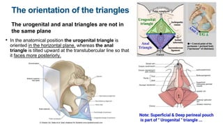
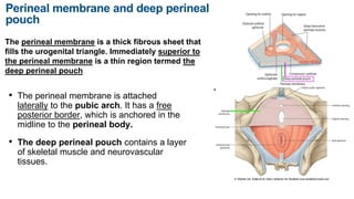
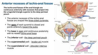
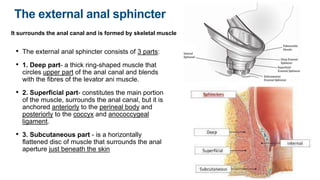
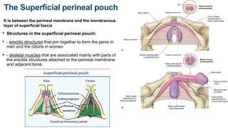
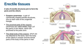
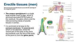
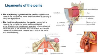
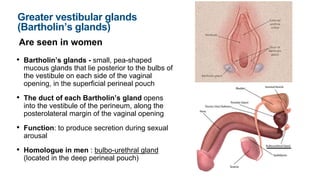
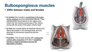
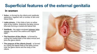
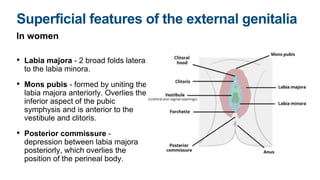
Anatomy VI
Anatomy of the Perineum. Triangles. Perineum. Anal triangle.
Urogenital triangle
Perineum
It is a diamond-shaped region positioned inferiorly to
the pelvic floor between the thighs
• Its peripheral boundary is the pelvic outlet
• Borders of the Perioneum:
• Anterior point- inferior border of the pubic
symphysis
• Posterior point - the tip of the coccyx
• Lateral points - ischial tuberosities
• Lateral margins anteriorly - ischiopubic rami
• Lateral margins posteriorly - sacrotuberous
ligaments
Division of the
Perineum
The perineum is divided into 2 triangles by an imaginary
line between the 2 ischial tuberosities. Anterior to the
line is the urogenital triangle and posterior to the line is
the anal triangle.
• The urogenital triangle is
associated with the openings of
the urinary system and the
reproductive systems and
functions to anchor the external
genitalia
• The anal triangle contains the
anus and the external anal
sphincter
The orientation of the triangles
The urogenital and anal triangles are not in
the same plane
• In the anatomical position the urogenital triangle is
oriented in the horizontal plane, whereas the anal
triangle is tilted upward at the transtubercular line so that
it faces more posteriorly.
The roof of the perineum
It is formed by the pelvic diaphragm (the levator ani
and coccygeus muscles) that separates the pelvic
cavity above from the perineum below
• The levator ani and coccygeus
muscles form a cone- or funnel-
shaped pelvic diaphragm, with the
anal aperture at its inferior apex in
the anal triangle.
• Anteriorly, in the urogenital
triangle, a U-shaped defect in the
muscles, the urogenital hiatus
allows passage of the urethra and
vagina.
Perineal membrane and deep perineal
pouch
The perineal membrane is a thick fibrous sheet that
fills the urogenital triangle. Immediately superior to
the perineal membrane is a thin region termed the
deep perineal pouch
• The perineal membrane is attached
laterally to the pubic arch. It has a free
posterior border, which is anchored in the
midline to the perineal body.
• The deep perineal pouch contains a layer
of skeletal muscle and neurovascular
tissues.
Perineal membrane and deep
perineal pouch
• Function:
• - Provide support for external genitalia,
attached to the inferior surface of the perineal
membrane
• - Provide support for the pelvic viscera above
in the part inferior to the urogenital hiatus in
the elevator ani muscle
• - The urethra and the vagina pass through the
deep perineal pouch and perineal membrane
Ischio-anal fossae
The ischioanal fossa (formerly called ischiorectal
fossa) is the fat-filled wedge-shaped space located
lateral to the anal canal and inferior to the pelvic
diaphragm.
• The lateral wall of each ischio-anal fossa:
ischium, obturator internus muscle,
sacrotuberous ligament
• The medial wall of each ischio-anal fossa: the
levator ani muscle
• The medial and lateral walls converge
superiorly where the levator ani muscle
attaches to the fascia overlying the obturator
internus muscle
• Function: The ischio-anal fossae allow
movement of the pelvic diaphragm and
expansion of the anal canal during defecation
Anterior recesses of ischio-anal fossae
The ischio-anal fossae of the anal triangle are
continuous anteriorly with recesses that project into
the urogenital triangle superior to the deep perineal
pouch
• The anterior recesses of the ischio-anal
fossae are shaped like three-sided pyramids:
• The apex of each pyramid is closed and
points anteriorly toward the pubis
• The base is open and continuous posteriorly
with its related ischio-anal fossa
• The inferior wall - the deep perineal pouch
• The superomedial wall - levator ani muscle
• The superolateral wall - obturator internus
muscle
Anal triangle of the perineum
It faces posteroinferiorly
• Borders of the anal triangle:
• - Laterally - medial margins of the sacrotuberous
ligaments
• - Anteriorly - horizontal line between 2 ischial
tuberosities
• - Posteriorly - the coccyx
• The ceiling of the anal triangle: the pelvic
diaphragm (levator ani and coccygeus muscles)
• The anal aperture occurs centrally in the anal
triangle and is related on either side to ischio-anal
fossa.
The external anal sphincter
It surrounds the anal canal and is formed by skeletal muscle
• The external anal sphincter consists of 3 parts:
• 1. Deep part- a thick ring-shaped muscle that
circles upper part of the anal canal and blends
with the fibres of the levator ani muscle.
• 2. Superficial part- constitutes the main portion
of the muscle, surrounds the anal canal, but it is
anchored anteriorly to the perineal body and
posteriorly to the coccyx and anococcygeal
ligament.
• 3. Subcutaneous part - is a horizontally
flattened disc of muscle that surrounds the anal
aperture just beneath the skin
Urogenital triangle
It is anterior part of the perineum and is oriented in the horizontal
plane
• Borders of the urogenital triangle:
• Laterally - the ischiopubic rami
• Posteriorly - the imaginary line between
ischial tuberosities
• Anteriorly - inferior margin of the pubic
symphysis
• The ceiling of the urogenital triangle: the
levator ani muscle
• The urogenital triangle contains: perineal
membrane and deep perineal pouch, which is
attached to the pubic arch. Also the roots of
the external genitalia and the openings of the
urogenital system
The Superficial perineal pouch
It is between the perineal membrane and the membranous
layer of superficial fascia
• Structures in the superficial perineal pouch:
• - erectile structures that join together to form the penis in
men and the clitoris in women
• - skeletal muscles that are associated mainly with parts of
the erectile structures attached to the perineal membrane
and adjacent bone
Erectile tissues
2 sets of erectile structures join to form the
penis and the clitoris
• Corpora cavernosa - a pair of
cylindrically shaped erectile structures,
one on each side of the urogenital
triangle.
• The proximal parts of the corpora
cavernosa are called crura (“legs”) of
the penis or the clitoris, which are
anchored to the pubic arch.
• The distal ends of the corpora, which are
not attached to the bone, form the body
of the clitoris in women and the dorsal
parts of the body of the penis in men
Erectile tissues (women)
The second set of erectile tissues surrounds
the openings of the urogenital system
• Bulbs of vestibule in women is a pair of
erectile structures situated one on each
side at the vaginal opening and are firmly
anchored to the perineal membrane.
• Glans clitoris in women is a single,
small, pea-shaped erectile mass, which
is positioned in the midline, at the end of
the body of the clitoris and anterior to the
opening of the urethra.
• Bulbs of the vestibule and the glans
clitoris are connected by small bands of
the erectile tissues.
Erectile tissues (men)
The second set of erectile tissues surrounds the openings of
the urogenital system
• The corpus spongiosum is a single
large erectile mass in men, which is
structural equivalent to the bulbs of
the vestibule, the glans clitoris and the
interconnecting bands of erectile
tissues in women.
• It is anchored at its base to the
perineal membrane. Its proximal end,
which is not attached, forms the
ventral part of the body of the penis
and expands over the end of the body
of the penis to form the glans penis.
Clitoris
It is composed of 2 corpora cavernosa and glans clitoris
• The root of the clitoris (the attached part) -
consists only of the 2 crura
• The body of the clitoris (the free part) - is formed
only by the unattached parts of the two corpora
cavernosa, angles posteriorly and is embedded in
the connective tissues of the perineum.
• Suspensory ligament of the clitoris - supports
the the body of the clitoris and attaches superiorly
to the pubic symphysis
• Glans clitoris - is attached to the distal end of the
body of the clitoris and is connected to the bulbs
of the vestibule by small bands of erectile tissue.
Penis
It is composed of the 2 corpora cavernosa and the
single corpus spongiosum, which contains the urethra
• The root of the penis (attached part) - consists of
2 crura, which are proximal parts of the corpora
cavernosa attached to the pubic arch and the bulb
of the penis, which is the proximal part of the corpus
spongiosum anchored to the perineal membrane.
• The body of the penis (free part)- is entirely
covered by skin and is formed by the tethering of
the 2 proximal free parts of the corpora cavernosa
and the related free part of the corpus spongiosum.
• Glans penis - is formed by expansion of the corpus
spongiosum over the distal ends of the corpora
cavernosa.
Ligaments of the penis
• The suspensory ligament of the penis - supports the
base of the body of the penis and is attached superiorly to
the pubic symphysis
• The fundiform ligament of the penis - supports the
base of the body of the penis and is positioned
superficially to the suspensory ligament. It is attached to
the linea alba of the anterior abdominal wall and is split
below into 2 bands that pass on each side of the penis
and unite inferiorly.
Greater vestibular glands
(Bartholin’s glands)
Are seen in women
• Bartholin’s glands - small, pea-shaped
mucous glands that lie posterior to the bulbs of
the vestibule on each side of the vaginal
opening, in the superficial perineal pouch
• The duct of each Bartholin’s gland opens
into the vestibule of the perineum, along the
posterolateral margin of the vaginal opening
• Function: to produce secretion during sexual
arousal
• Homologue in men : bulbo-urethral gland
(located in the deep perineal pouch)
Muscles of the superficial perineal pouch
There are 3 pairs of muscles: the ischiocavernosus,
bulbospongiosus and superficial transverse perineal
muscles
• Ischiocavernosus muscles - is
found in both males and females,
although it is significantly larger in
males. It is attached medially to the
ischial tuberosity and ischial rami.
The aponeurotic ending of the
muscle merges into the crus of the
penis (clitoris).
• Function: The primary action in
males is to aid in stabilizing the erect
penis; and in females it assists in
promoting clitoral erection.
Bulbospongiosus muscles
• Differ between males and females
• In women the muscle is superficial to the bulbs
of the vestibule and the associated glands. They
subsequently travel lateral to the vagina and
insert into the corpora cavernosa of the clitoris
along with the body of the glans clitoris itself.
• In men the muscle starts as identical halves and
fibers decussate in the perineal body and are
attached to transverse superficial perinei
muscles.
• Function: The muscle assists with voiding the
urethra after micturition and also in expelling
semen or vaginal secretions during the
ejaculatory process.
Superficial transverse perineal muscles
• As the name suggests, the
superficial transverse perineal
muscle courses transversely
across the superficial perineal
space with the anus posterior to it.
• It has attachments on the
anteromedial surface of the ischial
tuberosity and perineal body.
• Function: stabilise the perineal
body.
Superficial features of the external genitalia
In women
• Vulva - is formed by the clitoris and vestibular
apparatus, together with a number of skin and
tissue folds.
• Labia minora - 2 thin folds of skin on either
side of the midline. Anteriorly the labia minora
bifurcate forming a medial and lateral fold.
• Vestibule - the region enclosed between labia
minora, into which the urethra and vagina
open.
• The frenulum of the clitoris - is formed by
uniting the medial folds of the labia minora and
joins the glans clitoris.
• The prepuce of the clitoris (hood) - is formed
by uniting the lateral folds of the labia minora
over the glans clitoris and the body of the
clitoris
Superficial features of the external
genitalia
In women
• The frenulum of the labia minora (the fourchette) -
small transverse fold formed by uniting of the labia
minora, posterior to the vestibule.
• The hymen - a ring-like fold of membrane, that surrounds
the vaginal orifice, which may have a small central
perforation or may completely close the vaginal opening.
It is ruptured by first sexual intercourse or injury.
• Skene’s glands - para-urethral glands, the ducts of which
open into the vestibule, one on each side of the lateral
margin of the urethra.
• Bartholin’s glands - its ducts open adjacent to the
posterolateral margin of the vaginal opening, in the
crease between the vaginal orifice and remnants of the
hymen
Superficial features of the external genitalia
In women
• Labia majora - 2 broad folds lateral
to the labia minora.
• Mons pubis - formed by uniting the
labia majora anteriorly. Overlies the
inferior aspect of the pubic
symphysis and is anterior to the
vestibule and clitoris.
• Posterior commissure -
depression between labia majora
posteriorly, which overlies the
position of the perineal body.
Superficial features of the external genitalia
In men
• The scrotum - the male homologue of
the labia majora. It has a longitudinal
midline raphe that extends from the anus
over the scrotal sac and onto the inferior
aspect of the penis. The raphe is a
remnant of the line of fusion.
• The penis - consists of a root and a body
• The root of the penis - is palpable
posterior to the scrotum in the urogenital
triangle of the perineum
• The body of the penis - is entirely
covered by skin. The tip of the body is
covered by the glans penis
Superficial features of the
external genitalia
In men
• The external urethral orifice (meatus) - is a
sagittal slit at the tip of the glans penis.
• The raphe of the penis - a line of fusion formed in
the glans as the urethra develops in the fetus.
• The frenulum of the glans - a median fold of the
skin that attaches the glans to more loosely
attached skin proximal to the glans.
• The corona of the glans - the base of the glans,
which is expanded to form a raised circular margin.
• The neck of the glans - the depression posterior
to the corona.
• The prepuce - the fold of the skin extended
forward to cover the glans. It is removed during
male circumcision, leaving the glans exposed.
Superficial fascia of the urogenital triangle
It is continuous with similar fascia on the anterior abdominal wall
• The superficial perineal fascia has a membranous layer on its
deep surface (Colles' fascia). It is attached:
• - posteriorly to the perineal membrane and does not extend into
the anal triangle.
• - to the ischiopubic rami that form the lateral borders of the
urogenital triangle and therefore does not extend into the thigh.
• Anteriorly the membranous layer of fascia is continuous over the
pubic symphysis and pubic bones with the membranous layer of
fascia on the anterior abdominal wall.
• ! Because the membranous layer of fascia encloses the
superficial perineal pouch and continues up the anterior
abdominal wall, fluids or infectious materials that accumulate in
the pouch can track out of the perineum and onto the lower
abdominal wall.

Anatomy VI.pptx

  • 1.
    Anatomy VI Anatomy ofthe Perineum. Triangles. Perineum. Anal triangle. Urogenital triangle
  • 2.
    Perineum It is adiamond-shaped region positioned inferiorly to the pelvic floor between the thighs • Its peripheral boundary is the pelvic outlet • Borders of the Perioneum: • Anterior point- inferior border of the pubic symphysis • Posterior point - the tip of the coccyx • Lateral points - ischial tuberosities • Lateral margins anteriorly - ischiopubic rami • Lateral margins posteriorly - sacrotuberous ligaments
  • 3.
    Division of the Perineum Theperineum is divided into 2 triangles by an imaginary line between the 2 ischial tuberosities. Anterior to the line is the urogenital triangle and posterior to the line is the anal triangle. • The urogenital triangle is associated with the openings of the urinary system and the reproductive systems and functions to anchor the external genitalia • The anal triangle contains the anus and the external anal sphincter
  • 4.
    The orientation ofthe triangles The urogenital and anal triangles are not in the same plane • In the anatomical position the urogenital triangle is oriented in the horizontal plane, whereas the anal triangle is tilted upward at the transtubercular line so that it faces more posteriorly.
  • 5.
    The roof ofthe perineum It is formed by the pelvic diaphragm (the levator ani and coccygeus muscles) that separates the pelvic cavity above from the perineum below • The levator ani and coccygeus muscles form a cone- or funnel- shaped pelvic diaphragm, with the anal aperture at its inferior apex in the anal triangle. • Anteriorly, in the urogenital triangle, a U-shaped defect in the muscles, the urogenital hiatus allows passage of the urethra and vagina.
  • 6.
    Perineal membrane anddeep perineal pouch The perineal membrane is a thick fibrous sheet that fills the urogenital triangle. Immediately superior to the perineal membrane is a thin region termed the deep perineal pouch • The perineal membrane is attached laterally to the pubic arch. It has a free posterior border, which is anchored in the midline to the perineal body. • The deep perineal pouch contains a layer of skeletal muscle and neurovascular tissues.
  • 7.
    Perineal membrane anddeep perineal pouch • Function: • - Provide support for external genitalia, attached to the inferior surface of the perineal membrane • - Provide support for the pelvic viscera above in the part inferior to the urogenital hiatus in the elevator ani muscle • - The urethra and the vagina pass through the deep perineal pouch and perineal membrane
  • 8.
    Ischio-anal fossae The ischioanalfossa (formerly called ischiorectal fossa) is the fat-filled wedge-shaped space located lateral to the anal canal and inferior to the pelvic diaphragm. • The lateral wall of each ischio-anal fossa: ischium, obturator internus muscle, sacrotuberous ligament • The medial wall of each ischio-anal fossa: the levator ani muscle • The medial and lateral walls converge superiorly where the levator ani muscle attaches to the fascia overlying the obturator internus muscle • Function: The ischio-anal fossae allow movement of the pelvic diaphragm and expansion of the anal canal during defecation
  • 10.
    Anterior recesses ofischio-anal fossae The ischio-anal fossae of the anal triangle are continuous anteriorly with recesses that project into the urogenital triangle superior to the deep perineal pouch • The anterior recesses of the ischio-anal fossae are shaped like three-sided pyramids: • The apex of each pyramid is closed and points anteriorly toward the pubis • The base is open and continuous posteriorly with its related ischio-anal fossa • The inferior wall - the deep perineal pouch • The superomedial wall - levator ani muscle • The superolateral wall - obturator internus muscle
  • 11.
    Anal triangle ofthe perineum It faces posteroinferiorly • Borders of the anal triangle: • - Laterally - medial margins of the sacrotuberous ligaments • - Anteriorly - horizontal line between 2 ischial tuberosities • - Posteriorly - the coccyx • The ceiling of the anal triangle: the pelvic diaphragm (levator ani and coccygeus muscles) • The anal aperture occurs centrally in the anal triangle and is related on either side to ischio-anal fossa.
  • 12.
    The external analsphincter It surrounds the anal canal and is formed by skeletal muscle • The external anal sphincter consists of 3 parts: • 1. Deep part- a thick ring-shaped muscle that circles upper part of the anal canal and blends with the fibres of the levator ani muscle. • 2. Superficial part- constitutes the main portion of the muscle, surrounds the anal canal, but it is anchored anteriorly to the perineal body and posteriorly to the coccyx and anococcygeal ligament. • 3. Subcutaneous part - is a horizontally flattened disc of muscle that surrounds the anal aperture just beneath the skin
  • 14.
    Urogenital triangle It isanterior part of the perineum and is oriented in the horizontal plane • Borders of the urogenital triangle: • Laterally - the ischiopubic rami • Posteriorly - the imaginary line between ischial tuberosities • Anteriorly - inferior margin of the pubic symphysis • The ceiling of the urogenital triangle: the levator ani muscle • The urogenital triangle contains: perineal membrane and deep perineal pouch, which is attached to the pubic arch. Also the roots of the external genitalia and the openings of the urogenital system
  • 15.
    The Superficial perinealpouch It is between the perineal membrane and the membranous layer of superficial fascia • Structures in the superficial perineal pouch: • - erectile structures that join together to form the penis in men and the clitoris in women • - skeletal muscles that are associated mainly with parts of the erectile structures attached to the perineal membrane and adjacent bone
  • 16.
    Erectile tissues 2 setsof erectile structures join to form the penis and the clitoris • Corpora cavernosa - a pair of cylindrically shaped erectile structures, one on each side of the urogenital triangle. • The proximal parts of the corpora cavernosa are called crura (“legs”) of the penis or the clitoris, which are anchored to the pubic arch. • The distal ends of the corpora, which are not attached to the bone, form the body of the clitoris in women and the dorsal parts of the body of the penis in men
  • 17.
    Erectile tissues (women) Thesecond set of erectile tissues surrounds the openings of the urogenital system • Bulbs of vestibule in women is a pair of erectile structures situated one on each side at the vaginal opening and are firmly anchored to the perineal membrane. • Glans clitoris in women is a single, small, pea-shaped erectile mass, which is positioned in the midline, at the end of the body of the clitoris and anterior to the opening of the urethra. • Bulbs of the vestibule and the glans clitoris are connected by small bands of the erectile tissues.
  • 18.
    Erectile tissues (men) Thesecond set of erectile tissues surrounds the openings of the urogenital system • The corpus spongiosum is a single large erectile mass in men, which is structural equivalent to the bulbs of the vestibule, the glans clitoris and the interconnecting bands of erectile tissues in women. • It is anchored at its base to the perineal membrane. Its proximal end, which is not attached, forms the ventral part of the body of the penis and expands over the end of the body of the penis to form the glans penis.
  • 19.
    Clitoris It is composedof 2 corpora cavernosa and glans clitoris • The root of the clitoris (the attached part) - consists only of the 2 crura • The body of the clitoris (the free part) - is formed only by the unattached parts of the two corpora cavernosa, angles posteriorly and is embedded in the connective tissues of the perineum. • Suspensory ligament of the clitoris - supports the the body of the clitoris and attaches superiorly to the pubic symphysis • Glans clitoris - is attached to the distal end of the body of the clitoris and is connected to the bulbs of the vestibule by small bands of erectile tissue.
  • 20.
    Penis It is composedof the 2 corpora cavernosa and the single corpus spongiosum, which contains the urethra • The root of the penis (attached part) - consists of 2 crura, which are proximal parts of the corpora cavernosa attached to the pubic arch and the bulb of the penis, which is the proximal part of the corpus spongiosum anchored to the perineal membrane. • The body of the penis (free part)- is entirely covered by skin and is formed by the tethering of the 2 proximal free parts of the corpora cavernosa and the related free part of the corpus spongiosum. • Glans penis - is formed by expansion of the corpus spongiosum over the distal ends of the corpora cavernosa.
  • 21.
    Ligaments of thepenis • The suspensory ligament of the penis - supports the base of the body of the penis and is attached superiorly to the pubic symphysis • The fundiform ligament of the penis - supports the base of the body of the penis and is positioned superficially to the suspensory ligament. It is attached to the linea alba of the anterior abdominal wall and is split below into 2 bands that pass on each side of the penis and unite inferiorly.
  • 22.
    Greater vestibular glands (Bartholin’sglands) Are seen in women • Bartholin’s glands - small, pea-shaped mucous glands that lie posterior to the bulbs of the vestibule on each side of the vaginal opening, in the superficial perineal pouch • The duct of each Bartholin’s gland opens into the vestibule of the perineum, along the posterolateral margin of the vaginal opening • Function: to produce secretion during sexual arousal • Homologue in men : bulbo-urethral gland (located in the deep perineal pouch)
  • 23.
    Muscles of thesuperficial perineal pouch There are 3 pairs of muscles: the ischiocavernosus, bulbospongiosus and superficial transverse perineal muscles • Ischiocavernosus muscles - is found in both males and females, although it is significantly larger in males. It is attached medially to the ischial tuberosity and ischial rami. The aponeurotic ending of the muscle merges into the crus of the penis (clitoris). • Function: The primary action in males is to aid in stabilizing the erect penis; and in females it assists in promoting clitoral erection.
  • 24.
    Bulbospongiosus muscles • Differbetween males and females • In women the muscle is superficial to the bulbs of the vestibule and the associated glands. They subsequently travel lateral to the vagina and insert into the corpora cavernosa of the clitoris along with the body of the glans clitoris itself. • In men the muscle starts as identical halves and fibers decussate in the perineal body and are attached to transverse superficial perinei muscles. • Function: The muscle assists with voiding the urethra after micturition and also in expelling semen or vaginal secretions during the ejaculatory process.
  • 25.
    Superficial transverse perinealmuscles • As the name suggests, the superficial transverse perineal muscle courses transversely across the superficial perineal space with the anus posterior to it. • It has attachments on the anteromedial surface of the ischial tuberosity and perineal body. • Function: stabilise the perineal body.
  • 26.
    Superficial features ofthe external genitalia In women • Vulva - is formed by the clitoris and vestibular apparatus, together with a number of skin and tissue folds. • Labia minora - 2 thin folds of skin on either side of the midline. Anteriorly the labia minora bifurcate forming a medial and lateral fold. • Vestibule - the region enclosed between labia minora, into which the urethra and vagina open. • The frenulum of the clitoris - is formed by uniting the medial folds of the labia minora and joins the glans clitoris. • The prepuce of the clitoris (hood) - is formed by uniting the lateral folds of the labia minora over the glans clitoris and the body of the clitoris
  • 27.
    Superficial features ofthe external genitalia In women • The frenulum of the labia minora (the fourchette) - small transverse fold formed by uniting of the labia minora, posterior to the vestibule. • The hymen - a ring-like fold of membrane, that surrounds the vaginal orifice, which may have a small central perforation or may completely close the vaginal opening. It is ruptured by first sexual intercourse or injury. • Skene’s glands - para-urethral glands, the ducts of which open into the vestibule, one on each side of the lateral margin of the urethra. • Bartholin’s glands - its ducts open adjacent to the posterolateral margin of the vaginal opening, in the crease between the vaginal orifice and remnants of the hymen
  • 28.
    Superficial features ofthe external genitalia In women • Labia majora - 2 broad folds lateral to the labia minora. • Mons pubis - formed by uniting the labia majora anteriorly. Overlies the inferior aspect of the pubic symphysis and is anterior to the vestibule and clitoris. • Posterior commissure - depression between labia majora posteriorly, which overlies the position of the perineal body.
  • 29.
    Superficial features ofthe external genitalia In men • The scrotum - the male homologue of the labia majora. It has a longitudinal midline raphe that extends from the anus over the scrotal sac and onto the inferior aspect of the penis. The raphe is a remnant of the line of fusion. • The penis - consists of a root and a body • The root of the penis - is palpable posterior to the scrotum in the urogenital triangle of the perineum • The body of the penis - is entirely covered by skin. The tip of the body is covered by the glans penis
  • 30.
    Superficial features ofthe external genitalia In men • The external urethral orifice (meatus) - is a sagittal slit at the tip of the glans penis. • The raphe of the penis - a line of fusion formed in the glans as the urethra develops in the fetus. • The frenulum of the glans - a median fold of the skin that attaches the glans to more loosely attached skin proximal to the glans. • The corona of the glans - the base of the glans, which is expanded to form a raised circular margin. • The neck of the glans - the depression posterior to the corona. • The prepuce - the fold of the skin extended forward to cover the glans. It is removed during male circumcision, leaving the glans exposed.
  • 31.
    Superficial fascia ofthe urogenital triangle It is continuous with similar fascia on the anterior abdominal wall • The superficial perineal fascia has a membranous layer on its deep surface (Colles' fascia). It is attached: • - posteriorly to the perineal membrane and does not extend into the anal triangle. • - to the ischiopubic rami that form the lateral borders of the urogenital triangle and therefore does not extend into the thigh. • Anteriorly the membranous layer of fascia is continuous over the pubic symphysis and pubic bones with the membranous layer of fascia on the anterior abdominal wall. • ! Because the membranous layer of fascia encloses the superficial perineal pouch and continues up the anterior abdominal wall, fluids or infectious materials that accumulate in the pouch can track out of the perineum and onto the lower abdominal wall.