Case Review #19: 40 year old Male with Adult Idiopathic Scoliosis with Flatback Syndrome and Kyphosis
•
1 like•1,934 views

A 40 year old male presented after scoliosis surgery at age 14. He presented with Flatback Syndrome and increasing low back pain and required revision surgery.

Recommended
Recommended
More Related Content
What's hot
What's hot (20)
Similar to Case Review #19: 40 year old Male with Adult Idiopathic Scoliosis with Flatback Syndrome and Kyphosis
Similar to Case Review #19: 40 year old Male with Adult Idiopathic Scoliosis with Flatback Syndrome and Kyphosis (20)
More from Robert Pashman
More from Robert Pashman (15)
Recently uploaded
Recently uploaded (20)
Case Review #19: 40 year old Male with Adult Idiopathic Scoliosis with Flatback Syndrome and Kyphosis
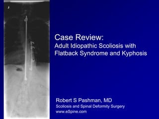
- 1. Case Review: Adult Idiopathic Scoliosis with Flatback Syndrome and Kyphosis Robert S Pashman, MD Scoliosis and Spinal Deformity Surgery www.eSpine.com
- 2. Patient History 40-year-old male Status post posterior fusion with Harrinton Rod instrumentation for adolescent idiopathic scoliosis at age 14. Increasing low back pain, radiating down both legs The patient has failed conservative therapy. On CT scan the patient has solid union from T2 down to T12, but has subadjacent severe spinal stenosis, multiple level foraminal stenosis, and clear flat back decompensation, with approximately 5 cm of forward decompensation on standing upright 36 x 14 scoliosis x-rays. The patient has multiple co-morbidities, including sleep apnea and brittle diabetes.
- 3. Pre-op X-rays The patient had distraction rod instrumentation, with compression rod over the convexity. He now has a thoracolumbar subadjacent flat back syndrome and severe stenosis on the moveable spine, with multiple level foraminal stenosis and degenerative disk disease due to stress riser and flat back syndrome.
- 4. Indications for Surgery Adult idiopathic scoliosis. Flat back syndrome, status post posterior instrumentation with iatrogenic distraction. Subadjacent kyphosis with critical spinal stenosis, thoracolumbar spine, status post posterior instrumented fusion for adolescent idiopathic scoliosis. Neuroforaminal stenosis with multiple level radiculopathy, lumbar spine, status post posterior instrumented fusion for adolescent idiopathic scoliosis. Failure to improve with conservative therapy. Severe low back pain. Retained hardware (Harrington rod instrumentation).
- 5. Surgical Strategy 1. Thoracic 2 to sacral pelvic fusion. This is a 16 level fusion for the above diagnoses, using a 1/4-inch stainless steel rod screw construct, with pelvic instrumentation. 2. Four level osteotomy, Smith-Peterson osteotomy, for correction of flat back deformity status post Harrington rod instrumentation at the thoracolumbar junction. 3. Re-exploration, decompression, laminectomy, T12-L1, for removal of adjacent segment compression status post Harrington rod instrumentation. 4. Multiple level osteotomy, lumbar spine, for recreation of lumbar lordosis, with radical facetectomy and Smith-Petersen osteotomy, L1- 2, 2-3, 3-4. 5. Posterior spinal fusion using locally harvested autogenous bone from fusion mass decompression with recombinant human bone morphogeneic protein, lumbar spine. 6. Removal of retained hardware (Harrington rod instrumentation, distraction rod, and compression rod instrumentation), thoracic spine. 7. Intraoperative somatosensory evoked potentials and motor evoked potential management. 8. Intraoperative fluoroscopy. 9. Interlaminar laminotomies, mesial facetectomies, lateral recess release for radiculopathy, L1-2, 2-3, 3-4, 4-5, and 5-1, under loupe magnification and high illumination fiberoptic light.
- 6. Post-Op Films The patient is doing very well post-operatively. His x-rays look good.