Case Review: 42 year old woman with Grade 3 Isthmic Spondylolisthesis
•
2 likes•2,812 views

A 42 year old very athletic woman presented with Grade 3 Isthmic Spondylolisthesis. She had a long history of back pain. Dr. Pashman performed an anterior and posterior spinal fusion.

Recommended
Recommended
More Related Content
What's hot
What's hot (20)
Viewers also liked
Viewers also liked (20)
Similar to Case Review: 42 year old woman with Grade 3 Isthmic Spondylolisthesis
Similar to Case Review: 42 year old woman with Grade 3 Isthmic Spondylolisthesis (20)
More from Robert Pashman
More from Robert Pashman (13)
Recently uploaded
Recently uploaded (20)
Case Review: 42 year old woman with Grade 3 Isthmic Spondylolisthesis
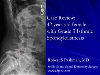
- 1. Case Review: 42 year old female with Grade 3 Isthmic Spondylolisthesis Robert S Pashman, MD Scoliosis and Spinal Deformity Surgery www.eSpine.com
- 2. Patient History 42-year-old female who is quite athletic. The patient was a competitive swimmer whose primary events were breaststroke and butterfly, both hyperextension events. The patient had some low back pain when she was a child, and she had this investigated, but in the intervening time she really had no problem until recently. Low back pain On physical examination, she has significant sacral promontory. The patient has significant rotation of the pelvis consistent with grade 3 Isthmic Spondylolisthesis. She has hamstring tightening and definite weakness of the dorsiflexors and extensor hallucis longus of left leg, paresthesias and numbness in the L5 distribution of the legs bilaterally.
- 3. Pre-op X-rays Grade 3 Isthmic Spondylolisthesis with significant slip angle degeneration, retrolisthesis of L4 on L5, bilateral pars fractures, compression of the L5 nerve root, lateral recess stenosis at L4-5, and significant slip angle and junctional lumbar kyphosis.
- 4. Flexion and Extension X-rays
- 5. Indications for Surgery 1. Grade 3 Isthmic Spondylolisthesis, L5-S1. 2. Degenerative disk disease, L4-5. 3. Evolving motor sensory deficit with weakness of extensor hallucis longus, tibialis anterior bilaterally indicating L5 nerve root crush. 4. Increasing low back pain due to significant progressive isthmic spondylolisthesis and hyperlordosis, lumbar spine. 5. Bilateral pars fractures pars interarticularis, L5.
- 6. Surgical Strategy Abdominal retroperitoneal approach to the lumbosacral spine. Subtotal vertebrectomy, L5, with removal of L5 overhang constituting greater than 1/3 of vertebra for entrance and reduction of spondylolisthesis, L5-S1. Radical diskectomy, L5-S1, including epidural decompression. Interbody fusion with 8 x small millimeter grafts with autogenous vertebrectomy bone centrally. Anterior screw fixation of fully threaded screw over a washer, L5- S1. Spondylolisthesis reduction. Intraoperative fluoroscopic interpretation.
- 7. Surgical Strategy – Part two Segmental spinal instrumentation L4 to S1 using the Allez Laguna titanium pedicle screw/rod construct, 5.5 titanium. Complete laminectomy Gill fragment of L5 lamina under loupe magnification and microscope. Neuroforaminotomy with removal of bilateral pars interarticularis callus, L5-S1, under the microscope. Posterolateral and posterior fusion, L4 to S1, using locally harvested autogenous bone, including laminectomy and anterior vertebrectomy bone. Intraoperative fluoroscopy management. Spondylolisthesis reduction at L-S1. Intraoperative O-arm neuronavigation and interpretation.
- 9. Pre-Op/Post-op Comparison The slip angle was reduced from a Grade 3 to a Grade 1