Case Review #17: 63 year old female with Denovo Scoliosis
•
1 like•740 views

63 year old female with Adult Idiopathic Scoliosis, Spondylolisthesis, and facet screws at L4-L5. Dr. Pashman treated the patient with a posterior spinal fusion from T10 to Pelvis. KIM/SRP Classification 2

Recommended
Recommended
More Related Content
What's hot
What's hot (20)
Viewers also liked
Viewers also liked (19)
Similar to Case Review #17: 63 year old female with Denovo Scoliosis
Similar to Case Review #17: 63 year old female with Denovo Scoliosis (20)
More from Robert Pashman
More from Robert Pashman (11)
Recently uploaded
Recently uploaded (20)
Case Review #17: 63 year old female with Denovo Scoliosis
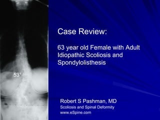
- 1. Case Review: 63 year old Female with Adult Idiopathic Scoliosis and Spondylolisthesis 53° Robert S Pashman, MD Scoliosis and Spinal Deformity www.eSpine.com
- 2. Patient History 63-year-old female Presented with failure to thrive. She cannot ambulate due to severe low back pain and radiculopathy. Status post attempted posterior instrumented fusion of L4 to S1 using facet screws Spondylolisthesis slippage. Chronic pain related to collapsing scoliosis proximally.
- 3. Pre-op X-rays Status post attempted posterior instrumented fusion of L4 to S1 using facet screws 53°
- 4. Indications for Surgery 1. Adult idiopathic scoliosis. 2. De novo scoliosis. 3. Status post posterior instrumented fusion for degenerative spondylolisthesis at 4-5. 4. L4 to S1 status post instrumented fusion for low back pain and spondylolisthesis. 5. Status post laminectomy, L4 to S1, for spinal stenosis. 6. Now with collapsing frontal and sagittal plane deformity and unremitting pain. 7. Multiple co morbidities.
- 5. Surgical Strategy 1. Segmental spinal instrumentation, T10 to the sacral pelvis, using CD Legacy 5.5 stainless steel screw-rod construct. 2. Re-exploration and repeat laminectomy, L3 to S1, using microscope and high power illumination. 3. Interlaminar decompression, L1 to L3 bilaterally, using the microscope and high intensity illumination for lateral recess stenosis and clearance of pseudarthrosis at L3-4 and L4-5. 4. Four level Smith-Petersen osteotomies for mobilization of the spine. 5. Repair of pseudarthrosis at L3-4 and L4-5. 6. Posterior spinal fusion with combination of locally harvested autogenous and rh-BMP graft, T10 to the sacral pelvis. 7. Intraoperative somatosensory and motor evoked potentials. 8. Intraoperative fluoroscopy. 9. Removal of retained hardware facet screws.
- 6. Findings during surgery The fusion mass of L5-S1 was solid but, at L4-5, there was a clear pseudarthrosis. The facet screws were intact, but not holding onto anything, as if maybe there was a pars fracture at L4-5 that went on to non-healing or went on to stress fracture. The adjacent segment at L3-4 was chronically degenerative and, as the patient tried to stand up and extend, the adjacent segment no doubt caused critical spinal stenosis with collapse and pseudarthrosis with critical compression of the exiting L3, L4 and L5 nerve roots bilaterally. The patient was stuck in partial flat back lumbar kyphosis from the previous spondylolisthesis attempted fusion, and this was necessary to be made up by multiple level Smith-Petersen osteotomies and takedown of the fusion mass so that hyperextension could be induced with correction of the coronal plane. The patient's bone was generally of moderate quality. There was significant dense scar over the laminectomy, a site that needed to be decompressed. This took a significant amount of time and effort to dig out the scar from around the edges of the previous laminectomy and to extricate the L5 and L4 nerve roots caught in the pseudarthrosis and underneath the fusion mass. There were no other abnormalities. There was no puss or sign of infection or fluid.
- 7. Post-Op Films The patient is completely relieved of her lower extremity symptoms and her back pain. Her x-rays look perfect with excellent sagittal and coronal plane balance.