1. Technical Support Questionnaire – IHC/ IF/ ICC
Name: CORO1B Antibody
Catalog #: NB100-61040
Lot Number: A1
PO/Order Number: 20120629001
Please upload an image of
your staining by clicking on
the center of the box.
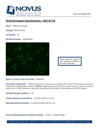
Species, Tissues or Cell Lines Tested: HEK293T
Test Sample Preparation: HEK293T cells were cultured on coverslip at 37°C under 5% CO2 and maintained in
Dulbecco’s modified Eagle’s medium (DMEM) supplemented with 10% fetal bovine serum, 1 mM non-essential
amino acids, 2 mM L-glutamine, penicillin-streptomycin (100 mg/ml), and amphotericin B (0.25 mg/ml).
Antibody Storage Conditions: 4℃
Fixation Solution and Conditions: 4% PFA at RT for 15 mins
Blocking Solution & Duration: 3% BSA/1X PBS at RT for 1 hr
Primary Antibody Diluent and Dilutions Tested: 1/100 in 1% BSA/1X PBS
2. Primary Antibody Incubation Time and Temperature: 4℃ overnight
Wash Solution Composition, Repetitions & Times: 0.5% Tween in 1X PBS three times, each for 10 minutes
Secondary Antibody Manufacturer, Host Species, Dilution, & Diluent: 1/300 in 1% BSA/1X PBS, Alexa Fluor
488 Goat Anti-Rabbit IgG (H+L) (molecular probes)
Secondary Antibody Incubation Time & Temperature: RT for 1 hr
Wash Solution Composition, Repetitions, & Times: 0.5% Tween in 1X PBS three times, each for 10 minutes
Detection System, Procedure & Development Time: Alexa Fluor 488 Goat Anti-Rabbit IgG (H+L) (molecular
probes)
Describe staining seen with this antibody: The major signal with this NOVUS CORO1B antibody is located in
nuclei.
Controls:
Observations: According to the literature, Coronin 1B should be mainly localized to the cytoplasm. However,
the major signal with this NOVUS CORO1B antibody is located in nuclei.
Ref 1: Coronin 1B Antagonizes Cortactin and Remodels Arp2/3-Containing Actin Branches in Lamellipodia.
Cell 134, 828–842, September 5, 2008.
Ref 2: Coronin 1B Coordinates Arp2/3 Complex and Cofilin Activities at the Leading Edge. Cell 128, 915–929,
March 9, 2007.