2. Calcium
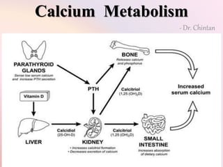
Extracellular calcium ion concentration is determined by,
interplay of calcium absorption from the intestine, renal
excretion of calcium, and bone uptake and release of calcium,
regulated by the hormones - vitamin D, parathyroid hormone
(PTH), and calcitonin
Phosphate homeostasis closely associated with Calcium
N Ca: 9 to 11 mg/dl
Contraction of skeletal, cardiac, and smooth muscles; blood
clotting; and transmission of nerve impulses, bone & teeth,
action of hormones, release of hormones
3. Calcium
Excitable cells, such as neurons, are very sensitive to changes
in calcium ion concentrations
Hypercalcemia cause progressive depression of the nervous
system - Hypocalcaemia cause the nervous system to become
more excited
only about 0.1 % of the total body calcium is in ECF,
about 1 % is in the cells,
the rest is stored in bones
the bones can serve as large reservoirs, releasing calcium
when extracellular fluid concentration decreases and storing
excess calcium
85 % of the body’s phosphate - bones, 15 % - cells, < 1 % - ECF
4.
5. Phosphate
Phosphate is found in ATP, DNA, RNA, cAMP, 2,3-DPG, many
proteins. Phosphorylation and dephosphorylation of proteins are
involved in the regulation of cell function – bone - buffer
Inorganic phosphate in the plasma is mainly in two forms: HPO4-
(1.05 mmol/L) and H2PO4- (0.26 mmol/L)
when the pH of the ECF becomes more acidic, there is a relative
increase in H2PO4- and a decrease in HPO4-, whereas the opposite
occurs when the ECF becomes alkaline
Decreasing the level of phosphate in the ECF from far below normal
does not cause major immediate effects on the body. In contrast,
even slight increases or decreases of calcium ion in the ECF can
cause extreme immediate physiologic effects.
chronic hypocalcemia or hypophosphatemia greatly decreases bone
mineralization
6. Hypo/Hyper calcemia
When the ECF Ca falls below normal, the nervous system
becomes progressively more excitable, because this
causes increased neuronal membrane permeability to Na
ions, allowing easy initiation of action potentials
Tetany – carpopedal spasm in hand - occasionally seizures
- 6 mg/dl
Hypercalcemia Depresses Nervous System and Muscle
Activity – shortened QT interval - loss of appetite and
constipation – above 12 mg/dl
9. Calcium Balance
1. Neutral
– normal healthy adults
– daily intake & excretion same
– bone entry & exit same
2. Positive
– growing children
– intestinal absorption > excretion
– bone entry > bone exit
3. Negative
– pregnant & lactating women
- intestinal absorption < excretion
– bone entry < bone exit
10. Bone Function
Protective – Rib cage, Skull
Mechanical – Support, Attachment
Body Movements – leverage effect
Metabolic – Ca, phosphate homeostasis
Hematopoietic – Blood cells from bone marrow
11. Bone Structure
Bone in children and adults is of two types: compact or cortical
bone, which makes up the outer layer of most bones and
accounts for 80% of the bone in the body;
and trabecular or spongy bones inside the cortical bone, which
make up the remaining 20% of bone in the body.
In compact bone - bone cells lie in lacunae - nutrients are
provided via Haversian canals, which contain blood vessels.
Around each Haversian canal, collagen is arranged in concentric
layers, forming cylinders called osteons or Haversian systems
Trabecular bone is made up of spicules or plates - many cells
sitting on the surface of the plates - Nutrients diffuse from
bone ECF into the trabeculae
12. Bone Physiology
Bone is composed of a tough organic matrix that is greatly
strengthened by deposits of calcium salts.
Average compact bone contains by weight about 30 % matrix
and 70 % salts. Newly formed bone have a higher % of matrix
in relation to salts.
Organic Matrix – 90 to 95 % collagen fibers - ground substance
(ECF plus proteoglycans, especially chondroitin sulfate and
hyaluronic acid)
Bone Salts - calcium and phosphate – major crystalline salt –
hydroxyapatite - Ca10 (PO4)6(OH)2
Magnesium, sodium, potassium, and carbonate – conjugated
to the hydroxyapatite crystals - osteogenic sarcoma
13. Bone Physiology
The concentrations of calcium and phosphate ions in ECF are
greater than those required to cause precipitation of
hydroxyapatite.
Inhibitors are present in almost all tissues of the body as well
as in plasma to prevent such precipitation – pyrophosphate
Bone Calcification - secretion of collagen molecules
(monomers) and ground substance by osteoblasts – collagen
monomers polymerize rapidly to form collagen fibers; the
resultant tissue becomes osteoid, a cartilage-like material
Dormant osteoblast - osteocytes
14. Bone Calcification
Within a few days after the osteoid is formed, calcium salts
begin to precipitate on the surfaces of the collagen fibers –
hydroxyapatite crystals
The initial calcium salts to be deposited are not hydroxyapatite
crystals but amorphous compounds - these amorphous salts
can be absorbed rapidly when there is need for extra Ca in the
ECF
Precipitation of Calcium in Nonosseous Tissues Under
Abnormal Conditions - they precipitate in arterial walls in the
condition called arteriosclerosis and cause the arteries to
become bonelike tubes
calcium salts frequently deposit in degenerating tissues or in
old blood clots - the inhibitor factors that normally prevent
deposition of calcium salts disappear from the tissues
15. Calcium Exchange
the bone contains a type of exchangeable calcium that
is always in equilibrium with the calcium ions in the
ECF
0.4 to 1 % of the total bone calcium
readily mobilizable salt such as CaHPO4
rapid buffering mechanism to keep the calcium ion
concentration in the extracellular fluids from rising to
excessive levels or falling to very low levels
16. Remodeling of Bone
Bone is continually being deposited by osteoblasts,
and it is continually being absorbed where
osteoclasts
Osteoblasts are found on the outer surfaces of the
bones and in the bone cavities
Osteoclasts – large phagocytic, multinucleated cells
– derivatives of monocytes - active on less than 1
per cent of the bone surfaces
17.
18. Remodeling of Bone
The osteoclasts send out villus-like projections toward the
bone - (1) proteolytic enzymes, released from the
lysosomes of the osteoclasts, (2) several acids, including
citric acid and lactic acid released from the mitochondria
and secretory vesicles
enzymes digest or dissolve the organic matrix of the bone,
and the acids cause solution of the bone salts -
phagocytosis of minute particles of bone matrix and
crystals
19.
20. Remodeling of Bone
the rates of bone deposition and absorption are equal - total
mass constant – growing bone exception
Osteoclasts eats away at the bone for about 3 weeks, creating
a tunnel that ranges in diameter from 0.2 to 1 millimeter - the
osteoclasts disappear and the tunnel is invaded by osteoblasts
- new bone begins to develop - Bone deposition then continues
for several months
The new bone laid down in successive layers of concentric
circles (lamellae) on the inner surfaces of the cavity until the
tunnel is filled. Deposition of new bone stops when the bone
begins to invade on the blood vessels supplying the area.
The canal through which these vessels run, called the
haversian canal, is all that remains of the original cavity -
osteon
21.
22.
23. Remodeling of Bone
bone ordinarily adjusts its strength in proportion to the
degree of bone stress - bones thicken when subjected to
heavy loads
the shape of the bone can be rearranged for proper
support of mechanical forces by deposition and absorption
of bone in accordance with stress patterns
old bone becomes relatively brittle and weak, new organic
matrix is needed as the old organic matrix degenerates -
the normal toughness of bone is maintained
Fragile bones in children
24. Remodeling of Bone
the bones of athletes become considerably heavier than
those of nonathletes
if a person has one leg in a cast but continues to walk on
the opposite leg, the bone of the leg in the cast becomes
thin and as much as 30 per cent decalcified within a few
weeks, whereas the opposite bone remains thick and
normally calcified
Fracture - massive numbers of new osteoblasts are formed
almost immediately from osteoprogenitor cells – callus –
bone stress to accelerate the rate of # healing
25. Vitamin D
Vitamin D has a potent effect to increase calcium
absorption from the intestinal tract
vitamin D must first be converted in the liver and the
kidneys to the final active product, 1,25-
dihydroxycholecalciferol - 1,25(OH)2D3
Vitamin D3 – cholecalciferol is formed in the skin as a
result of irradiation of 7-dehydrocholesterol, a substance
normally in the skin, by ultraviolet rays from the sun
Food – cholecalciferol
26. Vitamin D
The first step in the activation of cholecalciferol is to
convert it to 25-hydroxycholecalciferol in the liver. The 25-
hydroxycholecalciferol has a feedback inhibitory effect on
the conversion reactions
the intake of vitamin D3 can increase many times and yet
the concentration of 25-hydroxycholecalciferol remains
nearly normal - prevents excessive action of vitamin D
conserves the vitamin D stored in the liver for future use.
Once it is converted, it persists in the body for only a few
weeks, whereas in the vitamin D form, it can be stored in
the liver for many months.
27.
28. Vitamin D
the conversion in the proximal tubules of the kidneys of 25-
hydroxy cholecalciferol to 1,25 - dihydroxy cholecalciferol -
most active form of vitamin D
This conversion requires PTH
calcium ion itself has a slight effect in preventing the
conversion
calcium concentrations Below 9 mg/100 ml - PTH promotes
the conversion in the kidneys.
At higher calcium concentrations, when PTH is suppressed, the
25-hydroxycholecalciferol is converted to
24,25–dihydroxycholecalciferol — that has almost no vitamin D
effect
29. When the plasma calcium concentration is too high, the
formation of 1,25-dihydroxycholecalciferol is greatly
depressed – decreases the absorption of calcium from the
intestines, the bones, and the renal tubules
30.
31. Actions of Vitamin D
effects on the intestines, kidneys, and bones that increase
absorption of calcium and phosphate into the ECF
Increasing formation of a calcium-binding protein in the
intestinal epithelial at the brush border of these cells to
transport calcium into the cell cytoplasm, and the calcium then
moves through the basolateral membrane of the cell by
facilitated diffusion
protein remains in the cells for several weeks after the 1,25-
dihydroxycholecalciferol has been removed from the body, thus
causing a prolonged effect on calcium absorption
(1) a calcium-stimulated ATPase in the brush border of the
epithelial cells and
(2) an alkaline phosphatase in the epithelial cells
32. Actions of Vitamin D
Promotes Phosphate Absorption by the Intestines
increases calcium and phosphate absorption by the
epithelial cells of the renal tubules
extreme quantities of vitamin D causes absorption of
bone. In the absence of vitamin D, the effect of PTH in
causing bone absorption is greatly reduced or even
prevented
smaller quantities promotes bone calcification by
increasing calcium and phosphate absorption from the
intestines - enhances the mineralization of bone
33. Parathyroid Gland
4 parathyroid glands in humans; they are located immediately
behind the thyroid gland
difficult to locate during thyroid operations because they often
look like just another lobule of the thyroid gland - total or
subtotal thyroidectomy frequently resulted in removal of the
parathyroid glands
Removal of half the parathyroid glands usually causes no
major physiologic abnormalities - removal of three of the four
normal glands causes transient hypoparathyroidism
remaining parathyroid tissue - hypertrophy
34.
35. Parathyroid Hormone
Ribosomes – preprohormone - 110 amino acids
Prohormone - 90 amino acids
Hormone - 84 amino acids by ER and Golgi apparatus, and finally is
packaged in secretory granules in the cytoplasm of the cells -
molecular weight of about 9500
36. PTH - Bone
1st rapid phase (Osteolysis) that begins in minutes and increases
progressively for several hours - activation of osteocytes to promote
calcium and phosphate absorption
PTH causes removal of bone salts from two areas in the bone: (1)
from the bone matrix in the surrounding area of the osteocytes lying
within the bone
(2) in the vicinity of the osteoblasts along the bone surface
the osteoblasts and osteocytes form a system of interconnected
cells that spreads all through the bone and over all the bone
surfaces
long, transparent processes extend from osteocyte to osteocyte
throughout the bone structure, and these processes also connect
with the surface osteocytes and osteoblasts - osteocytic membrane
system - separates the bone from ECF
37. PTH - Bone
Between the osteocytic membrane and the bone is a small amount
of bone fluid
the osteocytic membrane pumps Ca ions from the bone fluid into
the ECF, creating a Ca ion concentration in the bone fluid only 1/3rd
that in the ECF
When the osteocytic pump becomes excessively activated, the bone
fluid Ca concentration falls even lower - calcium phosphate salts are
then absorbed from the bone – Osteolysis - occurs without
absorption of the bone’s fibrous and gel matrix
When the pump is inactivated, the bone fluid Ca concentration rises
to a higher level, and calcium phosphate salts are redeposited in the
matrix
38. PTH - Bone
cell membranes of both the osteoblasts and the osteocytes
have receptor proteins for binding PTH - activate the calcium
pump strongly, thereby causing rapid removal of calcium
phosphate salts
Increases the Ca permeability of the bone fluid side of the
osteocytic membrane, thus allowing calcium ions to diffuse
into the membrane cells from the bone fluid
The Ca pump on the other side of the cell membrane transfers
the calcium ions into the ECF
Actual bone – bone fluid – osteocytic membrane - ECF
39. PTH - Bone
2nd slower phase, requiring several days or even weeks to
become fully developed - proliferation of the osteoclasts,
followed by greatly increased osteoclastic reabsorption
osteoclasts do not themselves have membrane receptor
proteins for PTH
the activated osteoblasts and osteocytes send a secondary but
unknown “signal” to the osteoclasts, causing them to set about
their usual task of gulping up the bone over a period of weeks
or months
(1) immediate activation of the preformed osteoclasts
(2) formation of new osteoclasts
40. PTH - Bone
After a few months of excess PTH, osteoclastic resorption of bone
can lead to weakened bones and secondary stimulation of the
osteoblasts that attempt to correct the weakened state.
the late effect is actually to enhance both osteoblastic and
osteoclastic activity
Bone contains such great amounts of Ca in comparison with the
total amount in all the ECF (1000 times) that even when PTH causes
a great rise in Ca concentration in the fluids, it is impossible to
determine any immediate effect on the bones
Prolonged administration or secretion of PTH—over a period of
many months or years—finally results in very evident absorption in
all the bones and even development of large cavities filled with
large, multinucleated osteoclasts
41. PTH - Kidneys
Administration of PTH causes rapid loss of phosphate in the
urine owing to the effect of the hormone to diminish proximal
tubular reabsorption of phosphate ions
PTH increases renal tubular reabsorption of Ca - in the late
distal tubules, the collecting tubules, the early collecting ducts,
and possibly the ascending loop of Henle to a lesser extent
It increases the rate of reabsorption of Mg ions and H ions
it decreases the reabsorption of Na, K and amino acid
No PTH - continual loss of Ca into the urine would eventually
deplete both the ECF and the bones
42. PTH - Intestine
PTH greatly enhances both calcium and phosphate absorption from
the intestines by increasing the formation in the kidneys of 1,25-
dihydroxycholecalciferol from vitamin D
MOA – AC – cAMP - Within a few minutes after PTH administration,
the concentration of cAMP increases in the osteocytes, osteoclasts,
and other target cells
cAMP in turn is probably responsible for such functions as
osteoclastic secretion of enzymes and acids to cause bone
reabsorption and formation of 1,25- dihydroxycholecalciferol in the
kidneys
A local hormone, parathyroid hormone-related protein (PTHrP),
acts on one of the PTH receptors and is important in skeletal
development in utero
43. Control of PTH Secretion by Ca
Even the slightest decrease in Ca ion concentration in the ECF
causes the parathyroid glands to increase their rate of
secretion within minutes
parathyroid glands become greatly enlarged in rickets,
pregnancy, lactation
Reduced size of the parathyroid glands
(1) excess quantities of calcium in the diet,
(2) increased vitamin D in the diet,
(3) Bone absorption caused by disuse of the bones
44. Calcitonin
Calcitonin, a peptide hormone secreted by the thyroid gland,
tends to decrease plasma Ca concentration and, in general, has
effects opposite to those of PTH
Parafollicular cells, or C cells, lying in the interstitial fluid
between the follicles of the thyroid gland
32-amino acid peptide with a molecular weight of about 3400
The primary stimulus for calcitonin secretion is increased
plasma Ca ion concentration
calcitonin decreases blood Ca ion concentration rapidly,
beginning within minutes after injection of the calcitonin
45. Calcitonin
1. The immediate effect is to decrease the absorptive activities of
the osteoclasts and possibly the osteolytic effect of the osteocytic
membrane throughout the bone
Shifting the balance in favor of deposition of calcium in the
exchangeable bone calcium salts - especially significant in young
because of the rapid interchange of absorbed and deposited Ca
2. The second and more prolonged effect of calcitonin is to decrease
the formation of new osteoclasts. Also, because osteoclastic
resorption of bone leads secondarily to osteoblastic activity -
decreased numbers of osteoblasts
the effect on plasma calcium is mainly a transient one, lasting for a
few hours to a few days at most – Kidney, intestine ???
46. Calcitonin
Calcitonin Has a Weak Effect on Plasma Calcium
Concentration
in the Adult Human
Calcitonin - ↓ Ca - ↑ PTH
Thyroid removed – no calcitonin – no effect
The effect of calcitonin in children is much greater because
bone remodeling occurs rapidly in children, with
absorption and deposition of calcium as great as 5 grams
or more per day
47. Control of Ca ion
Buffer Function of the Exchangeable Calcium in Bones — the 1st Line
of Defense – rapid reaction
amorphous calcium phosphate compounds, probably mainly
CaHPO4 or some similar compound loosely bound in the bone and
in reversible equilibrium with the Ca and phosphate ions in the ECF
Because of the ease of deposition of these exchangeable salts and
their ease of resolubility, an increase in the concentrations of ECF
Ca and phosphate ions above normal causes immediate deposition
of exchangeable salt – vice versa – more blood flow
The mitochondria of many of the tissues of the body, especially of
the liver and intestine, contain a reasonable amount of
exchangeable Ca that provides an additional buffer system
48. Control of Ca ion
Hormonal Control of Calcium Ion Concentration — the 2nd
Line of Defense
Within 3 to 5 minutes after an acute increase in the calcium ion
concentration, the rate of PTH secretion Decreases – calcitonin
increases
In young animals and possibly in young children – the
calcitonin causes rapid deposition of calcium in the bones
Young children – hormonal control – 1st line
In prolonged calcium excess or prolonged calcium Deficiency
– only PTH important
49. Control of Ca ion
When a person has a continuing deficiency of calcium in the
diet, PTH often can stimulate enough calcium absorption from
the bones to maintain a normal plasma calcium ion
concentration for 1 year or more
eventually, the bones will run out of calcium
when the bone reservoir either runs out of calcium or,
oppositely, becomes saturated with calcium, the long-term
control of extracellular calcium ion concentration resides
almost entirely in the roles of PTH and vitamin D in controlling
calcium absorption from the gut and calcium excretion in the
urine
50. Applied - Hypoparathyroidism
When the parathyroid glands do not secrete sufficient PTH,
the osteocytic reabsorption of exchangeable calcium
decreases and the osteoclasts become inactive
As a result, calcium reabsorption from the bones is so
depressed that the level of calcium in the body fluids
decreases. Yet, because calcium and phosphates are not being
absorbed from the bone, the bone usually remains strong
the calcium level in the blood falls from the normal of 9.4
mg/dl to 6 to 7 mg/dl within 2 to 3 days, and the blood
phosphate concentration may double
Hypocalcemia – Tetany – laryngeal muscle spasm – respiratory
obstruction - death
51. Rx - Hypoparathyroidism
PTH is occasionally used for treating hypoparathyroidism -
because of the expense of this hormone - because its effect
lasts for a few hours at most - because the tendency of the
body to develop antibodies against it
the administration of extremely large quantities of vitamin D,
to as high as 100,000 units per day, along with intake of 1 to 2
grams of calcium
1,25-dihydroxycholecalciferol - much more potent and much
more rapid action
52. Primary Hyperparathyroidism
abnormality of the parathyroid glands causes inappropriate,
excess PTH
tumor of one of the parathyroid glands - more frequently in
women - pregnancy and lactation stimulate the parathyroid
glands
extreme osteoclastic activity in the bones – Hypercalcemia
Increase renal excretion of phosphate – hypophosphatemia
Mild - new bone can be deposited rapidly enough to
compensate
Severe – cant compensate – broken bone
53. Primary Hyperparathyroidism
Radiographs of the bone show extensive decalcification
and, occasionally, large punched-out cystic areas of the
bone that are filled with osteoclasts in the form of giant
cell osteoclast tumors
Multiple fractures of the weakened bones can result from only
slight trauma, especially where cysts develop. The cystic bone
disease of hyperparathyroidism is called osteitis fibrosa cystica
Osteoblastic activity in the bones also increases – secretion of
large quantities of alkaline phosphatase - diagnostic finding
54. Hypercalcemia
depression of the central and peripheral nervous systems,
muscle weakness,
constipation,
abdominal pain,
peptic ulcer,
lack of appetite,
Shortened QT interval
depressed relaxation of the heart during diastole
Systolic arrest
55. Parathyroid Poisoning
Metastatic Calcification
ECF phosphate concentration rises markedly instead of falling -
the kidneys cannot excrete rapidly enough all the phosphate
being absorbed from the bone.
the calcium and phosphate in the body fluids become greatly
supersaturated, so that calcium phosphate (CaHPO4) crystals
begin to deposit
alveoli of the lungs, the tubules of the kidneys, the thyroid
gland, the acid-producing area of the stomach mucosa, and the
walls of the arteries
Ca above 17 mg/dl + phosphate = Death
56. Kidney Stones
Most patients with mild hyperparathyroidism show few signs of
bone disease and few general abnormalities as a result of elevated
calcium, but they do have an extreme tendency to form kidney
stones
excess calcium and phosphate absorbed from the intestines or
mobilized from the bones in hyperparathyroidism must eventually
be excreted by the kidneys
crystals of calcium phosphate tend to precipitate in the kidney,
forming calcium phosphate stones - calcium oxalate stones develop
because even normal levels of oxalate cause calcium precipitation at
high calcium levels
solubility of most renal stones is slight in alkaline media - tendency
greater in alkaline urine - acidotic diets and acidic drugs for Rx