SlideShare a Scribd company logo
1 of 4
Download to read offline
THE PHYSICS TEACHER x Vol. 39, February 200118
Gorazd Planinsic, Department of Physics, University of Ljubljana, Jadranska 19, 1000 Ljubljana,
Slovenia; gorazd.planinsic@fiz.uni-lj.si
Due to a growing interest in biology
among science students, using demon-
stration experiments in physics classes that in-
volve observations of biological material or that
relate to plant, animal, or human physiology
can often raise student motivation. The experi-
ment described below involves observation of
microorganisms in a drop of pond water. An
explanation of the image formation in the
paraxial region is then given using geometrical
optics.
Beginning with the invention of the first mi-
croscope, humans have been fascinated by the
observation of microscopic life. In his spare
time, Dutch draper Antoni van Leeuwenhoek
(1632-1723) made his own single-lens micro-
scope using a small glass sphere. Keeping de-
Water-Drop Projector
tailed records of his observations, he made the
first drawing of a bacterium in 1683. Articles
describing construction of a simple variation of
Leeuwenhoek’s microscope (glass tubing micro-
scope) have appeared previously in TPT.1
However, it is easier still to build a single-lens
microscope by using a drop of clear water in-
stead of a glass lens. The small water drop
works as a spherical lens with a large magnifying
power.
Experiment
To collect your sample, fill a syringe with
water from a pond or large puddle with a lot of
decaying plants. If you live at the coast, use sea-
water. Try to catch some very small animals (0.2
mm to 0.5 mm) that move around in the water.
Many of them are found close to the bottom of
the pond.
Fix the syringe on a holder (Lego blocks or a
piece of wood works well) using adhesive tape as
shown in Fig. 1.
For a light source, fix a laser pointer on a
vertical positioner. An empty stick-deodorant
container will do, as shown in Fig. 1. To create
your lens, carefully push the syringe piston until
a water drop (about 2 mm in diameter) is
formed at the end of the nozzle.
Observation
Place the syringe holder on a table about two
meters away from a screen or white wall. Switch
on the laser pointer and adjust the beam to
point exactly through the middle of the water
drop and perpendicular to the screen. For fine
horizontal adjustment of the laser pointer, move
the deodorant container, and for vertical adjust-
ment use the built-in turn screw. With the right
adjustment of the laser, a bright spot extends in-
to a large round image on the screen about 2 m
Fig. 1. Water-drop projector: experiment setup.
19THE PHYSICS TEACHER x Vol. 39, February 2001
in diameter. Now, if your pond water is rich
enough with little animals, you should see their
magnified shadow images floating and moving
around on the screen. Small single-cell animals
like paramecium appear as dark spots surround-
ed with interference fringe contours. Some of
them exhibit movements, but no detailed struc-
ture can be recognized. Larger animals such as
mosquito larva (Anopheles species), cyclops (Cy-
clops strenuus), or water fleas (Daphnia species)
appear like real monsters on the screen (see Fig.
2). Here you can clearly identify individual
parts of their bodies and follow their move-
ments. In our case the images were projected on
the white wall and photographs were obtained
with a digital camera (Kodak DC260). You
may at first see nothing but the dark floating
spots surrounded by a few concentric circles
caused by light diffraction on small parts of de-
caying plants and single-cell protozoa. Be pa-
tient. The light attracts the little animals (a
green laser works best), and after some time they
swim down to the syringe nozzle and into the
water drop. You can also give them a little help
by dripping a few drops from the syringe.
How the Water-Drop Projector
Works
The drop at the end of the syringe, though
not a perfect sphere, can be treated as a small
spherical lens. The light beam that falls on the
drop refracts both times as it passes through the
water-air interface. Let’s follow the path of the
ray that enters the water drop just above an ob-
ject that floats in the water drop at a small dis-
tance x from the geometrical axis ( Fig. 3). The
ray will refract twice and reach the screen at the
distance y below the geometrical axis. The dis-
tance y is determined by the distance from the
drop to the screen d and the angle ␦, which can
be calculated using simple geometry:
␦ = ␣ – ЄRST
ЄRST = 2␤ – ␣
␦ = 2(␣ – ␤).
Using Snell's law,
Cyclops (Cyclops strenuus)
Mosquito larva (Anopheles species)
Water flea (Daphnia species)
Fig. 2. Photographs of projected images. The actual size of
each projected image is about 1 m x 1 m.
Author: are you certain of the taxonomic identity of this ani-
mal? If not, then perhaps “Cyclops (Cyclops species)”
would be a more appropriate caption for first photo
below.
THE PHYSICS TEACHER x Vol. 39, February 200120
ᎏ
s
s
i
i
n
n
␣
␤
ᎏ = ᎏ
n
n
0
ᎏ
(where for the water and air n =1.33 and n0 =
1.0, respectively), and with sin ␣ = x/r, the angle
␦ is given by the equation:
␦ = 2(arcsinᎏ
x
r
ᎏ – arcsin ᎏ
n
x
r
ᎏ).
For the rays close to the geometrical axes (parax-
ial region), all the angles in the calculation
above are very small; therefore, the expression
for ␦ can be simplified using the approximation
arcsin(z) Ϸ z:
␦ = 2
΂1 – ᎏ
n
1
ᎏ
΃.
The projected image on the screen is a magni-
fied shadow of the object (animal) with magni-
fication equal to
M = = ᎏ
dta
x
n␦
ᎏ Ϸ 2
΂1 – ᎏ
n
1
ᎏ
΃,
where the approximation (t an␦ Ϸ ␦) is valid in
the paraxial region. For the water drop 2 mm in
diameter, the shadow image on the screen 2 m
from the setup is about 1000 times larger than
the object. Note that in derivation we assumed
the object floats on the laser side of the drop. It
can be seen from Fig. 1 that the same shadow
d
ᎏ
r
y
ᎏ
x
x
ᎏ
r
height can be produced by a
smaller object placed on the
screen side of the drop (or
anywhere in between).
Clearly the magnification is
largest for the objects that
are floating in the screen
side of the drop. In this case
Mmax =
in the paraxial region. For
the parameters as given
above, the magnification
factor is 1985.
The results show that in
the paraxial region, the magnification depends
on the position of the object along the geomet-
rical axis but not on the object’s distance from
the axis. However, if the size of the object is
comparable to the size of the drop or the object
lies away from the geometrical axis, the angles
␣, ␤, and ␦ are not small any more. In this case,
the magnification is expressed using the exact
expression for the angle ␦:
M = ΄ᎏ
1
u
ᎏtan΂2(arsin u – arcsin ᎏ
n
u
ᎏ)΃΅,
where the distance of the incident ray from the
geometrical axis is measured in the units of wa-
ter drop radius (u = x/r) and can have any value
between 0 and 1. The magnification as the
function of parameter u is shown in Fig. 4 with
the set of parameters given earlier. We can de-
duce from the graph that the magnification is
reasonably constant within the region about
half radius from the geometrical axis. This gives
us a hint about the size of the drop for the given
laser-beam diameter if we want to produce
nondistorted shadow images.
The preceding derivation is based on the as-
sumption that the water drop is a perfect sphere.
Though the bottom part of the drop is spherical
to a good approximation, the upper part (where
the drop issues from the nozzle) is seriously dis-
torted. The light passing through this irregular-
ly curved surface causes a variety of patterns that
disturb our observation but are interesting sub-
d
ᎏ
r
n
ᎏ
2 – n
d
ᎏ
2r
Fig. 3. Optics of the water-drop projector.
21THE PHYSICS TEACHER x Vol. 39, February 2001
jects to explore themselves.2
However, there are
other ways to make a water drop lens that is
closer to a sphere than the one we suggest. For
example, wrap a copper wire (thickness of 0.2
mm or so) to make a small ring, then touch the
ring with the water drop hanging from the sy-
ringe. The surface tension will make the drop
jump onto the ring, where it will stay and form
an almost perfect sphere. Or simply touch the
drop from the syringe with a microscope glass
plate and you’ll get a nice plan-concave lens.
These alternatives can be used to observe the
small animals in the pond water, and the ob-
served shadow images may be even better than
those obtained with the hanging water drop
method. But the advantage of this method is
the simple construction and the fact that you
can have many animals inside the syringe. The
little animals attracted by the light swim down
into the drop by themselves. With the other
methods, getting the animals into the drop may
be difficult, and the observation time is limited
by the evaporation of the drop.
References
1. Wayne O. Williams, “Glass tubing microscope,”
Phys. Teach. 17, 204 (1979) .
2. J. Walker, “A drop of water becomes a gateway in-
to the world of catastrophe optics,” Sci. Am. 120
(1989).
Fig. 4. Water-drop magnification as the function of distance
from the optical axis.

More Related Content

What's hot (20)

RFID based Attendance System
RFID based Attendance SystemRFID based Attendance System
RFID based Attendance System
 
Study of Leach Protocol- A Review
Study of Leach Protocol- A ReviewStudy of Leach Protocol- A Review
Study of Leach Protocol- A Review
 
shashank soni sensors presentation
shashank soni   sensors presentationshashank soni   sensors presentation
shashank soni sensors presentation
 
RFID
RFIDRFID
RFID
 
Radar ppt
Radar pptRadar ppt
Radar ppt
 
Rfid security access control system
Rfid security access control systemRfid security access control system
Rfid security access control system
 
8.vsat
8.vsat8.vsat
8.vsat
 
Distance measurement using Ultrasonic sensor on Arduino Uno
Distance measurement using Ultrasonic sensor on Arduino UnoDistance measurement using Ultrasonic sensor on Arduino Uno
Distance measurement using Ultrasonic sensor on Arduino Uno
 
Radar Powerpoint
Radar PowerpointRadar Powerpoint
Radar Powerpoint
 
Smart Antenna
Smart AntennaSmart Antenna
Smart Antenna
 
Lecture 5
Lecture 5Lecture 5
Lecture 5
 
Micro Wave Band
Micro Wave Band Micro Wave Band
Micro Wave Band
 
RFID based access control ppt
RFID based access control pptRFID based access control ppt
RFID based access control ppt
 
radar
radarradar
radar
 
WSN-IEEE 802.15.4 -MAC Protocol
WSN-IEEE 802.15.4 -MAC ProtocolWSN-IEEE 802.15.4 -MAC Protocol
WSN-IEEE 802.15.4 -MAC Protocol
 
Distance Measurement by Ultrasonic Sensor
Distance Measurement by Ultrasonic SensorDistance Measurement by Ultrasonic Sensor
Distance Measurement by Ultrasonic Sensor
 
Land slide warning system
Land slide warning systemLand slide warning system
Land slide warning system
 
Radar
RadarRadar
Radar
 
Wireless Sensor Networks
Wireless Sensor NetworksWireless Sensor Networks
Wireless Sensor Networks
 
WSN IN IOT
WSN IN IOTWSN IN IOT
WSN IN IOT
 

Similar to Water drop projector

Phase contrast microscope
Phase contrast microscopePhase contrast microscope
Phase contrast microscopeRavi Kumar
 
1. Introduction to Microscopy.pdf
1.        Introduction to Microscopy.pdf1.        Introduction to Microscopy.pdf
1. Introduction to Microscopy.pdfMicrobiologyMicro
 
Ch 26 Light Refraction: Lenses and Optical Instruments
Ch 26 Light Refraction: Lenses and Optical InstrumentsCh 26 Light Refraction: Lenses and Optical Instruments
Ch 26 Light Refraction: Lenses and Optical InstrumentsScott Thomas
 
20131010 bigbangmanuscript
20131010 bigbangmanuscript20131010 bigbangmanuscript
20131010 bigbangmanuscriptHansOttoCarmesin
 
Solar limb darkening_function_from_baily_beads_observations
Solar limb darkening_function_from_baily_beads_observationsSolar limb darkening_function_from_baily_beads_observations
Solar limb darkening_function_from_baily_beads_observationsSérgio Sacani
 
Chapter 2 geometrical_optics_b
Chapter 2 geometrical_optics_bChapter 2 geometrical_optics_b
Chapter 2 geometrical_optics_bGabriel O'Brien
 
Riet ch 1 engineering physics optics main
Riet ch 1 engineering physics optics mainRiet ch 1 engineering physics optics main
Riet ch 1 engineering physics optics mainhasan ziauddin
 
To Study Principles of Microscopy: Light Microscope, Phase Contrast Microsco...
To Study Principles of Microscopy: Light Microscope, Phase Contrast  Microsco...To Study Principles of Microscopy: Light Microscope, Phase Contrast  Microsco...
To Study Principles of Microscopy: Light Microscope, Phase Contrast Microsco...Om Prakash
 
Periodic_Forcing.pdf
Periodic_Forcing.pdfPeriodic_Forcing.pdf
Periodic_Forcing.pdfluc faucheux
 
Refraction through a glass slab and the refractive index
Refraction through a glass slab and the refractive indexRefraction through a glass slab and the refractive index
Refraction through a glass slab and the refractive indexSharan Raj
 

Similar to Water drop projector (20)

Microscopy
Microscopy Microscopy
Microscopy
 
Telescopes
TelescopesTelescopes
Telescopes
 
Phase contrast microscope
Phase contrast microscopePhase contrast microscope
Phase contrast microscope
 
1. Introduction to Microscopy.pdf
1.        Introduction to Microscopy.pdf1.        Introduction to Microscopy.pdf
1. Introduction to Microscopy.pdf
 
Telescope
TelescopeTelescope
Telescope
 
optic
 optic optic
optic
 
Optic
OpticOptic
Optic
 
Optic
OpticOptic
Optic
 
1316 ditto[1]
1316 ditto[1]1316 ditto[1]
1316 ditto[1]
 
Ch 26 Light Refraction: Lenses and Optical Instruments
Ch 26 Light Refraction: Lenses and Optical InstrumentsCh 26 Light Refraction: Lenses and Optical Instruments
Ch 26 Light Refraction: Lenses and Optical Instruments
 
Microscope
Microscope Microscope
Microscope
 
Module No. 37
Module No. 37Module No. 37
Module No. 37
 
20131010 bigbangmanuscript
20131010 bigbangmanuscript20131010 bigbangmanuscript
20131010 bigbangmanuscript
 
Solar limb darkening_function_from_baily_beads_observations
Solar limb darkening_function_from_baily_beads_observationsSolar limb darkening_function_from_baily_beads_observations
Solar limb darkening_function_from_baily_beads_observations
 
Chapter 2 geometrical_optics_b
Chapter 2 geometrical_optics_bChapter 2 geometrical_optics_b
Chapter 2 geometrical_optics_b
 
Riet ch 1 engineering physics optics main
Riet ch 1 engineering physics optics mainRiet ch 1 engineering physics optics main
Riet ch 1 engineering physics optics main
 
To Study Principles of Microscopy: Light Microscope, Phase Contrast Microsco...
To Study Principles of Microscopy: Light Microscope, Phase Contrast  Microsco...To Study Principles of Microscopy: Light Microscope, Phase Contrast  Microsco...
To Study Principles of Microscopy: Light Microscope, Phase Contrast Microsco...
 
4.3
4.34.3
4.3
 
Periodic_Forcing.pdf
Periodic_Forcing.pdfPeriodic_Forcing.pdf
Periodic_Forcing.pdf
 
Refraction through a glass slab and the refractive index
Refraction through a glass slab and the refractive indexRefraction through a glass slab and the refractive index
Refraction through a glass slab and the refractive index
 

Recently uploaded

Is RISC-V ready for HPC workload? Maybe?
Is RISC-V ready for HPC workload? Maybe?Is RISC-V ready for HPC workload? Maybe?
Is RISC-V ready for HPC workload? Maybe?Patrick Diehl
 
Analytical Profile of Coleus Forskohlii | Forskolin .pdf
Analytical Profile of Coleus Forskohlii | Forskolin .pdfAnalytical Profile of Coleus Forskohlii | Forskolin .pdf
Analytical Profile of Coleus Forskohlii | Forskolin .pdfSwapnil Therkar
 
Natural Polymer Based Nanomaterials
Natural Polymer Based NanomaterialsNatural Polymer Based Nanomaterials
Natural Polymer Based NanomaterialsAArockiyaNisha
 
Artificial Intelligence In Microbiology by Dr. Prince C P
Artificial Intelligence In Microbiology by Dr. Prince C PArtificial Intelligence In Microbiology by Dr. Prince C P
Artificial Intelligence In Microbiology by Dr. Prince C PPRINCE C P
 
GFP in rDNA Technology (Biotechnology).pptx
GFP in rDNA Technology (Biotechnology).pptxGFP in rDNA Technology (Biotechnology).pptx
GFP in rDNA Technology (Biotechnology).pptxAleenaTreesaSaji
 
All-domain Anomaly Resolution Office U.S. Department of Defense (U) Case: “Eg...
All-domain Anomaly Resolution Office U.S. Department of Defense (U) Case: “Eg...All-domain Anomaly Resolution Office U.S. Department of Defense (U) Case: “Eg...
All-domain Anomaly Resolution Office U.S. Department of Defense (U) Case: “Eg...Sérgio Sacani
 
Recombination DNA Technology (Nucleic Acid Hybridization )
Recombination DNA Technology (Nucleic Acid Hybridization )Recombination DNA Technology (Nucleic Acid Hybridization )
Recombination DNA Technology (Nucleic Acid Hybridization )aarthirajkumar25
 
A relative description on Sonoporation.pdf
A relative description on Sonoporation.pdfA relative description on Sonoporation.pdf
A relative description on Sonoporation.pdfnehabiju2046
 
Disentangling the origin of chemical differences using GHOST
Disentangling the origin of chemical differences using GHOSTDisentangling the origin of chemical differences using GHOST
Disentangling the origin of chemical differences using GHOSTSérgio Sacani
 
Hubble Asteroid Hunter III. Physical properties of newly found asteroids
Hubble Asteroid Hunter III. Physical properties of newly found asteroidsHubble Asteroid Hunter III. Physical properties of newly found asteroids
Hubble Asteroid Hunter III. Physical properties of newly found asteroidsSérgio Sacani
 
Animal Communication- Auditory and Visual.pptx
Animal Communication- Auditory and Visual.pptxAnimal Communication- Auditory and Visual.pptx
Animal Communication- Auditory and Visual.pptxUmerFayaz5
 
Cultivation of KODO MILLET . made by Ghanshyam pptx
Cultivation of KODO MILLET . made by Ghanshyam pptxCultivation of KODO MILLET . made by Ghanshyam pptx
Cultivation of KODO MILLET . made by Ghanshyam pptxpradhanghanshyam7136
 
NAVSEA PEO USC - Unmanned & Small Combatants 26Oct23.pdf
NAVSEA PEO USC - Unmanned & Small Combatants 26Oct23.pdfNAVSEA PEO USC - Unmanned & Small Combatants 26Oct23.pdf
NAVSEA PEO USC - Unmanned & Small Combatants 26Oct23.pdfWadeK3
 
Call Girls in Munirka Delhi 💯Call Us 🔝9953322196🔝 💯Escort.
Call Girls in Munirka Delhi 💯Call Us 🔝9953322196🔝 💯Escort.Call Girls in Munirka Delhi 💯Call Us 🔝9953322196🔝 💯Escort.
Call Girls in Munirka Delhi 💯Call Us 🔝9953322196🔝 💯Escort.aasikanpl
 
Scheme-of-Work-Science-Stage-4 cambridge science.docx
Scheme-of-Work-Science-Stage-4 cambridge science.docxScheme-of-Work-Science-Stage-4 cambridge science.docx
Scheme-of-Work-Science-Stage-4 cambridge science.docxyaramohamed343013
 
Nightside clouds and disequilibrium chemistry on the hot Jupiter WASP-43b
Nightside clouds and disequilibrium chemistry on the hot Jupiter WASP-43bNightside clouds and disequilibrium chemistry on the hot Jupiter WASP-43b
Nightside clouds and disequilibrium chemistry on the hot Jupiter WASP-43bSérgio Sacani
 
Traditional Agroforestry System in India- Shifting Cultivation, Taungya, Home...
Traditional Agroforestry System in India- Shifting Cultivation, Taungya, Home...Traditional Agroforestry System in India- Shifting Cultivation, Taungya, Home...
Traditional Agroforestry System in India- Shifting Cultivation, Taungya, Home...jana861314
 
G9 Science Q4- Week 1-2 Projectile Motion.ppt
G9 Science Q4- Week 1-2 Projectile Motion.pptG9 Science Q4- Week 1-2 Projectile Motion.ppt
G9 Science Q4- Week 1-2 Projectile Motion.pptMAESTRELLAMesa2
 
Unlocking the Potential: Deep dive into ocean of Ceramic Magnets.pptx
Unlocking  the Potential: Deep dive into ocean of Ceramic Magnets.pptxUnlocking  the Potential: Deep dive into ocean of Ceramic Magnets.pptx
Unlocking the Potential: Deep dive into ocean of Ceramic Magnets.pptxanandsmhk
 

Recently uploaded (20)

Is RISC-V ready for HPC workload? Maybe?
Is RISC-V ready for HPC workload? Maybe?Is RISC-V ready for HPC workload? Maybe?
Is RISC-V ready for HPC workload? Maybe?
 
Analytical Profile of Coleus Forskohlii | Forskolin .pdf
Analytical Profile of Coleus Forskohlii | Forskolin .pdfAnalytical Profile of Coleus Forskohlii | Forskolin .pdf
Analytical Profile of Coleus Forskohlii | Forskolin .pdf
 
Natural Polymer Based Nanomaterials
Natural Polymer Based NanomaterialsNatural Polymer Based Nanomaterials
Natural Polymer Based Nanomaterials
 
Artificial Intelligence In Microbiology by Dr. Prince C P
Artificial Intelligence In Microbiology by Dr. Prince C PArtificial Intelligence In Microbiology by Dr. Prince C P
Artificial Intelligence In Microbiology by Dr. Prince C P
 
GFP in rDNA Technology (Biotechnology).pptx
GFP in rDNA Technology (Biotechnology).pptxGFP in rDNA Technology (Biotechnology).pptx
GFP in rDNA Technology (Biotechnology).pptx
 
All-domain Anomaly Resolution Office U.S. Department of Defense (U) Case: “Eg...
All-domain Anomaly Resolution Office U.S. Department of Defense (U) Case: “Eg...All-domain Anomaly Resolution Office U.S. Department of Defense (U) Case: “Eg...
All-domain Anomaly Resolution Office U.S. Department of Defense (U) Case: “Eg...
 
Recombination DNA Technology (Nucleic Acid Hybridization )
Recombination DNA Technology (Nucleic Acid Hybridization )Recombination DNA Technology (Nucleic Acid Hybridization )
Recombination DNA Technology (Nucleic Acid Hybridization )
 
The Philosophy of Science
The Philosophy of ScienceThe Philosophy of Science
The Philosophy of Science
 
A relative description on Sonoporation.pdf
A relative description on Sonoporation.pdfA relative description on Sonoporation.pdf
A relative description on Sonoporation.pdf
 
Disentangling the origin of chemical differences using GHOST
Disentangling the origin of chemical differences using GHOSTDisentangling the origin of chemical differences using GHOST
Disentangling the origin of chemical differences using GHOST
 
Hubble Asteroid Hunter III. Physical properties of newly found asteroids
Hubble Asteroid Hunter III. Physical properties of newly found asteroidsHubble Asteroid Hunter III. Physical properties of newly found asteroids
Hubble Asteroid Hunter III. Physical properties of newly found asteroids
 
Animal Communication- Auditory and Visual.pptx
Animal Communication- Auditory and Visual.pptxAnimal Communication- Auditory and Visual.pptx
Animal Communication- Auditory and Visual.pptx
 
Cultivation of KODO MILLET . made by Ghanshyam pptx
Cultivation of KODO MILLET . made by Ghanshyam pptxCultivation of KODO MILLET . made by Ghanshyam pptx
Cultivation of KODO MILLET . made by Ghanshyam pptx
 
NAVSEA PEO USC - Unmanned & Small Combatants 26Oct23.pdf
NAVSEA PEO USC - Unmanned & Small Combatants 26Oct23.pdfNAVSEA PEO USC - Unmanned & Small Combatants 26Oct23.pdf
NAVSEA PEO USC - Unmanned & Small Combatants 26Oct23.pdf
 
Call Girls in Munirka Delhi 💯Call Us 🔝9953322196🔝 💯Escort.
Call Girls in Munirka Delhi 💯Call Us 🔝9953322196🔝 💯Escort.Call Girls in Munirka Delhi 💯Call Us 🔝9953322196🔝 💯Escort.
Call Girls in Munirka Delhi 💯Call Us 🔝9953322196🔝 💯Escort.
 
Scheme-of-Work-Science-Stage-4 cambridge science.docx
Scheme-of-Work-Science-Stage-4 cambridge science.docxScheme-of-Work-Science-Stage-4 cambridge science.docx
Scheme-of-Work-Science-Stage-4 cambridge science.docx
 
Nightside clouds and disequilibrium chemistry on the hot Jupiter WASP-43b
Nightside clouds and disequilibrium chemistry on the hot Jupiter WASP-43bNightside clouds and disequilibrium chemistry on the hot Jupiter WASP-43b
Nightside clouds and disequilibrium chemistry on the hot Jupiter WASP-43b
 
Traditional Agroforestry System in India- Shifting Cultivation, Taungya, Home...
Traditional Agroforestry System in India- Shifting Cultivation, Taungya, Home...Traditional Agroforestry System in India- Shifting Cultivation, Taungya, Home...
Traditional Agroforestry System in India- Shifting Cultivation, Taungya, Home...
 
G9 Science Q4- Week 1-2 Projectile Motion.ppt
G9 Science Q4- Week 1-2 Projectile Motion.pptG9 Science Q4- Week 1-2 Projectile Motion.ppt
G9 Science Q4- Week 1-2 Projectile Motion.ppt
 
Unlocking the Potential: Deep dive into ocean of Ceramic Magnets.pptx
Unlocking  the Potential: Deep dive into ocean of Ceramic Magnets.pptxUnlocking  the Potential: Deep dive into ocean of Ceramic Magnets.pptx
Unlocking the Potential: Deep dive into ocean of Ceramic Magnets.pptx
 

Water drop projector

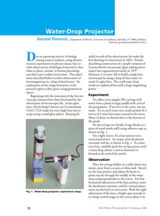
  • 1. THE PHYSICS TEACHER x Vol. 39, February 200118 Gorazd Planinsic, Department of Physics, University of Ljubljana, Jadranska 19, 1000 Ljubljana, Slovenia; gorazd.planinsic@fiz.uni-lj.si Due to a growing interest in biology among science students, using demon- stration experiments in physics classes that in- volve observations of biological material or that relate to plant, animal, or human physiology can often raise student motivation. The experi- ment described below involves observation of microorganisms in a drop of pond water. An explanation of the image formation in the paraxial region is then given using geometrical optics. Beginning with the invention of the first mi- croscope, humans have been fascinated by the observation of microscopic life. In his spare time, Dutch draper Antoni van Leeuwenhoek (1632-1723) made his own single-lens micro- scope using a small glass sphere. Keeping de- Water-Drop Projector tailed records of his observations, he made the first drawing of a bacterium in 1683. Articles describing construction of a simple variation of Leeuwenhoek’s microscope (glass tubing micro- scope) have appeared previously in TPT.1 However, it is easier still to build a single-lens microscope by using a drop of clear water in- stead of a glass lens. The small water drop works as a spherical lens with a large magnifying power. Experiment To collect your sample, fill a syringe with water from a pond or large puddle with a lot of decaying plants. If you live at the coast, use sea- water. Try to catch some very small animals (0.2 mm to 0.5 mm) that move around in the water. Many of them are found close to the bottom of the pond. Fix the syringe on a holder (Lego blocks or a piece of wood works well) using adhesive tape as shown in Fig. 1. For a light source, fix a laser pointer on a vertical positioner. An empty stick-deodorant container will do, as shown in Fig. 1. To create your lens, carefully push the syringe piston until a water drop (about 2 mm in diameter) is formed at the end of the nozzle. Observation Place the syringe holder on a table about two meters away from a screen or white wall. Switch on the laser pointer and adjust the beam to point exactly through the middle of the water drop and perpendicular to the screen. For fine horizontal adjustment of the laser pointer, move the deodorant container, and for vertical adjust- ment use the built-in turn screw. With the right adjustment of the laser, a bright spot extends in- to a large round image on the screen about 2 m Fig. 1. Water-drop projector: experiment setup.
  • 2. 19THE PHYSICS TEACHER x Vol. 39, February 2001 in diameter. Now, if your pond water is rich enough with little animals, you should see their magnified shadow images floating and moving around on the screen. Small single-cell animals like paramecium appear as dark spots surround- ed with interference fringe contours. Some of them exhibit movements, but no detailed struc- ture can be recognized. Larger animals such as mosquito larva (Anopheles species), cyclops (Cy- clops strenuus), or water fleas (Daphnia species) appear like real monsters on the screen (see Fig. 2). Here you can clearly identify individual parts of their bodies and follow their move- ments. In our case the images were projected on the white wall and photographs were obtained with a digital camera (Kodak DC260). You may at first see nothing but the dark floating spots surrounded by a few concentric circles caused by light diffraction on small parts of de- caying plants and single-cell protozoa. Be pa- tient. The light attracts the little animals (a green laser works best), and after some time they swim down to the syringe nozzle and into the water drop. You can also give them a little help by dripping a few drops from the syringe. How the Water-Drop Projector Works The drop at the end of the syringe, though not a perfect sphere, can be treated as a small spherical lens. The light beam that falls on the drop refracts both times as it passes through the water-air interface. Let’s follow the path of the ray that enters the water drop just above an ob- ject that floats in the water drop at a small dis- tance x from the geometrical axis ( Fig. 3). The ray will refract twice and reach the screen at the distance y below the geometrical axis. The dis- tance y is determined by the distance from the drop to the screen d and the angle ␦, which can be calculated using simple geometry: ␦ = ␣ – ЄRST ЄRST = 2␤ – ␣ ␦ = 2(␣ – ␤). Using Snell's law, Cyclops (Cyclops strenuus) Mosquito larva (Anopheles species) Water flea (Daphnia species) Fig. 2. Photographs of projected images. The actual size of each projected image is about 1 m x 1 m. Author: are you certain of the taxonomic identity of this ani- mal? If not, then perhaps “Cyclops (Cyclops species)” would be a more appropriate caption for first photo below.
  • 3. THE PHYSICS TEACHER x Vol. 39, February 200120 ᎏ s s i i n n ␣ ␤ ᎏ = ᎏ n n 0 ᎏ (where for the water and air n =1.33 and n0 = 1.0, respectively), and with sin ␣ = x/r, the angle ␦ is given by the equation: ␦ = 2(arcsinᎏ x r ᎏ – arcsin ᎏ n x r ᎏ). For the rays close to the geometrical axes (parax- ial region), all the angles in the calculation above are very small; therefore, the expression for ␦ can be simplified using the approximation arcsin(z) Ϸ z: ␦ = 2 ΂1 – ᎏ n 1 ᎏ ΃. The projected image on the screen is a magni- fied shadow of the object (animal) with magni- fication equal to M = = ᎏ dta x n␦ ᎏ Ϸ 2 ΂1 – ᎏ n 1 ᎏ ΃, where the approximation (t an␦ Ϸ ␦) is valid in the paraxial region. For the water drop 2 mm in diameter, the shadow image on the screen 2 m from the setup is about 1000 times larger than the object. Note that in derivation we assumed the object floats on the laser side of the drop. It can be seen from Fig. 1 that the same shadow d ᎏ r y ᎏ x x ᎏ r height can be produced by a smaller object placed on the screen side of the drop (or anywhere in between). Clearly the magnification is largest for the objects that are floating in the screen side of the drop. In this case Mmax = in the paraxial region. For the parameters as given above, the magnification factor is 1985. The results show that in the paraxial region, the magnification depends on the position of the object along the geomet- rical axis but not on the object’s distance from the axis. However, if the size of the object is comparable to the size of the drop or the object lies away from the geometrical axis, the angles ␣, ␤, and ␦ are not small any more. In this case, the magnification is expressed using the exact expression for the angle ␦: M = ΄ᎏ 1 u ᎏtan΂2(arsin u – arcsin ᎏ n u ᎏ)΃΅, where the distance of the incident ray from the geometrical axis is measured in the units of wa- ter drop radius (u = x/r) and can have any value between 0 and 1. The magnification as the function of parameter u is shown in Fig. 4 with the set of parameters given earlier. We can de- duce from the graph that the magnification is reasonably constant within the region about half radius from the geometrical axis. This gives us a hint about the size of the drop for the given laser-beam diameter if we want to produce nondistorted shadow images. The preceding derivation is based on the as- sumption that the water drop is a perfect sphere. Though the bottom part of the drop is spherical to a good approximation, the upper part (where the drop issues from the nozzle) is seriously dis- torted. The light passing through this irregular- ly curved surface causes a variety of patterns that disturb our observation but are interesting sub- d ᎏ r n ᎏ 2 – n d ᎏ 2r Fig. 3. Optics of the water-drop projector.
  • 4. 21THE PHYSICS TEACHER x Vol. 39, February 2001 jects to explore themselves.2 However, there are other ways to make a water drop lens that is closer to a sphere than the one we suggest. For example, wrap a copper wire (thickness of 0.2 mm or so) to make a small ring, then touch the ring with the water drop hanging from the sy- ringe. The surface tension will make the drop jump onto the ring, where it will stay and form an almost perfect sphere. Or simply touch the drop from the syringe with a microscope glass plate and you’ll get a nice plan-concave lens. These alternatives can be used to observe the small animals in the pond water, and the ob- served shadow images may be even better than those obtained with the hanging water drop method. But the advantage of this method is the simple construction and the fact that you can have many animals inside the syringe. The little animals attracted by the light swim down into the drop by themselves. With the other methods, getting the animals into the drop may be difficult, and the observation time is limited by the evaporation of the drop. References 1. Wayne O. Williams, “Glass tubing microscope,” Phys. Teach. 17, 204 (1979) . 2. J. Walker, “A drop of water becomes a gateway in- to the world of catastrophe optics,” Sci. Am. 120 (1989). Fig. 4. Water-drop magnification as the function of distance from the optical axis.