Sonographic evaluation of diaphragm excursion to strengthen non- invasive mechanical ventilation education program in a patient with chronic respiratory insufficiency and claustrophobia
•
0 likes•24 views

We use the Sonography of the diaphragm muscle as an educational tool to strengthen a cardio-respiratory pulmonary rehabilitation program of non invasive ventilation in patient's with problem's of Niv adaptation.
Recommended

Recommended
More Related Content
Similar to Sonographic evaluation of diaphragm excursion to strengthen non- invasive mechanical ventilation education program in a patient with chronic respiratory insufficiency and claustrophobia
Similar to Sonographic evaluation of diaphragm excursion to strengthen non- invasive mechanical ventilation education program in a patient with chronic respiratory insufficiency and claustrophobia (20)
Recently uploaded
Recently uploaded (20)
Sonographic evaluation of diaphragm excursion to strengthen non- invasive mechanical ventilation education program in a patient with chronic respiratory insufficiency and claustrophobia
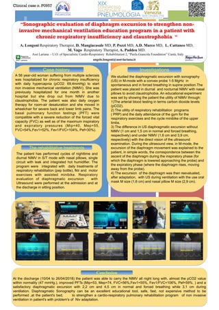
- 1. +“Sonographic evaluation of diaphragm excursion to strengthen non- invasive mechanical ventilation education program in a patient with chronic respiratory insufficiency and claustrophobia “ A. Longoni Respiratory Therapist, D. Mangiacasale MD, P. Pozzi MD, A.D. Marco MD, L. Cattaneo MD, M. Vago Respiratory Therapist, A. Paddeu MD. Asst Lariana - U.O. of Specialistic Cardio-Respiratory Rehabilitation 2, “Paola Giancola Foundation” Cantù, Italy angelo.longoni@asst-lariana.it A 56 year-old woman suffering from multiple sclerosis was hospitalized for chronic respiratory insufficiency with daily hypercapnia (pCO2: 69,4mmHg) to start non invasive mechanical ventilation (NIMV). She was previously hospitalized for one month in another hospital but she drop out from NIMV due to claustrophobia. The patient was also daily oxygen therapy for room-air desaturation and she moved in wheelchair for severe back and lower limb pains. The basal pulmonary function testings (PFT) were compatible with a severe reduction of the forced vital capacity (FVC) as well as of the maximum inspiratory and expiratory pressures (Mip=40, Mep=55, FVC=54%,Fev1=52%, Fev1/FVC=104%, Pef=30%). Clinical case n. P0957 Case history The rehabilitative treatments Conclusion At the discharge (10/04 to 26/04/2018) the patient was able to carry the NIMV all night long with, almost the pCO2 value within normality (47 mmHg.), improved PFTs (Mip=53, Mep=74, FVC=56%,Fev1=55%, Fev1/FVC=106%, Pef=59%. ) and a satisfactory diaphragmatic excursion with 2,2 cm and 4,5 cm in normal and forced breathing while 3,1 cm during ventilation. Diaphragmatic Sonography can be an excellent educational tool, safe, fast, not expensive method to be performed ,at the patient's bed, to strengthen a cardio-respiratory pulmonary rehabilitation program of non invasive ventilation in patient's with problem's of Niv adaptation. The patient has performed cycles of nighttime and diurnal NIMV in S/T mode with nasal pillows, single circuit with leak and integrated hot humidifier. The program were integrated with daily treatments of respiratory rehabilitation (pep bottle), fktr and motor exercises with assisted minibike. Respiratory evaluation of diaphragmatic excursion with Ultrasound were performed at the admission and at the discharge in sitting position. We studied the diaphragmatic excursion with sonography (US) in M-mode with a convex probe 1-5 MgHz in spontaneous and in forced breathing in supine position.The patient was placed in diurnal and nocturnal NIMV with nasal pillows to avoid claustrophobia. An educational experiment was set by showing the patient the utility of NIMV through: 1)The arterial blood testing in terms carbon dioxide levels (pCO2). 2) The utility of respiratory rehabilitation programs ( PRP) and the daily attendance of the gym for the respiratory exercises and the cycle minibike of the upper limbs. 3) The difference in US diaphragmatic excursion without NIMV (1 cm and 1,5 cm in normal and forced breathing, respectively) and under NIMV (1,6 cm and 3,9 cm, respectively) with the direct vision of the ultrasound examination. During the ultrasound view, in M-mode, the excursion of the diaphragm movement was explained to the patient, in simple words, the correspondence between the ascent of the diaphragm during the inspiratory phase (for which the diaphragm is lowered approaching the probe) and the expiratory phase (where the diaphragm rises, moving away from the probe). 4) The excursion of the diaphragm was then reevaluated, after adaptation, with US during ventilation with the use oral mask M size (1,6 cm) and nasal pillow M size (2,9 cm). Investigations