
Recommended
More Related Content
What's hot
What's hot (20)
Similar to Retained placenta by dr alka mukherjee & dr apurva mukherjee
Similar to Retained placenta by dr alka mukherjee & dr apurva mukherjee (20)
More from alka mukherjee
More from alka mukherjee (20)
Recently uploaded
Recently uploaded (20)
Retained placenta by dr alka mukherjee & dr apurva mukherjee
- 1. RETAINED PLACENTA DR ALKA MUKHERJEE DR APURVA MUKHERJEE NAGPUR M.S.DR ALKA MUKHERJEE 1
- 2. DR ALKA MUKHERJEE MBBS DGO FICOG FICMCH PGDCR PGDMLS MA(PSY) Director & Consultant At Mukherjee Multispecialty Hospital MMC ACCREDITATED SPEAKER MMC OBSERVER MMC MAO – 01017 / 2016 Present Position Director of Mukherjee Multispecialty Hospital Hon.Secretary INTERNATIONAL COUNCIL FOR HUMAN RIGHTS Hon.Secretary NARCHI NAGPUR CHAPTER (2018-2020) Hon.Secretary AMWN (2018-2021) Hon.Secretary ISOPARB (2019-2021) Life member, IMA, NOGS, NARCHI, AMWN & Menopause Society, India, Indian medico-legal & ethics association(IMLEA), ISOPRB, HUMAN RIGHTS Founder Member of South Rapid Action Group, Nagpur. On Board of Super Specialty, GMC, IGGMC, AIIMS Nagpur, NKPSIMS, ESIS and Treasury, Nagpur for “ WOMEN SEXUAL HARASSMENT COMMITTEE.” mukherjeehospital@yahoo.com www.mukherjeehospital.com https://www.facebook.com/ Mukherjee Multispeciality https://www.instagram.com/ Achievement Winner of NOGS GOLD MEDAL – 2017-18 Winner of BEST COUPLE AWARD in Social Work - 2014 APPRECIATION Award IMA - MS Past Position Organizing joint secretary ENDO-GYN 2019 Vice President IMA Nagpur (2017-2018) Vice President of NOGS(2016-2017) Organizing joint secretary ENDO-GYN Organizing secretary AMWICON – 2019
- 3. INTRODUCTION • Retained placenta is a condition in which all or part of the placenta or membranes remain in the uterus during the third stage of labour. • Retained placenta can be broadly divided into: 1. failed separation of the placenta from the uterine lining 2. placenta separated from the uterine lining but retained within the uterus • A retained placenta is commonly a cause of postpartum haemorrhage, both primary and secondary. • Retained placenta is generally defined as a placenta that has not undergone placental expulsion within 30 minutes of the baby’s birth where the third stage of labor has been managed actively. DR ALKA MUKHERJEE 3
- 4. RISKS OF RETAINED PLACENTA • Risks of retained placenta: 1. Hemorrhage and 2. Infection. • After the placenta is delivered, the uterus should contract down to close off all the blood vessels inside the uterus. • If the placenta only partially separates, the uterus cannot contract properly, so the blood vessels inside will continue to bleed. • A retained placenta thereby leads to hemorrhage DR ALKA MUKHERJEE 4
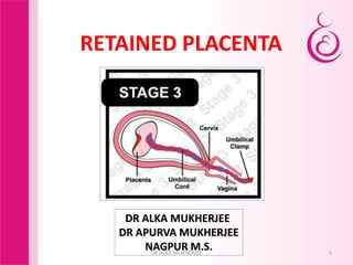
- 5. PLACENTAL SEPARATION & EXPULSION Normally the placenta is expelled in three stage 1.It first separates from the uterine muscle, 2. Then it descends into lower uterine segment of uterus & vagina 3. Then it is expelled outside. Problems can occur at any of these stages DR ALKA MUKHERJEE 5
- 6. SIGNS OF PLACENTAL SEPARATION 1. Lengthening of the visible portion of the umbilical cord. 2. Increased bleeding from the vagina. 3. Change in shape of the uterus from flat (discoid) to round (globular). 4. The placenta being expelled from the vagina. DR ALKA MUKHERJEE 6
- 7. RISK FACTORS Previous retained placenta Previous injury or surgery to the uterus Preterm delivery Induced labor Multiparity DR ALKA MUKHERJEE 7
- 8. CAUSES • Placenta separated but not expelled out • Simple adherent placenta • Morbidly adherent placenta – placenta accreta • – placenta increta • – placenta percreta DR ALKA MUKHERJEE 8
- 9. DR ALKA MUKHERJEE 9
- 10. INITIAL MANAGEMENT Drugs, such as intraumbilical or intravenous oxytocin, are often used in the management of placental retention. It is useful ensuring the bladder is empty. Avoid Ergometrine as it causes tonic uterine contractions which may delay placental expulsion. Controlled cord traction has been recommended as a second alternative after more than 30 minutes have passed after stimulation of uterine contractions, provided the uterus is contracted. Manual extraction may be required if cord traction also fails, or if heavy ongoing bleeding occurs. Very rarely a curettage is necessary to ensure that no remnants of the placenta remain (in rare conditions with very adherent placenta such as a placenta accreta. However, in birth centers and attended home birth environments, it is common for licensed care providers to wait for the placenta's birth up to 2 hours in some instances. DR ALKA MUKHERJEE 10
- 11. MANAGEMENT • Depend on severity of bleeding. • The retained or partially detached placenta interferes with uterine contraction and retraction and leads to bleeding. • Bleeding may be visible or may manifest only by the increasing size of the uterus. • In the absence of any evidence of placental detachment, consider the diagnosis of complete placenta accreta or a variant. • This condition may be present with bleeding if only a portion of the placenta is abnormally implanted. DR ALKA MUKHERJEE 11
- 12. MANAGEMENT DETAILS If the placenta is undelivered after 30 minutes Consider As retained placenta Ensuring that the bladder is empty {may speed the delivery of the placenta and at least aid in the assessment and control of the uterus}. Breastfeeding or nipple stimulation Change of position - encourage an upright position DR ALKA MUKHERJEE 12
- 13. IN STABLE WOMEN WITH MINIMAL BLEEDING • In stable women with minimal bleeding while preparations for a manual removal are being made. • Injection into the umbilical cord vein (Carroli, 2002). Saline, oxytocin and saline, prostaglandin and saline, and dextran 70. • The studies comparing injection of oxytocin (commonly, 10 IU) and saline (commonly, 20 mL) with expectant management or saline injection alone suggest that this practice indeed reduces the need for manual removal of the placenta. DR ALKA MUKHERJEE 13
- 14. Manual removal of the placenta if the above maneuvers have failed to deliver the placenta or if significant bleeding occurs. a. Anesthesia (regional or general) {manual removal can cause considerable abdominal cramping}. b. Sometimes, IV narcotic analgesia will prove helpful in relieving this discomfort. c. Nb: The cessation of an oxytocin infusion or the administration of uterine relaxants to promote uterine exploration and manual removal is of questionable value and may lead to increased bleeding. d. Ultrasound may be useful in select cases. MANUAL REMOVAL OF THE PLACENTA DR ALKA MUKHERJEE 14
- 15. 1. An elbow-length glove is worn and attention is paid to asepsis. 2. The perineum and vagina must be prepared. 3. The vaginal hand may be immersed in povidone- iodine solution to facilitate easier entry. 4. The hand is passed into the vagina through the cervix and into the lower segment following the umbilical cord. 5. Care is taken to minimize the profile of the hand as it enters, keeping the thumb and fingers together in the shape of a cone to avoid damage. 6. Control of the uterine fundus with the nonvaginal hand is essential. 7. If the placenta is encountered in the lower segment, it is removed. If the placenta is not encountered, the placental edge is sought 8. Separate the placenta from the uterus with a sweeping motion 9. After the placenta is mostly separated, curl your palm around the bulk of it. MANUAL REMOVAL OF THE PLACENTA - PROCEDURE DR ALKA MUKHERJEE 15
- 16. 10. Continue to grasp the placenta as you remove it from the uterine cavity. 11. Once found, the fingers gently develop the space between the placenta and uterus and shear off the placenta. The placenta is pushed to the palmar aspect of the hand and wrist; when it is entirely separated, the hand is withdrawn. 12. an oxytocin infusion is running rapidly as the hand is withdrawn {encourage strong uterine contraction}, and then perform uterine massage. Care must be taken to tease out the membranes. 13. Once uterine contraction is established, examine the placenta and membranes to determine whether further exploration or curettage is necessary. 14. Antibiotics DR ALKA MUKHERJEE 16
- 17. Placenta Accreta and Percreta I. partial and focal: a. the attachments can be manually broken and the placenta removed. b. b. It may be necessary to curette the placental bed to reduce bleeding. Recovery is usually satisfactory, although more than the usual amount of post partum bleeding will be noted. DR ALKA MUKHERJEE 17
- 18. II. extensive or complete: • you probably won't be able to remove the placenta in other than handfuls of fragments. • Bleeding from this problem will be considerable, and the patient will likely end up with multiple blood transfusions while you prepare her for a life- saving, post partum uterine artery ligation or hysterectomy. • If surgery is not immediately available, consider tight uterine and/or vaginal packing to slow the bleeding until surgery is available. DR ALKA MUKHERJEE 18
- 19. A retained placenta with absent sonolucent area between the placenta and uterine wall suggestive of placenta increta. DR ALKA MUKHERJEE 19
- 20. DR ALKA MUKHERJEE 20
- 21. Morbid adhesion of the placenta: Morbid adhesion of the placenta can occur wh en the placenta is implanted deeply into the uterine muscles and thus fails to separate. The placenta can burrow upto different depths in the uterine muscle. In simple cases, it is only attached firmly to muscle and can be stripped off by hand. In severe morbid adhesion, the placenta can b urrow through the full thickness of the muscle In this case, the uterus may be needed to be removed ('hysterectomy') to control the bleedi ng. DR ALKA MUKHERJEE 21
- 22. DR ALKA MUKHERJEE 22
- 23. Three types of morbid adhesion of the placenta 1.Placenta Accreta: In this condition, the placenta pen etrates deep into the uterine endometrium and reach es the muscles but does not penetrate into the muscl es. 2.Placenta Increta: Here, the placenta reaches even de eper into the uterine wall and penetrates into the ute rine muscle. 3.Placenta Percreta: In this condition, the placenta not only penetrates through the full thickness of the uter ine muscles but also Reaches to another organ such as the bladder or the rectum. Placenta percreta is very rareDR ALKA MUKHERJEE 23
- 24. DR ALKA MUKHERJEE 24
- 25. CALL FOR HELP Inform Anesthetist Insert large bore 18 no cannula Catheterize patient IV 20UNITS OXYTOCIN DRIP @ 60 DROPS/MT ESTIMATE BLOOD LOSS COAGULATION PROFILE ALONG WITH CBC ARRANGE FOR BLOOD PREPARE PATIENT FOR MRP, SOS HYSTERECTOMY TAKE INFORMED CONSENT 1.If bleeding profusely DR ALKA MUKHERJEE 25
- 26. Risks of Retained Placenta There may be severe bleeding which may be life- threatening. Attempts at manual removal of the placenta can cause multiple injuries to the mother such as like vulvar hematoma, perineal tears, cervical tears and vaginal wall tears. Complications of a Retained Placenta Uterine inversion Shock (hypovolemic) Postpartum hemorrhage Puerperal Sepsis Subinvolution Hysterectomy DR ALKA MUKHERJEE 26
- 27. POST-PROCEDURE CARE 1. Observe the woman closely until the effect of IV sedation has worn off. 2. Monitor the vital signs (pulse, blood pressure, respiration) every 30 minutes for the next 6 hours or until stable. 3. Palpate the uterine fundus to ensure that the uterus remains contracted. 4. Check for excessive lochia. 5. Continue infusion of IV fluids. 6. Transfuse as necessary. DR ALKA MUKHERJEE 27
- 28. DR ALKA MUKHERJEE 28