
Recommended
More Related Content
What's hot
What's hot (20)
Similar to Sutures emad
Similar to Sutures emad (20)
Recently uploaded
Recently uploaded (20)
Sutures emad
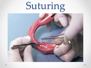
- 1. Suturing 1
- 2. SUTURE: sutures are the stitches that doctors and especially surgeons use to hold skin, internal organs, blood vessels and all other tissues of the human body together, after they have been severed by injury or surgery. 2
- 3. FEATURES OF IDEAL SUTURE MATERIALS: 1. STRONG (don’t break). 2. NON TOXIC & HYPO-ALLERGIC (avoid adverse reaction in the body). 3. FLEXIBLE (so they can be tied and knoted easily). 4. They must be LACK the so called “wick effect“ which means that suture must not allow fluid to penetrate the body through them from out side . 5. PREDICTABLE ABSORPTION. 3
- 4. Purpose of suturing Closing the wounds by means of suturing promotes healing and prevents complication, because an open wound is subjected to entrance of saliva loaded with food debris and bacteria. Suturing also prevents post-operative hemorrhage To hold a wound together in good position until natural healing process is sufficiently well established 4
- 5. Function of suturing 1. Coapt wound margins 2. Aids in hemostasis 3. Help to hold soft tissue flap over bone 4. Helps in maintaining blood clots in the tooth socket wounds 5
- 6. Advantages of suturing :- Promotes healing. Prevents complications: Infection. Hemorrhage. Tissue necrosis. Preserve the normal contour and shape of tissues. 6
- 7. Suturing armamentarium 1. Needle holder: -There are many different needle holders the most commonly used needle holder has a rocking handle and short beaks and are about 6 inches (15cm) long. 2. Suture needles: -The suture needles are supplied in different patterns and shapes to maintain accessibility to the sutured area and avoid complications . - the size or length of the needle varies according to the field of application . - nowadays the suture needle are already threaded with the suture materials and supplied in sealed and sterile packs this is for time saving and very practical suture needles 3. Tissue holding forceps 4. Suture cutting scissor 5. Suture material 7
- 8. Suturing Needles: • They may differ in shape, diameter, cross-sectional view, and size • They are usually made of stainless steel, which is a strong and flexible material. • The needles are atraumatic disposable needles with pre-attached sutures on their posterior ends. • Needles that may be used and sterilized many times are also available, with an eye or groove in the needle, through which the suture is passed. 8
- 9. Patterns of suture needles according to accessibility 1. Straight suture needles : used for skin suturing and require large space for manipulation and are of little importance in oral surgery 1. Curved suture needles : - used for intraoral and deep suturing . - it’s the most commonly used shape . - its easier to use in small spaces 9
- 10. • there are various curved needles :1/4 circle ,3/8 circle, ½ circle and 5/8 circle • In the oral cavity the most frequently used needles are 3/8 or ½ circle 10
- 11. patterns of suture needles according to cross section 1. Tapered (round) suture needles : − The needle is round in cross section through its whole length − The needle maybe straight , curved of half-circle to be accessible to the sutured area − This type is adequate for suturing fragile , delicate or thinned tissues such as mucous membrane and oral mucosa − Their advantage is that there’s no cutting , damage and laceration to the tissue − Their disadvantage is that great pressure is required when passing through the tissues, which may make suturing the wound harder 11
- 12. 2. Cutting (triangular) suture needles; - Has triangular cross section. - Has sharp edges that allow the needle to penetrate tough tissue like gingiva and skin - No excessive pressure is needed for the needle to penetrate the tissue - Easy and sharp penetration by needle through dense tissues such as the oral mucoperiosteoum - If applied to delicate tissue the thin oral epithelium or the mucosa of the lips, will case excessive cuts and lacerations of those tissues 12
- 13. 13
- 14. Eye NeedleSawged Needle 1. Reusable 2. May have burs that could weaken the suture material 1. Prepackaged and pre-sterilized 2. Needle attached to suture material 3. It’s the most favorable for intraoral use 4. Its expensive 5. Not reusable 6. Minimum tissue trauma than eye needles 7. Immediately ready for use 8. Always a new and sharp needle 9. No time for wasting threading 14
- 15. 15
- 16. Suture materials: • Are available in different types and gauges (thickness)to be suitable for different tissues • The thickness of the suture materials is expressed in the terms of zeros • The thickness is inversely proportional to the number of zeros i.e. (0) is the thickest and (00) is thinner etc.. • The less the number of zeros the more is the thickness of the suture material • That is to say that 2-0 is thicker that 3-0 is thicker than 4- 0 etc.. • The suture material commonly used in the oral cavity is 3-0 or 4-0 thickness 16
- 17. suture gauge: Gauge is the caliber of the suture and is expressed in no., the gauge used depend on the strength required and no. of sutures. • 9-0 or 10-0 is used for micro-surgery • 5-0 or 6-0 is used for facial skin closure • 3-0 or 4-0 is used for muscle, deep skin and intra- oral mucosal closure 17
- 18. 18
- 19. Types of suture materials: I. Natural suture materials 1) Absorbable suture materials Will break down harmlessly in the body ever time without intervention. • Catgut : maybe plain or chromic Plain Cutgut: Made from submucosal layer of sheep intestine or from serosal layer of beef intestines Mono-filament, Supplied in foil packages that prevent desiccation Digested completely within 5-7 weeks and full tensile strength remains for at least 7 days Disadvantage: - maybe local reaction that may arise form the tissue during digestion and absorption that may delay healing or even break down of the tissue - Absolut sterilization of catgut without causing impairing of its tensile strength is not possible 19
- 20. Plain catgut is used in suturing of deep facia or muscles Plain catgut packaged in a solution of 89% isopropanol, 10% water and 1% triethanolmine Plain catgut generally causes more severe tissue reaction than chromic. 20
- 21. • Chromic catgut Is treated with chromium to decrease tissue reactivity and to provide greater resistance to absorption. • Indicated for use in general soft tissue approximation and/or ligation, including use in ophthalmic procedures, but not for use in cardiovascular and neurological tissues . • Chromic gut is processed to provide greater resistance to absorption • will provide effective wound support for 10-21 days, but don't truly dissolve for 90 days . 21
- 22. 2) Natural Non absorbable suture materials • Silk : - it is made from the cocoon of silk worm. - It is braided, has good handling and knot-tying - Indicated for use in general soft tissue approximation and/or ligation, including use in cardiovascular, ophthalmic, and neurological procedures. - Its made in black and white silk - The black silk can easily be differentiated form the oral tissue - Well tolerated by the oral tissue 22
- 23. - Not expensive - Can be autoclaved or boiled - Has great tensile strength so that very fine strands - Available only in a multi-felamintous form - The most suitable type used in the oral cavity are gauge 3-0 and 4-0 black silk 23
- 24. 24
- 25. II. Synthetic suture materials 1) Absorbable suture materials: - Polyglycolic acid (Dexon): • Absorbtion occurs between 15-30 days • The physical properties of dexon are superior to those catgut sutures • Braided multifilament form - Polyglactin (Vicryl): • Digested within 60-90 days • Less stiff than catgut sutures, so that the sutures knots are more likely to remain tight • Maintains its strength for longer than other absorbable suture, • More costly that catgut sutures • Multifilament form 25
- 26. Absorbable suture materials cont. - Polydioxone (PDS) - Is a sterile synthetic absorbable monofilament suture . - Made from the polyester (p-dioxanone.). - Intended for use in general soft tissue approximation, including use in paediatric cardiovascular tissue, in microsurgery and in ophthalmic surgery. - These sutures are particularly useful where the combination of an absorbable suture and extended wound support (up to six weeks) is desirable. - These sutures, being absorbable, should not be used where prolonged (beyond 6 weeks) approximation of tissues under stress is required or in conjunction with prosthetic devices, for example, heart valves or synthetic grafts. 26
- 27. 1) Absorbable suture materials CONT - Polyglyconate (Maxon) - Prepared from a copolymer of glycolic acid and trimethylene carbonate - Synthetic monofilament absorbable sutures . - Used in general soft tissue approximation and/or ligation, including use in pediatric cardiovascular tissue, where growth is expected to occur, and in peripheral vascular surgery - Absorption is minimal until about the 60th day after implantation and is essentially complete within six months. 27
- 28. 2) Synthetic Non absorbable suture materials • The most commonly used nonabsobable suture in oral and maxillofacial surgery are; Nylon, polyester and polypropylene. • Nylon: 1. Good for skin closure 2. If used intra-orally it may cause irritation to the oral tissues due to high hardness 3. available in mono-filamint form • Polyester 1. Remains in the body 2. Available in multi-filament form 28
- 29. • Synthetic Non absorbable suture materials CONT • Prolene ( polypropylene) - available in mono-filament form - Has less tissue reaction than with other suture materials • Metal sutures - Rustless steel wire ( stainless steel, nickel, chromium,aluminium,silver, titanium ) - Smooth, strong, playable - Non-corrosive - Non irritant to the tissue - Occasionally used by surgeons extra-orally 29
- 30. Suture materials according to filament composition 1. Monofilament - Made of single strand - Resists harboring micro-organisms Examples: plain catgut, chromic catgut, nylon and prolene 2. Poly Filament or Multi-filamentous - Consists of several fibers - Either braded or twisted - Gives good handling and tighing qualities. Example :silk, dexon, vicryl, polyester 30
- 31. Characteristics of suture materials • The ideal suture materials would be; 1. Sterile 2. High tensile strength, holding the wound securely through the healing period by rapid absorption 3. Non-allergic and non-cancerogenic 4. Easy to handle, ties down well , provides optimum knot security 5. Minimally reactive in tissue and not predisposed to bacterial growth 6. Resistant to shrinking in tissues 7. Absorbed completely with minimal tissue reaction after serving its purpose 31
- 32. The strength of the sutures varies according to their size, which can be determined by a uniformly applied number. Example :- 6-0 suture is more delicate and has less strength than a 4-0 suture. 32
- 33. Basic requirements for suturing techniques: • The needle should be od suitable size and shape and the material of suitable thickness • Grasp needle ¾ the distance away form the point 33
- 34. • Place needle perpendicular to tissue surface on entry 34
- 35. • Suture start first through mobile then non-mobile tissue (from the loose to the fixed) • The needle should be passed from the thinner layer • The insertion of the needle should at an equal distance on both sides of 2-3mm away form the free edge of the soft tissue, to avoid laceration • Suture should not be placed under tension • Too tight suture cause strangulation of the blood vessels and delayed healing 35
- 36. • The knots should be away from the incision line • Suture should be removed 5-7days after surgery, otherwise foreign body reaction may develop 36
- 37. • When removing the suture it should be cut as near to the mucosa as possible, this is to avoid introducing the infection into the tissue when the suture is withdrawn 37
- 38. removal of sutures: The removal time is balance between strength of healthy and cosmetic result. Some areas are better vascularised, under less tension, and there fore heal quicker than others . •The following is arrange of removal of different areas: face and neck : 3-4 days. scalp : 5-7 days. limbs : 5-7 days. Hands and feet : 10-14 days. Abdomen : 8-10 days. 38
- 39. 39
- 40. Suture techniques • There are many suture techniques that are used intra orally as well as extra-orally : 1. Simple Interrupted suture The most commonly used in oral surgery Advantage : - Simplicity of application - if one knot united other sutures will maintain the strength of the suture line Disadvantages : - Time consuming because of the high number of knots and consuming of large amount of suture material. 40
- 41. 2. Continuous sutures Advantages : - its simplicity - can be performed rapidly, and consumption of lesser amount of suture material. - Running one continuous length of material Disadvantages: when one stitch is untied, the strength of the suture line can’t be maintained. 41
- 42. A. Simple continuous suturing A. Interlocking continuous suture 42
- 43. 3. Figure of eight Used to hold materials (Gel foam or pack in the socket) 43
- 44. • horizontal mattress - With stents and Without stents Advantages: - - It can be used in areas where much tension is placed on the skin. - Strength of closure Disadvantages: - It interferes with blood supply to the skin and interferes with healing. 44
- 45. • Vertical mattress Advantages: • It provides closure for both deep and superficial layers . • Allows perfect eversion and vertical opposition of the superficial skin edges. Disadvantages: - It consumes much suture material and more time. - A relatively high propensity to dig into skin and cause prominent stitch mark scars 45
- 46. 46
- 48. Principles of suturing The curved needle is held approximately two thirds of the distance between the tip and the base of the needle. 48
- 49. The needle should enter tissue at right angles to the tissue. Do not place sutures closer than 2 mm to 3 mm from the flap edges to prevent tearing through the flap during suturing, or if postoperative swelling occurs. 49
- 50. The suture needle should be inserted and pulled through the tissue in line with the curve of the needle. 50
- 51. Principles of suturing CONT. First insert sutures through the more mobile tissue flap (usually facial), for better precision, it is best to use two passes in most situations. The needle should pass through the deeper to the superfitial side. The needle should pass from the thinner to the thicker side. The knot should be positioned to the side of the incision. When suturing, flaps should be approximated without blanching of the tissue. Each suture should be placed 3-4 mm apart . 51
- 52. The needle holder is held by using the thumb and the ring finger in rings and the first and second fingers to control the instrument . 52
- 54. 54