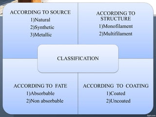
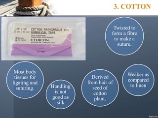
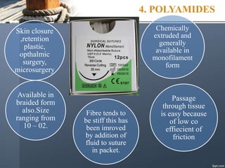
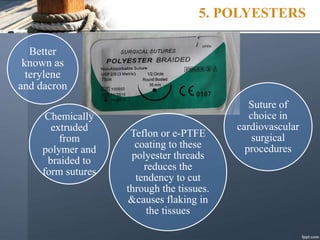
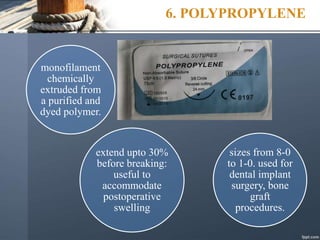
SUTURE MATERIALS
AND
TECHNIQUES
DEFINITION
• A suture is a strand or thread of material used to approximate
tissues and also to ligate blood vessels to maintain flap in
desired position until healing progress to a point where sutures
are not needed.
• Suturing is defined as a process of holding severed tissue in
close approximation until the healing process provides wound
with sufficient strength to withstand stress without the need for
support.
HISTORICAL BACKGROUND
• The first description of sutures used in operative procedures is
recorded by Edwin Smith Papyrus dated 16th century B.C.The
first great Arabian, Rhazes started his life as a story teller and
later on he turned to medicine in 900 A.D.
• He was credited with first employing “kitgut” to suture
abdominal wounds, which was the natural material for a lute
player to choose.(The Arabic word kit means a dancing
master’s fiddle, the musical strings of which were made up of
sheep intestines).
• The word gradually evolved into “catgut” or “surgical gut.”
• Many different materials have been used as ligatures and
sutures through centuries, e.g. horse hair strands, pig`s bristles,
gold/ silver wires, silk, silkworm gut, linen, cotton (flax), and
the tendons and intestinal tissues of various animals.
• By 1900, the catgut industry was firmly established in
Germany, due to the use of sheep intestine in their sausage
industry.
• In 1902, Claudius established iodine sterilization of the
suturing material.
• The development of synthetic absorbable sutures began in
1931, with the production of an absorbable synthetic fibre of
polyvinyl alcohol.
REQUISITES
Tensile strength Tissue
biocompatibility
Low capillarity
Good handling and
knotting properties
Sterilization
without
deterioration of
properties
ACCORDING TO SOURCE
1)Natural
2)Synthetic
3)Metallic
ACCORDING TO
STRUCTURE
1)Monofilament
2)Multifilament
ACCORDING TO FATE
1)Absorbable
2)Non absorbable
ACCORDING TO COATING
1)Coated
2)Uncoated
CLASSIFICATION
PRINCIPLES OF SELECTION
• Rate of healing of tissues
• Tissue contamination
• Cosmetic results
• Suture size
PARTS OF SUTURE NEEDLE
SUTURE NEEDLE
The surgical needles are of two types:
1. Eyed and
2. Eyeless
Suture needles usually are also classified
according to their curvature, radius and shape.
The surgical needle comprises of three parts:
1. Needle eye or swaged end
2. Needle body
3. Needle point
BASED ON EYE TYPES
CLOSED EYE FRENCH EYE SWAGED
EYE
Needle eye or swaged end
(i) Eyed suturing needle
(ii) Eyeless (swagged) suturing
needle
Advantages of eyeless needle.
(A) Tissue disruption caused by
double suture strand with eyed
needle.
(B) Minimum tissue disruption by
single suture strand swaged to
needle
New needle
every time
No chances of
accidental
unthreading
Faster, efficient
procedure.
high quality
steel; Tru-
tempering
process -
uniform
strength.
Code Number-
type, size, length
of the suture as
well as the details
of the needle.
no need of
ordering,
cleaning,
sterilising and
threading the
eyed needles.
BASED ON NEEDLE SHAPE
Needle point geometry
Taper-Point
Suited to soft tissue
Dilates rather than cuts
Reverse cutting
Very sharp
Ideal for skin
Cuts rather than dilates
Conventional
Cutting
Very sharp
Cuts rather than dilates
Creates weakness allowing suture
tearout
Taper-cutting
Ideal in tough or calcified tissues
Mainly used in Cardiac & Vascular
procedures.
NEEDLE POINT GEOMETRY
Blunt
Also known as “Protect Point”
Mainly used to prevent needle stick
injuries i.e. for abdominal wall closure.
Premium point
spatula
Ophthalmic Surgery
Spatula
Ophthalmic Surgery
DermaX*
NEW: ½ The Penetration force
Superior Cosmetic Effect
CHOOSING SUTURE
NEEDLE
• "One basic assumption must be made in considering
the ideal surgical needle for a given application,
namely, that the tissue being sutured should be altered
as little as possible by the needle since the only
purpose of the needle is to introduce the suture into
the tissue for apposition."
SUTURE SIZE
Thick
2/0..3/0..4/0..5/0..6/0..7/0..8/0..9/0..10/0..11/0
Thin
NEEDLE HOLDER
PROPER NEEDLE HOLDER
 Longer handle & shorter beak Mech. Adv
 Width of beak should not be greater than 30-35% of needle
radius .
Too wide beaks Straightening of needle
Too narrow beaks Rotation of needle
along the long axis of
needle holder.
SUTURE SCISSOR
PACKAGING
• All sutures are doubled wrapped in peel- apart envelops.
• Outer wrapper non-sterile on the outside but sterile on the
inner-side.
• Inner wrapper is sterile both inside and outside.
• Data printed on inner wrapper : type, size, length, needle
type & size, date of manufacture and expiration.
• Inner wrappers are color coded, according to type of suture.
SUTURE PACKAGING
STRAND
SIZE
MATERIAL
STRAND
LENGTH
COLOUR
PRODUCT
CODE
NEEDLE CODE
WITH LIFE
SIZE PICTURE
OF NEEDLE
NEEDLE CIRCLE POINT TYPE
NEEDLE LENGTH
NON-ABSORBABLE SUTURES
• These surgical threads are fabricated either from natural,
metallic or synthetic materials.
1.PERMA-HAND SURGICAL SILK
Derived
from
cocoon of
silk worm
larvae
Protein like
keratin of hair
and skin
covered
initially by
albuminous
layer
Suture is
braided round
a core and
coated with
wax to reduce
capillary
action
Available as
eyeless
needled sterile
suture sized 7-
0 to 1-0.
High tensile
strength; lost
gradually after
2 years.
2. LINEN
Made from
flax and is
cellulose
material.
It is
natural
cellulose
polymer.
Twisted to
form a fibre to
make a suture.
Gains 10% in
tensile
strength when
wet and fairly
unique in this
respect.
Ligation of
superficial
vessels ;
mucosal
suturing
without stress.
3. COTTON
Most body
tissues for
ligating and
suturing.
Derived
from hair of
seed of
cotton
plant.
Weaker as
compared
to linen
Handling
is not
good as
silk
Twisted to
form a fibre
to make a
suture.
4. POLYAMIDES
Skin closure
,retention
plastic,
opthalmic
surgery,
microsurgery
Chemically
extruded and
generally
available in
monofilament
form
Passage
through tissue
is easy because
of low co
effiecient of
friction
Fibre tends to
be stiff this has
been imroved
by addition of
fluid to suture
in packet.
Available in
braided form
also.Size
ranging from
10 – 02.
5. POLYESTERS
Better
known as
terylene
and dacron
Chemically
extruded
from
polymer and
braided to
form sutures
Suture of
choice in
cardiovascular
surgical
procedures
Teflon or e-PTFE
coating to these
polyester threads
reduces the
tendency to cut
through the tissues.
&causes flaking in
the tissues
6. POLYPROPYLENE
monofilament
chemically
extruded from
a purified and
dyed polymer.
extend upto 30%
before breaking:
useful to
accommodate
postoperative
swelling
sizes from 8-0
to 1-0. used for
dental implant
surgery, bone
graft
procedures.
7. STAINLESS STEEL
strength
and low
tissue
reaction.
it can pull or
tear out the
tissues and
necrosis can
result due to
too tight a
suture
Barbs
on the
end.
Used
In
Orthopaedic
and
neurosurgery.
NATURALABSORBABLE SUTURES
submucosa of
sheep intestines
or the serosa of
beef cattle
intestines and is
over 99% pure
collagen
absorbed by a
process of
enzymatic digestion
by proteolytic
enzymes derived
from lysozymes
contained within
polymorphs and
macrophages.
Absorption
rate-70
days
loses its
tensile
strength
in 15 days
it is coated with a
chromic compound
that extends
absorption up to 7
to 10 days and
extends loss of
tensile strength for
up to 5 days. Absorption
rate-90
days
loses
tensile
strength
within 30
days
• Surgical gut sutures may break more rapidly in patients with a
very low intraoral pH. A decrease in intraoral pH may be
found in metabolic disorders, Sjögren’s syndrome,
chemotherapy, radiation therapy, some medications like
antipsychotics, diuretics, antihypertensive agents, antipsoriasis
medications and steroid inhalers. Surgical gut is
contraindicated in these situations
• available with eyeless needles as well as standard sutures, in
sizes 5-0 to 2-0.
SYNTHETIC ABSORBABLE
SUTURES
Superior to natural absorbable sutures and more reliable in their
performance.
Manufactured in the laboratory and from carbohydrates
These are polymers. A polymer is made up of a large number of
molecules called monomers, linked together to form a chain. In
synthetic absorbable sutures, the linkage is broken by hydrolysis.
Therefore, allergic reactions are eliminated and early absorption in
hypoproteinemic conditions is possible.
These can be either monofilament or braided, coated or
noncoated.
They are twice as strong as compared to natural absorbable
sutures and lose their tensile strength slowly; thus helping to hold
the tissues together during the critical period of wound healing.
 They do not leave behind any foreign body after total absorption,
which is by a simple process of hydrolysis and evoke minimal
tissue reaction.
 They have excellent handling characteristics, but require a
special square knotting procedure.
 They are sterilised by Ethylene Oxide. Their shelf life is five
years and should be stored away from heat.
 They can be suitably selected for different tissue layers in
various surgical procedures, depending on whether they are
required for short, medium or long-term wound healing.
 They are available in full range, in varying sizes from 10-0 to 1-0
and swagged to different types of needles
1.Coated vicryl - polyglactin
910 braided suture material
lactic acid
monomer has
been introduced
in a ratio of one
part to every
nine parts of
glycolic acid.
90% glycolide,
10% lactide, while
Polyglactin coating
is 35% glycolide
and 65% lactide.
Calcium stearate is
an absorbable
organic lubricant.
sizes ranging
from 10-0 to 2-0
on different types
of needles. Shelf
life 5 years
2.Vicryl plus:synthetic absorbable
antibacterial suture.
world’s first
and only
Antibacterial
suture
contains
Triclosan
which is a
broad
spectrum
antibacterial
agent
defence against
unwanted
bacteria by a
zone of
inhibition that
stops bacterial
growth
3.Vicryl rapide
It is
identical to
coated
vicryl
suture
The rapid
absorption quality is
achieved by
exposure of coated
vicryl to gamma
irradiation, resulting
in material with low
molecular weight
than coated vicryl.
loses all its
strength
between 10th
and 12th days
and gets totally
absorbed within
42 days.
Shelf-life of
vicryl Rapide
sutures is five
years. It is
available in
sizes 3-0 to
1-0.
4. Monocryl-
polyglecaprone 25
Monofilament
synthetic
absorbable
suture.
co-polymer
of 75%
glycolide
and 25%
caprolactone
most pliable,
flexible
monofilament
suture with
excellent
handling
properties
predictable loss
of tensile strength
over three weeks
period. This is
due to systematic
hydrolysis of the
suture material
It is absorbed
in 90-120
days.Available
in sizes from
5-0 to 1-0.
5. PDS And PDS II
formed by
polymerising the
monomer-para-
dioxanone, in the
presence of a catalyst
to form a strong but
soft, pliable filament,
which is broken
down by hydrolysis
in the presence of
moisture.
PDS II sutures are
an improved
version of the
initial PDS sutures,
which give wound
support beyond the
four weeks period.
Total mass is
absorbed
between 180-
210 days. It is
available in
sizes 1-0 to 6-0.
PRINCIPLES OF SUTURING
NEEDLE GRASP
– 1/3rd from eye
– 2/3rd from point
ENTRY OF NEEDLE
- The needle should enter the tissues perpendicular to the
tissue surface.
PASSING OF NEEDLE
THROUGH TISSUES
Passed through tissues along the curve
DEPTH OF NEEDLE
Equal depth & distance from incision on both sides.
Sutures should be placed at a greater depth than the
distance from the incision, so as to evert the wound
margins.
Sutures on the skin are usually removed in 5 days and intraoral
sutures in 7 days. If there is tension while suturing, the sutures
may be kept for 10 days.
movable
tissue to
the fixed
tissue.
through the
thinner
tissue to the
thicker
tissue.
knot should
never lie on
the incision
line.
should be
tied only to
approximate
the tissues,
not to blanch
from the
deeper
tissue to the
superficial
tissue.
BIOLOGIC RESPONSE
• The first response of the body as in any healing process is
invasion of the tissue site by neutrophils.
• If uncomplicated by infection or trauma, the acute cellular
tissue response to the suture material changes in about three
days after implantation.
• The original population of neutrophils is replaced
predominantly by monocytes,plasma cells, and lymphocytes.
• Small sprouts of fragile vessels infiltrate the area, and
eventually fibroblasts and connective tissue proliferate.
Natural
Absorbable
• Proteolytic
degradation
• Intense tissue
response
Synthetic
Absorbable
• Hydrolysis
• Less intense
Non Absorbable
• Encapsulation
• Acellular
response
Response to all the sutures is relatively the same for the first five to
seven days. After this the response is more related to the type of
suture material.
RAILROAD SCAR
FORMATION
• All sutures passing through the mucous membrane or skin
provide a “wick” or pathway through which bacteria can track
down and gain access to the underlying tissues.
• The longer the suture remains, the deeper the epithelial
invasion of the underlying tissues. When the suture is removed
the epithelial tract remains.
• These cells may eventually disappear or remain to form
keratin. The epithelial pathway may also result in the typical
“railroad scar” formation.
Simple interrupted suture
Figure of eight suture
Horizontal mattress sutures
Continuous independent
sling suture
Anchor suture
Closed anchor suture
Periosteal suture
CONCLUSION
• sutures in attempt primary wound closure and the role of
suturing Is providing wound stabilization.
• The primary objective of suturing is to position and secure
surgical flaps to promote optimal healing.
• When used properly, surgical sutures should hold flap edges in
apposition until the wound has healed or has enough strength
to withstand normal functional stresses.
• With the sophisticated surgical procedures applied today, there
is a greater need for knowledge with regard to the various
types of suturing techniques and materials currently available.
REFERENCES
Clinical Periodontology – Carranza 11th Edition
Rino burkhardt & Niklaus p. Lang. Influence of suturing on
wound healing
Periodontology 2000, Vol. 68, 2015, 270–281
Textbook of Oral and Maxillofacial Surgery –Neelima Anil
Malik 2nd edition.
Suture Material and Needle Options in Oral and Periodontal
Surgery. Ellen D. Domnick, DVM

SUTURING TECHNIQUE.pptx

  • 1.
  • 2.
    DEFINITION • A sutureis a strand or thread of material used to approximate tissues and also to ligate blood vessels to maintain flap in desired position until healing progress to a point where sutures are not needed. • Suturing is defined as a process of holding severed tissue in close approximation until the healing process provides wound with sufficient strength to withstand stress without the need for support.
  • 3.
    HISTORICAL BACKGROUND • Thefirst description of sutures used in operative procedures is recorded by Edwin Smith Papyrus dated 16th century B.C.The first great Arabian, Rhazes started his life as a story teller and later on he turned to medicine in 900 A.D. • He was credited with first employing “kitgut” to suture abdominal wounds, which was the natural material for a lute player to choose.(The Arabic word kit means a dancing master’s fiddle, the musical strings of which were made up of sheep intestines). • The word gradually evolved into “catgut” or “surgical gut.”
  • 4.
    • Many differentmaterials have been used as ligatures and sutures through centuries, e.g. horse hair strands, pig`s bristles, gold/ silver wires, silk, silkworm gut, linen, cotton (flax), and the tendons and intestinal tissues of various animals. • By 1900, the catgut industry was firmly established in Germany, due to the use of sheep intestine in their sausage industry. • In 1902, Claudius established iodine sterilization of the suturing material. • The development of synthetic absorbable sutures began in 1931, with the production of an absorbable synthetic fibre of polyvinyl alcohol.
  • 5.
    REQUISITES Tensile strength Tissue biocompatibility Lowcapillarity Good handling and knotting properties Sterilization without deterioration of properties
  • 6.
    ACCORDING TO SOURCE 1)Natural 2)Synthetic 3)Metallic ACCORDINGTO STRUCTURE 1)Monofilament 2)Multifilament ACCORDING TO FATE 1)Absorbable 2)Non absorbable ACCORDING TO COATING 1)Coated 2)Uncoated CLASSIFICATION
  • 7.
    PRINCIPLES OF SELECTION •Rate of healing of tissues • Tissue contamination • Cosmetic results • Suture size
  • 8.
  • 9.
    SUTURE NEEDLE The surgicalneedles are of two types: 1. Eyed and 2. Eyeless Suture needles usually are also classified according to their curvature, radius and shape. The surgical needle comprises of three parts: 1. Needle eye or swaged end 2. Needle body 3. Needle point
  • 10.
    BASED ON EYETYPES CLOSED EYE FRENCH EYE SWAGED EYE
  • 11.
    Needle eye orswaged end (i) Eyed suturing needle (ii) Eyeless (swagged) suturing needle Advantages of eyeless needle. (A) Tissue disruption caused by double suture strand with eyed needle. (B) Minimum tissue disruption by single suture strand swaged to needle
  • 12.
    New needle every time Nochances of accidental unthreading Faster, efficient procedure. high quality steel; Tru- tempering process - uniform strength. Code Number- type, size, length of the suture as well as the details of the needle. no need of ordering, cleaning, sterilising and threading the eyed needles.
  • 13.
  • 14.
    Needle point geometry Taper-Point Suitedto soft tissue Dilates rather than cuts Reverse cutting Very sharp Ideal for skin Cuts rather than dilates Conventional Cutting Very sharp Cuts rather than dilates Creates weakness allowing suture tearout Taper-cutting Ideal in tough or calcified tissues Mainly used in Cardiac & Vascular procedures.
  • 15.
    NEEDLE POINT GEOMETRY Blunt Alsoknown as “Protect Point” Mainly used to prevent needle stick injuries i.e. for abdominal wall closure. Premium point spatula Ophthalmic Surgery Spatula Ophthalmic Surgery DermaX* NEW: ½ The Penetration force Superior Cosmetic Effect
  • 16.
    CHOOSING SUTURE NEEDLE • "Onebasic assumption must be made in considering the ideal surgical needle for a given application, namely, that the tissue being sutured should be altered as little as possible by the needle since the only purpose of the needle is to introduce the suture into the tissue for apposition."
  • 17.
  • 18.
  • 19.
    PROPER NEEDLE HOLDER Longer handle & shorter beak Mech. Adv  Width of beak should not be greater than 30-35% of needle radius . Too wide beaks Straightening of needle Too narrow beaks Rotation of needle along the long axis of needle holder.
  • 20.
  • 21.
    PACKAGING • All suturesare doubled wrapped in peel- apart envelops. • Outer wrapper non-sterile on the outside but sterile on the inner-side. • Inner wrapper is sterile both inside and outside. • Data printed on inner wrapper : type, size, length, needle type & size, date of manufacture and expiration. • Inner wrappers are color coded, according to type of suture.
  • 22.
    SUTURE PACKAGING STRAND SIZE MATERIAL STRAND LENGTH COLOUR PRODUCT CODE NEEDLE CODE WITHLIFE SIZE PICTURE OF NEEDLE NEEDLE CIRCLE POINT TYPE NEEDLE LENGTH
  • 23.
    NON-ABSORBABLE SUTURES • Thesesurgical threads are fabricated either from natural, metallic or synthetic materials.
  • 24.
    1.PERMA-HAND SURGICAL SILK Derived from cocoonof silk worm larvae Protein like keratin of hair and skin covered initially by albuminous layer Suture is braided round a core and coated with wax to reduce capillary action Available as eyeless needled sterile suture sized 7- 0 to 1-0. High tensile strength; lost gradually after 2 years.
  • 25.
    2. LINEN Made from flaxand is cellulose material. It is natural cellulose polymer. Twisted to form a fibre to make a suture. Gains 10% in tensile strength when wet and fairly unique in this respect. Ligation of superficial vessels ; mucosal suturing without stress.
  • 26.
    3. COTTON Most body tissuesfor ligating and suturing. Derived from hair of seed of cotton plant. Weaker as compared to linen Handling is not good as silk Twisted to form a fibre to make a suture.
  • 27.
    4. POLYAMIDES Skin closure ,retention plastic, opthalmic surgery, microsurgery Chemically extrudedand generally available in monofilament form Passage through tissue is easy because of low co effiecient of friction Fibre tends to be stiff this has been imroved by addition of fluid to suture in packet. Available in braided form also.Size ranging from 10 – 02.
  • 28.
    5. POLYESTERS Better known as terylene anddacron Chemically extruded from polymer and braided to form sutures Suture of choice in cardiovascular surgical procedures Teflon or e-PTFE coating to these polyester threads reduces the tendency to cut through the tissues. &causes flaking in the tissues
  • 29.
    6. POLYPROPYLENE monofilament chemically extruded from apurified and dyed polymer. extend upto 30% before breaking: useful to accommodate postoperative swelling sizes from 8-0 to 1-0. used for dental implant surgery, bone graft procedures.
  • 30.
    7. STAINLESS STEEL strength andlow tissue reaction. it can pull or tear out the tissues and necrosis can result due to too tight a suture Barbs on the end. Used In Orthopaedic and neurosurgery.
  • 31.
    NATURALABSORBABLE SUTURES submucosa of sheepintestines or the serosa of beef cattle intestines and is over 99% pure collagen absorbed by a process of enzymatic digestion by proteolytic enzymes derived from lysozymes contained within polymorphs and macrophages. Absorption rate-70 days loses its tensile strength in 15 days
  • 32.
    it is coatedwith a chromic compound that extends absorption up to 7 to 10 days and extends loss of tensile strength for up to 5 days. Absorption rate-90 days loses tensile strength within 30 days
  • 33.
    • Surgical gutsutures may break more rapidly in patients with a very low intraoral pH. A decrease in intraoral pH may be found in metabolic disorders, Sjögren’s syndrome, chemotherapy, radiation therapy, some medications like antipsychotics, diuretics, antihypertensive agents, antipsoriasis medications and steroid inhalers. Surgical gut is contraindicated in these situations • available with eyeless needles as well as standard sutures, in sizes 5-0 to 2-0.
  • 34.
    SYNTHETIC ABSORBABLE SUTURES Superior tonatural absorbable sutures and more reliable in their performance. Manufactured in the laboratory and from carbohydrates These are polymers. A polymer is made up of a large number of molecules called monomers, linked together to form a chain. In synthetic absorbable sutures, the linkage is broken by hydrolysis. Therefore, allergic reactions are eliminated and early absorption in hypoproteinemic conditions is possible. These can be either monofilament or braided, coated or noncoated. They are twice as strong as compared to natural absorbable sutures and lose their tensile strength slowly; thus helping to hold the tissues together during the critical period of wound healing.
  • 35.
     They donot leave behind any foreign body after total absorption, which is by a simple process of hydrolysis and evoke minimal tissue reaction.  They have excellent handling characteristics, but require a special square knotting procedure.  They are sterilised by Ethylene Oxide. Their shelf life is five years and should be stored away from heat.  They can be suitably selected for different tissue layers in various surgical procedures, depending on whether they are required for short, medium or long-term wound healing.  They are available in full range, in varying sizes from 10-0 to 1-0 and swagged to different types of needles
  • 36.
    1.Coated vicryl -polyglactin 910 braided suture material lactic acid monomer has been introduced in a ratio of one part to every nine parts of glycolic acid. 90% glycolide, 10% lactide, while Polyglactin coating is 35% glycolide and 65% lactide. Calcium stearate is an absorbable organic lubricant. sizes ranging from 10-0 to 2-0 on different types of needles. Shelf life 5 years
  • 37.
    2.Vicryl plus:synthetic absorbable antibacterialsuture. world’s first and only Antibacterial suture contains Triclosan which is a broad spectrum antibacterial agent defence against unwanted bacteria by a zone of inhibition that stops bacterial growth
  • 38.
    3.Vicryl rapide It is identicalto coated vicryl suture The rapid absorption quality is achieved by exposure of coated vicryl to gamma irradiation, resulting in material with low molecular weight than coated vicryl. loses all its strength between 10th and 12th days and gets totally absorbed within 42 days. Shelf-life of vicryl Rapide sutures is five years. It is available in sizes 3-0 to 1-0.
  • 39.
    4. Monocryl- polyglecaprone 25 Monofilament synthetic absorbable suture. co-polymer of75% glycolide and 25% caprolactone most pliable, flexible monofilament suture with excellent handling properties predictable loss of tensile strength over three weeks period. This is due to systematic hydrolysis of the suture material It is absorbed in 90-120 days.Available in sizes from 5-0 to 1-0.
  • 40.
    5. PDS AndPDS II formed by polymerising the monomer-para- dioxanone, in the presence of a catalyst to form a strong but soft, pliable filament, which is broken down by hydrolysis in the presence of moisture. PDS II sutures are an improved version of the initial PDS sutures, which give wound support beyond the four weeks period. Total mass is absorbed between 180- 210 days. It is available in sizes 1-0 to 6-0.
  • 41.
  • 42.
    NEEDLE GRASP – 1/3rdfrom eye – 2/3rd from point
  • 43.
    ENTRY OF NEEDLE -The needle should enter the tissues perpendicular to the tissue surface.
  • 44.
    PASSING OF NEEDLE THROUGHTISSUES Passed through tissues along the curve
  • 45.
    DEPTH OF NEEDLE Equaldepth & distance from incision on both sides. Sutures should be placed at a greater depth than the distance from the incision, so as to evert the wound margins.
  • 46.
    Sutures on theskin are usually removed in 5 days and intraoral sutures in 7 days. If there is tension while suturing, the sutures may be kept for 10 days.
  • 47.
    movable tissue to the fixed tissue. throughthe thinner tissue to the thicker tissue. knot should never lie on the incision line. should be tied only to approximate the tissues, not to blanch from the deeper tissue to the superficial tissue.
  • 48.
    BIOLOGIC RESPONSE • Thefirst response of the body as in any healing process is invasion of the tissue site by neutrophils. • If uncomplicated by infection or trauma, the acute cellular tissue response to the suture material changes in about three days after implantation. • The original population of neutrophils is replaced predominantly by monocytes,plasma cells, and lymphocytes. • Small sprouts of fragile vessels infiltrate the area, and eventually fibroblasts and connective tissue proliferate.
  • 49.
    Natural Absorbable • Proteolytic degradation • Intensetissue response Synthetic Absorbable • Hydrolysis • Less intense Non Absorbable • Encapsulation • Acellular response Response to all the sutures is relatively the same for the first five to seven days. After this the response is more related to the type of suture material.
  • 50.
    RAILROAD SCAR FORMATION • Allsutures passing through the mucous membrane or skin provide a “wick” or pathway through which bacteria can track down and gain access to the underlying tissues. • The longer the suture remains, the deeper the epithelial invasion of the underlying tissues. When the suture is removed the epithelial tract remains. • These cells may eventually disappear or remain to form keratin. The epithelial pathway may also result in the typical “railroad scar” formation.
  • 51.
  • 52.
  • 53.
  • 54.
  • 55.
  • 56.
  • 57.
  • 58.
    CONCLUSION • sutures inattempt primary wound closure and the role of suturing Is providing wound stabilization. • The primary objective of suturing is to position and secure surgical flaps to promote optimal healing. • When used properly, surgical sutures should hold flap edges in apposition until the wound has healed or has enough strength to withstand normal functional stresses. • With the sophisticated surgical procedures applied today, there is a greater need for knowledge with regard to the various types of suturing techniques and materials currently available.
  • 59.
    REFERENCES Clinical Periodontology –Carranza 11th Edition Rino burkhardt & Niklaus p. Lang. Influence of suturing on wound healing Periodontology 2000, Vol. 68, 2015, 270–281 Textbook of Oral and Maxillofacial Surgery –Neelima Anil Malik 2nd edition. Suture Material and Needle Options in Oral and Periodontal Surgery. Ellen D. Domnick, DVM

Editor's Notes

  • #25 Albuminous layer removed by degumming;most univerasslay used as inexpensive,moderate tissue reaction,Natural multifilament nonabsorbable
  • #26 1.Minimal tissur reaction. Poor tensile strenth cant be used for suturing under tension,Natural multifilament nonabsorbable
  • #27 Minimal tissue reaction,Natural multifilament nonabsorbable
  • #28 Synthetic,Muti/monofilament or nonabsorbable.tissue reaction extremely low,Has memory low knot security. Tensile strength loss after one year is aprox 25%
  • #29 Therefore, Ethicon introduced Ethibond sutures which are polyester coated with polybutylate. It provides excellent bonding and does not flake in the tissues and does not increase the suture diameter. extremely high tensile strength
  • #30 extremely low tissue reactivity It slides through the tissues readily and is sterilised by ethylene oxide. It is non-biodegradable.
  • #32 When used intraorally, it loses most of its tensile strength in 24 to 48 hours, unless
  • #37  The chemical similarity between the two materials ensures that the coating will bond permanently on to the basic braiding.
  • #39 These sutures are intended for use in closing mucosa and skin, where only short term wound support (less than 10 days) is required
  • #40 ideal for subcutaneous closure.
  • #41 Adhesive should not be allowed to enter the wound, otherwise foreign body reaction will delay the wound healing.
  • #51 jj