
Recommended
More Related Content
Similar to scalp_1.pdf
Similar to scalp_1.pdf (20)
More from abdulrazaq583901
More from abdulrazaq583901 (18)
Recently uploaded
Recently uploaded (20)
scalp_1.pdf
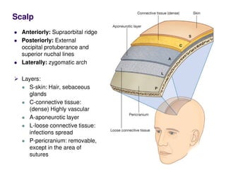
- 1. Anteriorly: Supraorbital ridge Posteriorly: External occipital protuberance and superior nuchal lines Laterally: zygomatic arch Layers: S-skin: Hair, sebaceous glands C-connective tissue: (dense) Highly vascular A-aponeurotic layer L-loose connective tissue: infections spread P-pericranium: removable, except in the area of sutures Scalp
- 2. Muscles of the Scalp Occipitofrontalis: Origin: Frontal belly: Skin and superficial fascia of eyebrows Occipital belly: Highest nuchal line of occipital bone Insertion: Epicranial aponeurosis NS: Facial nerve Temporal branches Posterior auricular branch Action: Moves scalp on skull and raises eyebrows
- 4. Sensory Nerve Supply Anterior to the ears and the vertex: (Trigeminal) Ophthalmic Supratrochlear nerve Supraorbital nerve Maxillary division: Zygomaticotemporal nerve Mandibular division: Auriculotemporal nerve Posterior to the ears and the vertex: (cervical plexus) Lesser occipital nerve: (C2) Greater occipital nerve: cervical plexus (C2)
- 5. Remember foramina from which cranial nerves exit the Skull
- 6. Trigeminal Nerve Ophthalmic Frontal: Superior orbital fissure Supratrochlear nerve Supraorbital nerve Maxillary Zygomatic: Pteyrgopalatine fossa Inferior orbital fissure Zygomaticotemporal nerve Mandibular Auriculotemporal nerve
- 7. Frontal Nerve Between the periosteum of the orbit and the levator palpebrae superioris Supratrochlear nerve Supraorbital nerve Both supply as far as the vertex of the skull
- 8. Auriculotemporal nerve Deep to the parotid gland, Anterior to the ear
- 10. Sensory Nerve Supply of the Scalp Supratrochlear nerve: (Ophthalmic division) as far as the vertex of the skull Supraorbital nerve: (Ophthalmic division) as far as the vertex of the skull Zygomaticotemporal nerve (maxillary division): supplies the scalp over the temple Auriculotemporal nerve (mandibular division) Lesser occipital nerve: cervical plexus (C2) Greater occipital nerve: cervical plexus (C2)
- 11. Arterial Supply of the Scalp Branches from external and internal carotid arteries
- 12. common carotid artery Right common carotid: from the brachiocephalic artery Left common carotid artery: from arch of the aorta Begins: sternoclavicular joint Ends: upper border of the thyroid cartilage. Divisions: External and internal carotid arteries
- 13. The external carotid artery Medial to the internal carotid artery, then passes backward and lateral to it.
- 14. Terminates: in the substance of the parotid gland behind the neck of the mandible .