
Recommended
More Related Content
What's hot
What's hot (20)
Similar to pharynx.pdf
Similar to pharynx.pdf (20)
More from abdulrazaq583901
More from abdulrazaq583901 (19)
Recently uploaded
Recently uploaded (20)
pharynx.pdf
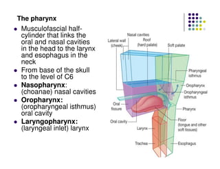
- 1. The pharynx Musculofascial half- cylinder that links the oral and nasal cavities in the head to the larynx and esophagus in the neck From base of the skull to the level of C6 Nasopharynx: (choanae) nasal cavities Oropharynx: (oropharyngeal isthmus) oral cavity Laryngopharynx: (laryngeal inlet) larynx
- 2. Attachments of the pharynx Base of the skull: irregular C-shaped The open part of the C faces the nasal cavities. Each arm of the C begins at the posterior margin of the medial plate of the pterygoid process of the sphenoid bone, just inferior to the cartilaginous part of the pharyngotymp anic tube. The line crosses inferior to the pharyngotympanic tube and then passes onto the petrous part of the temporal bone where it is just medial to the roughening for the attachment of levator veli palatini From here, the line swings medially onto the occipital bone and joins the line from the other side at a prominent elevation of bone in the midline (the pharyngeal tubercle).
- 3. Anterior vertical line of attachment for the lateral pharyngeal walls (1st) Begins superiorly on the posterior edge of the medial pterygoid plate of the sphenoid bone just inferior to where the pharyngotympanic tube lies against this plate. Continues inferiorly along the edge of the medial plate of the pterygoid process and onto the pterygoid hamulus. Descends along the pterygomandibular raphe to the mandible where this part of the line terminates
- 4. Anterior vertical line of attachment for the lateral pharyngeal walls (2nd) Begins on the lower aspect of the stylohyoid ligament, The line continues onto the lesser horn and then turns and runs posteriorly along the entire upper surface of the greater horn of the hyoid
- 5. Anterior vertical line of attachment for the lateral pharyngeal walls (3rd) Begins superiorly on the superior tubercle of the thyroid cartilage Descends along the oblique line to the inferior tubercle. Continues over the cricothyroid muscle along a tendinous thickening of fascia to the cricoid cartilage where it terminates
- 6. Muscles of the pharynx Superior constrictor: Origin: Pharyngeal raphe Insertion: Pterygomandibular raphe and adjacent bone on the mandible and pterygoid hamulus NS: pharyngeal branch of vagus Action: Constriction of pharynx - Palatopharyngeal sphincter: A special band of muscle originates from the anterolateral surface of the soft palate and circles the inner aspect of the pharyngeal wall, blending with the inner aspect of the superior constrictor
- 7. Muscles of the pharynx Middle constrictor: Origin: Pharyngeal raphe Insertion: Upper margin of greater horn of hyoid bone and adjacent margins of lesser horn and stylohyoid ligament NS: pharyngeal branch of vagus Action: Constriction of pharynx
- 8. Muscles of the pharynx Inferior constrictor: Origin: Pharyngeal raphe Insertion: Cricoid cartilage, oblique line of thyroid cartilage, and a ligament that spans between these attachments and crosses the cricothyroid muscle NS: pharyngeal branch of vagus Action: Constriction of pharynx
- 9. Pharyngeal raphe: From the pharyngeal tubercle on the base of the skull to the level of C6 where the raphe blends with connective tissue in the posterior wall of the esophagus - Fascia: Buccopharyngeal fascia: thin layer coats the outside of the muscular part of the wall (component of the pretracheal layer of cervical fascia) Pharyngobasilar fascia: a thick layer lines the inner surface of pharynx
- 10. Muscles of the pharynx Stylopharyngeus: Origin: Medial side of base of styloid process Insertion: Pharyngeal wall NS: Glossopharyngeal nerve Action: Elevation of the pharynx
- 11. Muscles of the pharynx Salpingopharyngeus: Origin: Inferior aspect of pharyngeal end of pharyngotympanic tube Insertion: Pharyngeal wall NS: Vagus nerve Action: Elevation of the pharynx
- 12. Muscles of the pharynx Palatopharyngeus: Origin: Upper surface of palatine aponeurosis Insertion: Pharyngeal wall NS: Vagus nerve Action: Elevation of the pharynx; closure of the oropharyngeal isthmus
- 13. Gaps in the pharyngeal wall
- 14. The nasopharynx Behind the posterior apertures (choanae) of the nasal cavities Above the level of the soft palate Ceiling: base of the skull and consists of the posterior part of the body of the sphenoid bone and the basal part of the occipital bone Continuous below with the cavity of the oropharynx at the pharyngeal isthmus. (marked on the pharyngeal wall by a mucosal fold caused by the underlying palatopharyngeal sphincter) Pharyngeal tonsils: large collection of lymphoid tissue in the mucosa covering the roof of the nasopharynx. enlargement of this tonsil, is known as adenoids
- 15. The nasopharynx Opening of the pharyngotympanic tube: posterior to and slightly above the level of the hard palate, and lateral to the top of the soft palate. Torus tubarius: posterior rim of the opening of the pharyngotympanic tube (elevation or bulge on the pharyngeal wall). Pharyngeal recess: (deep) Posterior to torus tubarius Salpingopharyngeal fold: small vertical, descends from the tubal elevation and overlies salpingopharyngeus muscle Torus levatorius: a broad fold or elevation that emerge from just under the opening of the pharyngotympanic tube, overlies the levator veli palatini muscle
- 16. The oropharynx Posterior to the oral cavity, Inferior to the level of the soft palate Superior to the upper margin of the epiglottis Oropharyngeal isthmus: boundary between the oral cavity and the oropharynx (palatoglossal muscles) Palatopharyngeal arches: posterior and medial to palatoglossal arches The anterior wall of the oropharynx inferior to the oropharyngeal isthmus is posterior one-third or pharyngeal part of the tongue (lingual tonsils) The palatine tonsils: large ovoid collection of lymphoid tissue in the mucosa lining the superior constrictor muscle and between the palatoglossal and palatopharyngeal arches on the lateral walls of the oropharynx
- 17. The laryngopharynx Extends from the superior margin of the epiglottis to the top of the esophagus at the level of C6 The laryngeal inlet opens into the anterior wall of the laryngopharynx. Inferior to the laryngeal inlet, the anterior wall consists of the posterior aspect of the larynx Valleculae: pair of mucosal pouches anterior to the cavity of the laryngopharynx (between the base of the tongue and epiglottis).
- 18. The laryngopharynx The valleculae are depressions formed between a midline mucosal fold and two lateral folds that connect the tongue to the epiglottis Piriform fossae: pair of mucosal recesses between the central part of the larynx and the more lateral lamina of the thyroid cartilage. - Form channels that direct solids and liquids from the oral cavity around the raised laryngeal inlet and into the esophagus
- 20. Blood supply of the pharynx Ascending pharyngeal artery Ascending palatine and tonsillar branches of the facial artery Branches of the maxillary and the lingual arteries Pharyngeal branches from the inferior thyroid artery
- 21. Pharyngeal plexus Formed by: Pharyngeal branch of the vagus nerve Branches from the external laryngeal nerve from the superior laryngeal branch of the vagus Pharyngeal branches of the glossopharyngeal nerve Motor: All muscles of the pharynx are innervated by the vagus nerve [X], except for the stylopharyngeus, which is innervated by glossopharyngeal nerve Sensory: Oropharynx and laryngopharynx: glossopharyngeal nerve Nasopharynx: pharyngeal branch of maxillary Nerve supply of the pharynx