Appendix In Femoral Hernia.Pdf
•
1 like•3 views

Academic Paper Writing Service http://StudyHub.vip/Appendix-In-Femoral-Hernia.Pdf 👈

Recommended
Recommended
More Related Content
Similar to Appendix In Femoral Hernia.Pdf
Similar to Appendix In Femoral Hernia.Pdf (20)
More from Zaara Jensen
More from Zaara Jensen (20)
Recently uploaded
Recently uploaded (20)
Appendix In Femoral Hernia.Pdf
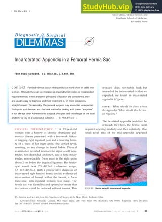
- 1. 3 5 w w w . c o n t e m p o r a r y s u r g e r y . c o m © 2003 Dow den Health M edia | V O L 5 9 , N O 1 / J A N 2 0 0 3 ■ revealed clear, non-turbid fluid; but instead of the incarcerated fat that we expected, we found an incarcerated appendix (Figure). D I L EM M A : What should be done about the appendix? How should this hernia be repaired? The herniated appendix could not be reduced; therefore, the hernia canal required opening medially and then anteriorly. One small focal area of the mid-appendix appeared C LI N I C AL P R E S E N T AT I O N | A 75-year-old woman with a history of chronic obstructive pul- monary disease presented with a two-week history of nagging right inguinal pain and a four-day histo- ry of a mass in her right groin. She denied fever, vomiting, or any change in bowel habits. Physical examination revealed normal vital signs, a soft, non- tender, non-distended abdomen, and a firm, mildly tender, non-reducible 3-cm mass in the right groin about 2 cm below the inguinal ligament. Her leuko- cyte count was 7.9x10³ /mL (reference range, 3.5–10.5x10³ /mL). With a preoperative diagnosis of incarcerated right femoral hernia and no evidence of incarceration of bowel within the hernia, a 5-cm transverse, infra-inguinal incision was made. The hernia sac was identified and opened to ensure that its contents could be reduced without trauma. This context Femoral hernias occur infrequently but more often in older, thin women. Although they can be mistaken as inguinal lymph nodes or incarcerated inguinal hernias, when anatomic principles of location are considered, they are usually easy to diagnose and their treatment is, on most occasions, straightforward. Occasionally, the general surgeon may encounter unexpected findings in such hernias, and the ideal method of dealing with these “ surprises” is not always clear. Adherence to surgical principles and knowledge of the local anatomy is key to a successful outcome. — D. FARLEY, M D ■ ■ D E P A R T M E N T E D I T O R ■ DAVID R. FARLEY, M D Mayo Clinic, Medical School, and Graduate School of Medicine, Rochester, Minn | DILEM M AS | Incarcerated Appendix in a Femoral Hernia Sac Diagno stic &Surgical DILEMMAS FERN A N D O CORD ERA , M D ; M I CH A EL G. SA RR, M D Drs Cordera and Sarr are from the Department of Surgery at the Mayo Clinic, Rochester, Minn. Correspondence: Fernando Cordera, MD; Mayo Clinic, 200 First Street SW, Rochester, MN 55905; telephone (407) 284-2511; fax (507) 536-7151 (e-mail: cordera.fernando@mayo.edu). F I G URE . Hernia sacwith incarcerated appendix.
- 2. 3 6 ■ V O L 5 9 , N O 1 / J A N 2 0 0 3 c o n t e m p o r a r y s u r g e r y Diagno stic &Surgical DILEMMAS mildly ischemic, and thus an appendectomy was performed. The hernia sac was ligated and reduced above the femoral canal, and the hernia defect was closed with a polypropylene mesh plug. Two days later, erythema was noted around her incision. She was taken back to the operating room, the mesh was removed, and the defect was closed primarily. Discharge was uneventful two days later. D I S C U S S I O N | Femoral hernias occur through a space bounded superiorly by the iliopubic tract, inferiorly by Cooper’s ligament, laterally by the femoral vein, and medially by the insertion of the iliopubic tract to Cooper’s ligament. Femoral her- nias account for up to 4% of all groin hernias and are much more common in women.1 Herniation of the intestinal tract into femoral hernias occurs in one third of patients, and the rate of incarceration is higher (14%–56%) than in inguinal hernias (6%–10%). Small bowel, colon, Meckel’s diverticu- lum, and even gastric herniation have been described. Herniation of the appendix into a femoral sac, however, is rare. In a series of 655 femoral hernia repairs, Wakeley2 reported the inci- dence of herniation of the appendix into a femoral sac as less than 1%. An incarcerated appendix in a femoral hernia was first reported by De Garengot in 1731.3 By 1974, 242 patients had been reported with an appendix herniating into a femoral hernia, 59 of whom presented with acute appendicitis.4 Incarceration is not an indication for appendec- tomy; however, if the appendix shows signs of strangulation or inflammation, it should be removed.5 If the appendix appears viable, not inflamed, and is easily reduced, resection is unnec- essary and may be counterproductive because it can increase the rate of wound infection. In this particular patient, the appendix appeared to have a small area of mild ischemia at the incarceration site and was thus removed. There are four different anatomic approaches to a femoral hernia: infra-inguinal; anterior inguinal (through the posterior inguinal floor); preperi- toneal; and transperitoneal via an endoscopic, min- imal access approach. There is no consensus on which approach is the best, and each has its poten- tial advantages and disadvantages. The lack of any preoperative symptoms of gut incarceration led the senior surgeon (MGS) to approach the hernia in this older female patient from the least debilitating approach, i.e. infra-inguinally, fully expecting to find incarcerated preperitoneal fat. Finding the appendix instead proved to be a surprise. When incarceration is suspected preoperatively, a “high” or more rostral approach provides advantages over other incisions.6-7 Once the appendectomy has been performed and the anatomy of the femoral region has been identified, the surgeon must decide how the defect should be closed. Although the fluid in the hernia sac was clear, the appendix was clearly viable, and the mildly ischemic focus was non-inflamed, the decision to “plug” the defect to reduce the herniat- ed content with mesh was in retrospect probably ill- advised (the senior surgeon agrees!). Prosthetic material should be avoided whenever possible in procedures in which contamination is likely. Other options would have included a primary repair by sewing the pectineus fascia to the remnants of the inguinal ligament (this choice was deemed less opti- mal at the time of surgery because of the enlarge- ment of the femoral canal needed to mobilize the appendix) or to make a separate counter-incision and repair the hernia “from above.” Probably the former would have been most advisable. This case, though describing an uncommon problem, underscores how adherence to surgical principles should be observed to obtain a successful outcome. Understanding the clinical problem, potential complications, and knowledge of the regional anatomy are key in every patient. Editor’s comment | What a fantastic specialty! General surgery allows one to help patients that harbor a diversity of pathophysiologic abnormalities that oth- Prosthetic material should be avoided whenever possible in procedures in which contamination is likely.
- 3. References 1. Bay-Nielsen M, Kehlet H, Strand L, et al. Quality assessment of 26,304 herniorrhaphies in Denmark: a prospective nationwide study. Lancet. 2001;358:1124-1128. 2. Wakeley CPG. Hernia of the Vermiform appendix. In: Maingot R, ed, Abdominal Operations. New York: Appleton Century-Crofts; 1969:1288. 3. Garland EA. Femoral appendicitis. J Indiana Med Assoc. 1955;48:1292- 1294. 4. Voitk AJ, MacFarlane JK, Estrada RL. Ruptured appendicitis in femoral hernias: report of two cases and review of the literature. Ann Surg. 1974;179:24-26. 5. Naude GP, Ocon S, Bongard F. Femoral hernia: the dire consequences of a missed diagnosis. Am J Emerg Med. 1997;15:680-682. 6. Wyatt JP, Varma JS. Femoral hernia appendix causing small intestinal obstruction. Postgrad Med J. 1992;68:223-224. 7. Khatib CM. Strangulated femoral hernia containing acute gangrenous appendicitis: case report and review of the literature. Can J Surg. 1987;30:50. ers can only dream of… an appendix incarcerated in a femoral hernia. The specialty rewards hard- working young men and women with bountiful opportunities in clinical and research realms. The junior author makes good and moves from interna- tional medical student to general surgery prelim to categorical trainee at an “ivory tower institution.” The field generates, cultivates, and perpetuates the creation of men and women of honor, class, and humility. The senior author willingly offers up this dilemma so others may avoid the pitfalls inherent in contemplating or breaking with surgical dictum. Bravo! My hat is off to general surgeons everywhere! —D. FARLEY, MD Dilemmas: Incarcerated Appendix