The Impact of Lysogenic and Tail Assembly Chaperone Proteins on the Life Cycle and Virulence of Mycobacteriophage Emma
•
1 like•112 views

Recommended

Recommended
More Related Content
What's hot
What's hot (20)
Similar to The Impact of Lysogenic and Tail Assembly Chaperone Proteins on the Life Cycle and Virulence of Mycobacteriophage Emma
Similar to The Impact of Lysogenic and Tail Assembly Chaperone Proteins on the Life Cycle and Virulence of Mycobacteriophage Emma (20)
Recently uploaded
Recently uploaded (20)
The Impact of Lysogenic and Tail Assembly Chaperone Proteins on the Life Cycle and Virulence of Mycobacteriophage Emma
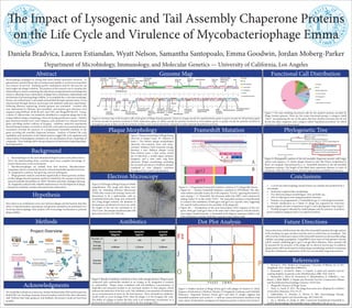
- 1. References • Bertani G. 1951. Studies on Lysogenesis. University of Illinois. 62: 23-295. DrugBank. 2013. Ampicillin (DB00415). • Krumsiek J., Arnold R., Rattei, T. Gepard: A rapid and sensitive tool for creating dotplots on genome scale. Bioinformatics 2008; 23(8) 1026-8. • Marianelli, L., Piuri, M., Swinonova, Z., Balachandran, A., Oldfield, L., van Kessel, J. 2008. BRED: A Simple and Powerful Tool for Construction Mutant and Recombinant Bacteriophage Genomes. PLOS One. • PhagesDB, Glossary of Phage Terms. • Stern, A., Sorek, R. 2011. The phage-host arms-race: Shaping the evolution of microbes. Bioessays; 33(1) 43-51. • Sulakvelidze, A., Alavidze, Z., Morris, G. 2001. Bacteriophage Therapy. Antimicrobial Agents and Chemotherapy; 48(3) 649-659. • Xu, J., Hendrix, R., Duda, R. 2004. Conserved Translational Frameshift in dsDNA Bacteriophage Tail Assembly Genes. Molecular Cell, 16: 11-21 Future Directions Future directions will determine the role of the frameshift mutation through analysis of the resulting two gene products and the ratios in which they are translated. This will reveal the evolutionary impact of the frameshift mutation on tail assembly, phage viability, and phage propagation. Using BRED, three types of viral genome mutants will be created, exhibiting gp12, gp13, and gp12&13 deletions. These mutants will be assessed for the presence of the phage tail via electron microscopy. In addition, plaque assays will be performed to analyze plaque morphology and draw conclusions about the evolutionary implications of the two tail assembly chaperone proteins. Conclusions • A novel mycobacteriophage named Emma was isolated and purified from a soil sample. • Emma has a siphoviridae morphology. • Two observed plaque morphologies: clear and bulls-eye. • 36% of called genes were assigned putative function. • Presence of a programmed +1 frameshift for gp 13, a tail chaperone protein. • Emma’s classification as a Cluster F1 phage was supported by restriction enzyme digests, dot plot comparisons of gp13, and a phylogenetic map of gp13. • Possible lysogeny indicated by turbid plaques and the presence of putative genes coding for integrase and a Cro repressor protein. Phylogenetic Tree Functional Call Distribution Dot Plot Analysis Frameshift Mutation Figure 4: +1 Programmed frameshift mutation common in F1 phages like Emma. • Figure 4A — Emma’s Frameshift Mutation visualized in DNAMaster. The ribo- some stutters and adds Lysine (K) at the sequence “AAAA,” ignoring the fourth A and causing a “+1” frameshift. The ribosome shifts into ORF 3 and continues by adding Valine (V) at the codon “GTG.” The frameshift mutation is hypothesized to conserve the translation of both gp12 and gp13 in a specific ratio, suggesting the need for both proteins in the assembly of the phage tail. • Figure 4B — Visualization of the post-transcription RNA sequence of the two genes. Both gp12 and gp13 start at the same start codon (green), but the transla- tion of gp13 results from the +1 frameshift at the slippery sequence (yellow), and both translations continue until their respective stop codons (red). Antibiotics Electron Microscopy Plaque Morphology Genome Map Acknowledgments We would like to thank our instructors, Jordan Moberg Parker PhD and Krisanavane Reddi PhD, our Teaching Assistant, Emma Goodwin, and all of the other laboratory staff. Without their help, guidance, and feedback, this project would not have been possible. Methods Hypothesis Since there is an evolutionary arms-race between phages and the bacteria that they infect, if characterization experiments and genome annotation are performed on a novel mycobacteriophage, then analysis will reveal phage mechanisms to promote phage viability. Background • Bacteriophages are the most abundant biological units on the planet (Gross, 2012). By understanding them, scientists gain more complete knowledge of system interactions in the environment. • Mycobacteriophages are isolated from host bacteria, Mycobacterium smegmatis, which is commonly found in soil and mammalian genital secretions. M. smegmatis is common, fast-growing, and non-pathogenic. • Phage genomic analysis contributes significantly to future genomic analysis of other phages because genomic analysis is limited by the amount of previous research available (Risenfeld et al., 2004). Therefore, the study of phages infecting M. smegmatis could provide valuable implications in the development of phage therapy for antibiotic-resistant Mycobacterium tuberculosis. Abstract Bacteriophages propagate by seizing their host’s limited replication resources. To gainprimarycontrolofthese,thetwoentitiesmustrapidlyco-evolvetoincreasetheir own chances of survival. Studying specific mechanisms used for this process can lend insight into phage evolution. The purpose of this research was to examine this relationshipinasystemcontainingMycobacteriumsmegmatisandanovelphagethat infects it, allowing closer examination of phage-host evolutionary relationships and mechanisms of promoting phage viability. To accomplish this, a mycobacteriophage, Emma, was isolated from a soil sample and purified through repeated assays. It was characterized through electron microscopy and antibiotic induction experiments. Following Illumina sequencing, Emma’s genome was annotated. Location calls were determined by Glimmer and GeneMark, and putative gene functions were assigned using HHPred, NCBI BLAST and CDD, and PhagesDB BLAST. Emma, a cluster F1 siphoviridae, was tentatively identified as a temperate phage due to the unique bullseye plaque morphology observed during purification assays. Putative gene functions include LysA, LysB, integrase, and a potential Cro-repressor protein genes, providing further evidence of lysogeny. Antibiotic induction experiments showed Emma’s tendency to switch to a lytic life-cycle under environmental stresses. Annotation revealed the presence of a programmed frameshift mutation in the genes encoding tail assembly chaperone proteins. Analysis of Emma’s life cycle capabilities and annotation of tail-related proteins suggest life cycle regulation and conservation of advantageous mutations are two mechanisms of out-competing M. smegmatis. These findings add to the growing pool of information regarding phage- host interactions. Daniela Bradvica, Lauren Estiandan, Wyatt Nelson, Samantha Santopoalo, Emma Goodwin, Jordan Moberg-Parker Department of Microbiology, Immunology, and Molecular Genetics — University of California, Los Angeles Figure 6: Genome map of the location calls of the genes of Phage Emma’s genome. Genes in orange encode for capsid proteins; genes in green encode for tail proteins; genes in brown encode for proteins involved in DNA replication; genes in yellow encode for proteins involved in transcription; genes in purple encode for proteins involved in propagation; genes in blue encode for proteins of other, more narrow functions; and genes in pink represent hypothetical proteins. Figure 8: Phylogenetic analysis of the tail assembly chaperone protein with longer amino acid sequence. F1 cluster phages (boxed in red), like Emma (underlined in blue), are uniquely characterized by a +1 frameshift mutation in the tail assembly chaperone proteins. The length of the black lines represents distance in units of mutations, while the red numbers represent approximate probability of accuracy. Figure 7: Pie chart detailing functional calls for the putative proteins encoded by Phage Emma’s genome. There are five major functional groups, a category called “other” encompassing the rest of the genes that have putative functions but do not fit into the other categories, and a category called “hypothetical proteins” for which functions could not be determined. The Impact of Lysogenic and Tail Assembly Chaperone Proteins on the Life Cycle and Virulence of Mycobacteriophage Emma B Figure1:PlaquemorphologyofPhageEmma observed in plaque assay, 10-5 dilution of lysate 1. Two distinct plaque morphologies observed, less-common clear and more- common “bullseye”, both consistent average diameter 3 mm. Bullseye plaques reveal three distinct zones: a clear center, (initial site of infection), a turbid region (putative lysogeny), and a clear outer ring (lytic infection). Plaque morphology (including the increased prevalence of bullseye plaques compared to clear plaques) consistent throughout four rounds of purification. Figure 2: EM Image of Phage Emma at 67000x magnification. This image and others were taken via Tunneling Electron Microscope (TEM) after uranyl acid staining. Emma was determined to be a siphoviridae, with an icosahedral head and a long, non-contractile tail. Using ImageJ software, the diameter of the head was calculated to be 62±4 nm, while the length of the tail was calculated to be 182±13 nm. The sample visualized under EM had a titer of 9.45 x 10¹⁰ PFU/mL. Figure 3: Results of antibiotic interference tests, with example pictures. Plaque assays conducted with carbenicillin resulted in no change, as M. smegmatis is resistant to carbenicillin. Plaque assays completed with sub-inhibitory concentrations of ampicillin and isoniazid resulted in an increased number of clear plaques, which indicates the induction of the lytic cycle. Sub-inhibitory concentrations of antibiotics are an environmental of stress for the host bacteria. Host cell death by antibiotics would result in a loss of phage DNA when the phage is in the lysogenic life cycle. The ability of a phage to induce the lytic cycle is an evolutionary mechanism, as it prevents host cell lysis which would result in the loss of phage DNA. Figure 5: Dotplot analysis of Phage Emma gp13 with phages of Clusters F1 (Bobi, Seagreen,Kimberlium),J(Redno2,Wanda),O(Yungjamal,Catdawg),andP(Malithi, Phayonce). Alignment between Emma gp13 and other F1 phages suggests that frameshift mutations such as the F1 +1 shift are conserved between members of the same cluster. Kimberlium's analogous tail chaperone protein is shorter than Emma's. A gp12 gp13 S T A R T S T A R T S T O P S T O P S L I P S L I P