SlideShare a Scribd company logo
1 of 26
Download to read offline
Minimal Access Robotic Surgery
INTRODUCTION
On July 11, 2000, the Food and Drug Administration (FDA)
approved the first completely robotic surgery device, the
da Vinci surgical system from Intuitive Surgical (Mountain
View, CA). The system enables surgeons to remove
gallbladders and perform other general surgical procedures
while seated at a computer console and three-dimensional
(3D) video imaging system across the room from the patient.
The surgeons operate controls with their hands and fingers
to direct a robotically controlled laparoscope (Fig. 1).
This system and other robotic devices developed or
under development by companies such as Computer Motion
(Santa Barbara, CA) and Integrated Surgical Systems (Davis,
CA) have the potential to revolutionize surgery and the
operating room. They provide surgeons with the precision
and dexterity necessary to perform complex, minimally
invasive surgery (MIS) procedures, such as beating-heart
single-ordouble-vesselbypassandneurological,orthopedic,
and plastic surgery, among many other future applications.
Manufacturers believe that their products will broaden the
scope and increase the effectiveness of MIS; improve patient
outcomes; and create a safer, more efficient, and more cost-
effective operating room. It is the vision of these companies
that robotic systems will one day be applicable to all surgical
specialties, although it is too early to tell the full extent to
which they will be used.
The first generation of surgical robots is already being
installed in a number of operating rooms around the world.
These are not true autonomous robots that can perform
surgicaltasksontheirown,buttheyarelendingamechanical
helping hand to surgeons. These machines still require
a human surgeon to operate them and input instructions.
Remote control and voice activation are the methods by
which these surgical robots are controlled. Robotics are
being introduced to minimal access surgery because they
allow for unprecedented control and precision of surgical
instruments. So far, these machines have been used to
position an endoscope, perform gallbladder surgery, and
correct gastroesophageal reflux and heartburn. The ultimate
goal of the robotic surgery field is to design a robot that can
be used to perform closed-chest, beating-heart surgery.
Recently, transatlantic surgery between USA and
Strasbourg is a revolution in transatlantic minimal access
surgery. In this surgery, there was slight delay (66 ms) in
transfer of data but in future, this delay can easily minimized.
In the future, remote handling technology will overcome the
manipulative restriction in the current instruments. There
is no doubt 10 years from now that some surgeons will be
operating exclusively via a computer interface controlling a
master-slave manipulation. If computer-controlled machinery
can mimic the awareness, adaptability, and knowledge of a
human surgeon, such a takeover in the operating theater is
actually realistic.
In the operating room of the future, physicians will use
tiny high-tech tools to travel inside the body with dexterity
and precision beyond imaging. The future of any new
technology depends upon the training.
Three surgical robots that have been recently developed are:
1. da Vinci surgical system
2. ZEUS robotic surgical system
3. Automated Endoscopic System for Optimal Positioning
(AESOP) robotic system.
The “da Vinci” system has seven degree of freedom
movement, so it can perform more complex task. The
“da Vinci” system has been used to perform a number of
general surgical procedures such as cholecystectomy and
fundoplication.Roboticfundoplicationallowseasierpassage
around and behind the esophagus during its dissection and
Fig. 1: Robotic surgery port placement for nephrectomy.
Prof. Dr. R. K. Mishra
592 SECTION 6: Miscellaneous
easier mobilization of the curvature of the stomach. Suturing
the wrap and the crural approximation are also easier with
the help of these robots.
The $1 million da Vinci system consists of two primary
components:
1. A viewing and control console
2. A surgical arm unit (Figs. 2A to C).
In using da Vinci for gallbladder surgery, three incisions,
no larger than the diameter of a pencil, are made in the
patient’s abdomen, which allows for three stainless-steel
rods to be inserted. The rods are held in place by three
robotic arms. One of the rods is equipped with a camera,
while the other two are fitted with surgical instruments that
are able to dissect and suture the tissue of the gallbladder.
Unlike in conventional surgery, these instruments are not
directly touched by the doctor’s hands.
Effect or tips of the da Vinci surgical system incorporate
miniature wrists that allow them to mimic any movement
made by the surgeon at the control console. Sitting at the
control console, a few feet from the operating table, the
surgeon looks into a viewfinder to examine the 3D images
being sent by the camera inside the patient. The images
show the surgical site and the two surgical instruments
mounted on the tips of two of the rods. Joystick-like controls,
located just underneath the screen, are used by the surgeon
to manipulate the surgical instruments. Each time one of the
joysticks is moved, a computer sends an electronic signal
to one of the instruments, which moves in synchronization
with the movements of the surgeon’s hands.
Another robotic system is the ZEUS system, made by
Computer Motion, which is already available in Europe.
However, both the da Vinci and ZEUS systems must receive
governmental approval for each procedure that a surgeon
plans to use it for. The $750,000 ZEUS has a similar setup to
that of the da Vinci. It has a computer workstation, a video
display, and hand controls that are used to move the table-
mounted surgical instruments. While the ZEUS system has
not yet been cleared for American use beyond clinical trials,
German doctors have already used the system to perform
coronary bypass surgery.
The ZEUS system employs the assistance of the AESOP
robotic system. Released by Computer Motion in 1994,
AESOP was the first robot to be cleared by the FDA for
assisting surgery in the operating room. AESOP is much
simpler than the da Vinci and ZEUS systems. It is basically
just one mechanical arm, used by the physician to position
the laparoscope. Foot pedals or voice-activated software
allows the surgeon to position the camera, leaving his or
her hands free to continue operating on the patient (Fig. 3).
The use of a computer console to perform operations from
a distance opens up the idea of telesurgery, which would
involve a doctor performing delicate surgery thousands of
Figs. 2A to C: (A and B) Robotic arms of da Vinci surgical system; (C) Effector tips of the da Vinci surgical system.
A B
C
593
CHAPTER 47: Minimal Access Robotic Surgery
Fig. 3: Robotic surgery. (1) Surgeon console; (2) Image processing
equipment; (3) EndoWrist instruments; (4) Surgical arm cart; and (5) High-
resolution three-dimensional (3D) endoscope.
miles away from the patient. If it were possible to use the
computer console to move the robotic arms in real time, then
it would be possible for a doctor in New Delhi to operate on
a patient in New York. A major obstacle in telesurgery has
been the time delay between the doctor moving his or her
hands to the robotic arms responding to those movements.
Currently, the doctor must be in the room with the patient
for robotic systems to react instantly to the doctor’s hand
movements.
Having fewer personnel in the operating room and
allowing doctors the ability to operate on a patient, long
distance could lower the cost of health care. In addition to
cost-efficiency, robotic surgery has several other advantages
over conventional surgery, including enhanced precision
and reduced trauma to the patient. For instance, heart
bypass surgery now requires that the patient’s chest 30.48 cm
long incision. However, with the da Vinci or ZEUS systems,
it is possible to operate on the heart by making three small
incisions in the chest, each only 10 mm in diameter. The
patient would experience less pain and less bleeding, which
means a faster recovery.
Robotics also decreases the fatigue that doctors experi-
ence during surgeries that can last several hours. Surgeons
can become exhausted during those long surgeries and can
experience hand tremors as a result. Even the steadiest of
human hands cannot match those of a surgical robot. The
da Vinci system has been programmed to compensate for
tremors, so if the doctor’s handshakes, the computer ignores
it and keeps the mechanical arm steady. While surgical
robots offer some advantages over the human hand, we are
still a long way from the day when autonomous robots will
operate on people without human interaction. But, with
advances in computer power and artificial intelligence, it
could be that in this century a robot will be designed that can
locate abnormalities in the human body, analyze them, and
operate to correct those abnormalities without any human
guidance.
Fig. 4: Robotic surgery via master-slave manipulator.
The fallopian tube reconnection procedure, referred
to as tubal reanastomosis, was performed by Dr Tommaso
Falcone, who is Head of the Reproductive Endocrinology
and Infertility Section at the Cleveland Clinic. Dr Falcone
used Computer Motion’s ZEUS robotic surgical system as
part of a clinical trial, approved by the United States FDA.
The patient, a 38-year-old woman, and her healthy
10-day-old son are both in excellent condition and have
returned to their Cleveland-area home. The mother had
originally undergone a tubal ligation sterilization operation
in her 20s. She and her partner later wished to have a child
together and began preparing for a reversal operation.
The patient saw an advertisement for the ZEUS study
and consulted with trial leader Dr Falcone. The lady was
informed in detail of the investigational protocol and agreed
to have the robotically-assisted procedure.
In addition to the ZEUS System, Computer Motion
markets the AESOP 3000, a voice-controlled endoscope
positioning system, and the HERMES Control Center, a
centralized system, which enables the surgeon to voice
control a network of “smart” medical devices. Currently,
the ZEUS system is under an FDA-approved investigational
device exemption and is also CE marked for commercial sale
in Europe.
Robotics is rapidly developing in surgery, although the
word is slightly misused in this connection. None of the
systems under development involves a machine acting
autonomously. Instead, the machine acts as a remote
extension of the surgeon. The correct term for such a system
is a “master-slave manipulator,” although it seems unlikely
that this term will gain general currency.
Minimal invasive surgery is itself a form of telemani-
pulation because the surgeon is physically separated from
the workspace. Telerobotics is an obvious tool to extend the
surgeons capabilities. The goal is to restore the tactile cues
and intuitive dexterity of the surgeon, which are diminished
by MIS. A slave manipulator, controlled through a spatially
consistent and intuitive master with a force feedback system,
could replace the tactile sensibilities and restore dexterity
(Fig. 4).
594 SECTION 6: Miscellaneous
Fig. 5: Master-slave manipulator.
Fig. 6: Robotic console. Fig. 7: Robotic arm.
Fig. 8: Ports in cardiac minimal access surgery.
Althoughthepotentialofroboticsurgeryisjustbeginning
but progress may come quickly. Laparoscopic gallbladder
surgery was first done in 1987, but it became standard within
5 years. Just think about a surgeon! He picks up this black box
and waves it over your body and you are fixed. How is that
going to happen? One day, a surgeon may use robotic devices
to enter the body through its own orifices. They could carry
medical instruments inside the body, where they would be
manipulated by simple computer commands (Figs. 5 to 7).
The robotic arm after addition of wrists permits the sur-
geon to mimic his own movements, rather than experience
limitations of the rigid long cylindrical laparoscopic instru-
ment and has obvious advantages in terms of dexterity and
complexity of instrument (Fig. 8).
INSTRUMENTS OF ROBOTIC SURGERY
da Vinci Surgical System (Figs. 9 to 20)
Using the most advanced technology available today, the da
Vinci surgical system enables surgeons to perform delicate
and complex operations through a few tiny incisions with
increased vision, precision, dexterity, and control. The da
Vinci surgical system consists of several key components,
including: an ergonomically designed console where the
surgeon sits while operating, a patient-side cart where the
patient lays during surgery, four interactive robotic arms, a
high-definition 3D vision system, and proprietary EndoWrist
instruments.
595
CHAPTER 47: Minimal Access Robotic Surgery
Fig. 9: da Vinci high-definition surgical robot.
Fig. 10: da Vinci surgeon console. Fig. 11: Patient cart.
Fig. 12: Seven-degree movement is possible. Fig. 13: da Vinci vision cart.
596 SECTION 6: Miscellaneous
Fig. 14: Robotic EndoWrist instruments.
Figs. 15A and B: Robotic scissors and bipolar dissector. Figs. 16A and B: Robotic tenaculum and grasper.
Figs. 17A and B: Robotic needle holder. Figs. 18A and B: Robotic atraumatic grasper.
Figs. 19A and B: Robotic clip applicator. Figs. 20A and B: Robotic scissor and Maryland.
da Vinci is powered by state-of-the-art robotic techno-
logy that allows the surgeon’s hand movements to be scaled,
filtered, and translated into precise movements of the
EndoWrist instruments working inside the patient’s body.
Core Technology
	
■ System components
	
■ Three-dimensional high-definition (HD) vision.
A B A B
A B
A B A B
A B
597
CHAPTER 47: Minimal Access Robotic Surgery
Instrumentation
Using the da Vinci surgical system, the surgeon operates
seated comfortably at a console while viewing an HD, 3D
image inside the patient’s body.
	
■ The surgeon’s fingers grasp the master controls below
the display with hands and wrists naturally positioned
relative to his or her eyes
	
■ The system seamlessly translates the surgeon’s hand,
wrist, and finger movements into precise, real-time
movements of surgical instruments
	
■ The patient-side cart is where the patient is positioned
during surgery. It includes either three or four robotic
arms that carryout the surgeon’s commands
	
■ The robotic arms move around fixed pivot points, which
reduce trauma to the patient, improve the cosmetic
outcome, and increase overall precision
	
■ The system requires that every surgical maneuver be
under the direct control of the surgeon. Repeated safety
checks prevent any independent movement of the
instruments or robotic arms
	
■ A full range of EndoWrist instruments is available to the
surgeon while operating
	
■ The instruments are designed with seven degrees of
motion—a range of motion even greater than the human
wrist
	
■ Each instrument has a specific surgical mission such as
clamping, suturing, and tissue manipulation
	
■ Quick-release levers speed instrument changes during
surgery
	
■ The vision system is equipped with an HD, 3D endoscope
(flexible tube with a camera and light at the tip) and
image processing equipment that provides true-to-life
images of the patient’s anatomy
	
■ A view of the operating field is available to the entire
operative room (OR) team on a large viewing monitor
(vision cart). This widescreen view provides the surgical
assistant at the patient’s side with a broader perspective
and visualization of the procedure.
INSTRUMENTS AND ACCESSORIES
Intuitive surgical’s exclusive EndoWrist instruments are
designed to provide surgeons with natural dexterity and full
range of motion for precise operation through tiny incisions.
Modeled after the human wrist, EndoWrist instruments
can offer an even greater range of motion than the human
hand. They truly allow the da Vinci®
system to take surgical
precision and technique beyond the limits of the human
hand. Similar to human tendons, an EndoWrist instrument’s
internal cables provide maximum responsiveness,
allowing rapid and precise suturing, dissection, and tissue
manipulation. EndoWrist instruments provide enhanced
dexterity, precision, and control:
	
■ 7° of freedom
	
■ 90° of articulation
	
■ Intuitive motion and finger-tip control
	
■ Motion scaling and tremor reduction.
The wrist-like movement, responsiveness, and robotic
control afforded by the da Vinci system and its exclusive
EndoWristinstrumentsprovidesurgeonsfluidambidexterity
and unparalleled precision. The EndoWrist instruments
are available in a wide selection of specialized tip designs
to enable a broad range of da Vinci procedures. As da Vinci
surgery is adopted in new specialties, our engineers work
side-by-side with surgeons to develop new EndoWrist
instruments to address new clinical needs. The following
is a list of instrument categories for which intuitive offers
specialized tip designs.
Energy instruments are used by the da Vinci surgeon
to provide coagulation, cutting, and dissection of tissues.
These include monopolar and bipolar cautery instruments
(electrical energy), the Harmonic™ ACE (mechanical
energy), the PK™ dissecting forceps (advanced bipolar),
and laser.
Grasping instruments allow for manipulation of a wide
range of tissues, from fine, thin tissues such as peritoneum
to dense, fibrous tissues such as uterus. Needle drivers
provide the ability to suture with the finest of needles used in
cardiovascular surgery as well as the thickest needles used in
repair of uterine defects. Suture Cut™ needle drivers include
an integral cutting blade for efficient cutting of suture after
knot tying and increased surgeon autonomy and efficiency.
Retracting instruments allow the surgeon to dynamically
provide exposure of the surgical field. This minimizes
dependency on the patient-side assistant, providing the da
Vinci surgeon with full control of the operative field.
Clip appliers, probe graspers, and cardiac stabilizers
are available to allow the da Vinci surgeon to perform
specialized procedures such as vessel clipping, cryoablation,
and beating-heart surgery.
A selection of 5 mm EndoWrist instruments provides the
surgeon with the ability to use smaller access ports. Some
surgeons prefer smaller access ports when performing
pediatric, thoracic, bariatric, gynecologic, and general
surgery.
OPERATING ROOM CONFIGURATION, PORT
PLACEMENT, AND DOCKING
Before any procedure, the robot has to be prepared for
surgery. It includes the connection of all necessary parts
such as sterile drapes and connectors needed for surgery
and the calibration process. These steps are conducted by
a core team of scrub nurses specifically trained in handling
the robot while the patient is in preparation for surgery.
Currently, the only available system for laparoscopic surgery
is the da Vinci surgical system, developed by Intuitive
Surgical (Sunnyvale, CA, USA).
Anyroboticprocedureisperformedbyateamofsurgeons
and nurses. It includes the console surgeon, patient-side
598 SECTION 6: Miscellaneous
assistant surgeon, and the scrub nurse. Although console
surgeon is the leader of the team, a trained assistant
surgeon is of paramount importance as he/she is the
person responsible for robot docking, instrument change,
manipulation of laparoscopic instruments, application
of hemostatic instruments/clips, lavage and aspiration,
specimen extraction, drainage, and closure of abdominal
wall. A trained scrub nurse is also important in draping the
robotic arms, attaching the optics, instrument changes, and
undocking of the robot.
Operating Room Configuration
This topic includes the optimum positioning of the following
components in the operating room (OR) so as to allow for
maximum functionality:
	
■ Surgeon console positioning
	
■ Patient cart positioning
	
■ Vision cart positioning.
Surgeon Console Positioning
The surgeon console is placed outside of the sterile field. It
should be oriented in such a position so that the surgeon
has a clear view of the operative field, vision cart, and able
to communicate directly with the assistant surgeon and the
scrub nurse.
For moving or positioning the surgeon console, only the
handle (Fig. 21) on the back of the console is used. It should
never be pushed or pulled by the console body or armrest to
maneuver the console into place. Wheel locks located on the
rear wheels of the surgeon console should be locked after
positioning of console for the surgery.
Patient Cart Positioning
The patient cart is placed in the sterile field. It should be
draped in a separate area in the room prior to moving it into
place for surgery. This should be an area of the room where
it will not easily come into contact with nonsterile objects or
impedetraffic.Oncethepatientcartisdrapedandthepatient
is positioned, prepared, and draped, ports are placed. Then,
use the patient cart motor drive to help move the cart into
the sterile field.
The patient cart brakes are designed to automatically
engage when the motor drive is not in use.
Motor drive operation: The motor drive interface consists of
the following components (Fig. 22):
	
■ Throttle
	
■ Throttle enable switch
	
■ Shift switches.
To operate the motor drive:
	
■ Ensure that the patient cart is powered on
	
■ Ensure the shift switches are in the drive position
(Figs. 23A and B)
	
■ Throttle enable switch is to be held and throttle rotated
away from the operator or toward him/her depending on
the intended direction of movement
	
z The cart power light-emitting diode (LED) will flash
green whenever the throttle enable switch is activated
	
z The drive speed of the cart can be controlled by
rotating the throttle to different extent in each
direction.
The motor drive will not engage whenever cannulas or
instruments are installed on the system. A yellow LED on
motor drive interface labeled “cannula installed: cart drive
disabled” will indicate when cannulas or instruments are
installed and motor drive is nonoperational. This has been
done for safety purpose.
Shift switches: The patient cart can be moved without the
use of the motor drive (for example, during a power loss)
by rotating the shift switches (Fig. 23B) to the neutral (N)
position. The cart can then be moved manually. When the
cart has been moved, shift switches should be placed in the
drive (D) position to set the patient cart brakes.
Fig. 21: Surgeon console handle. Fig. 22: Location of throttle, throttle enable switch, and shift switches.
599
CHAPTER 47: Minimal Access Robotic Surgery
Figs. 23A and B: (A) Drive motor engaged; (B) Drive motor disengaged.
(D: drive; N: neutral)
For patient safety, the shift switches must be kept in the
drive (D) position so that the motor drive remains engaged
during surgery (Fig. 23A).
Vision Cart Positioning
The vision cart is placed adjacent to the patient cart, just
outside of the sterile field, to allow the patient cart operator
to see the component displays (Fig. 24).
	
■ The vision cart should be close enough to the patient cart
to allow unrestricted camera cable movement during
surgery
	
■ Wheel locks are located on the rear wheels of the vision
cart. These should be locked after the cart is positioned
for surgery.
Steps of docking:
	
■ Position patient and OR table, including table tilt
	
■ Position patient cart over patient
	
■ Set patient cart brakes
	
■ Docking the camera arm
	
■ Docking the instrument arms
	
■ Check system setup.
Fig. 24: Position of vision cart.
Fig. 25: Camera port positioning.
The patient table should be positioned according to
surgeon preference (depending on the contemplated
procedure) before docking the robotic arms. Once the arms
are docked to the ports and instruments are placed, patient
position should not be changed. The pneumoperitoneum is
created and the ports are inserted by either the lead surgeon
or the patient-side assistant surgeon.
Port Placement
The port positions vary from patient-to-patient, procedure-
to-procedure, and surgeon-to-surgeon. It is very difficult
to form guidelines-specific position of ports, but broad
guidelines are framed to maximize endoscopic view,
instrument reach, and to minimize external arm clashing.
Camera port is inserted keeping following principles in
mind (Fig. 25):
	
■ Should be in line with the target anatomy (TA)
	
■ Should beat 10–20 cm distance from the TA
A B
600 SECTION 6: Miscellaneous
Figs. 26A and B: (A) Disposable 12 mm trocar cannula assembly for camera port; (B) Camera port being mounted on the camera arm.
Figs. 27A and B: Port placement.
	
■ Should be in line with the center column of the patient
cart. A diagnostic laparoscopy is done after insertion of
the camera port to look for safe entry, any adhesions, and
surgical feasibility.
A disposable 12 mm trocar cannula assembly is used as
the camera port (Figs. 26A and B). There are specialized
camera arm cannula mounts (on the patient cart camera
arm) corresponding to each validated third-party cannula.
Working Ports
An 8 mm da Vinci ports are inserted for robotic arms that are
inserted keeping in mind the following principles:
	
■ >8 cm distance between the da Vinci ports (Fig. 27A)
	
■ 10–20 cm distance should be maintained between the da
Vinci ports and TA (Fig. 27B).
A 10 cm distance from TA is good but 20 cm distance is
better. Ports placed closed to the TA impede the view of the
surgical site and make the operation technically challenging.
Conversely, ports placed >20 cm from TA make it difficult to
see or reach with robotic instruments.
Assistant port, if needed, is inserted 5–10 cm away from
the da Vinci ports in the desirable position. 5 or 10 mm ports
can be used according to the intended function of the port.
The da Vinci provides 8 mm reusable cannulas with
disposable seals for the robotic arms. They come with
bladeless obturator for insertion.
These come in two lengths (Fig. 28):
1. Short (11 cm cannula)
2. Long (16 cm cannula) for high body mass index (BMI)
patients.
Remote Center Technology
Remote center is the fixed point in the space around which
surgical arm and cannula move. It helps in maneuvering
A B
A B
601
CHAPTER 47: Minimal Access Robotic Surgery
instruments/endoscopes in surgical site while exerting
minimal force on the abdominal or thoracic wall (Figs. 29A
and B).
It is marked on the da Vinci cannulas at a point to
minimizestresstothepatient.Remotecentercanbeadjusted
on patient side at the patient cart using the clutch button.
Positioning the Patient Cart
Once the patient is positioned and the ports are inserted, it
is time to attach patient cart instrument arms to the patient
in a process called docking. Before moving patient cart into
position over the patient, it is important to align the OR table
and the patient. Then, push the patient cart over the patient
using the motor drive on the cart.
Motor drive can be used in integrated mode when patient
cart is connected to the rest of the system or in standalone
mode as well. The shift switches on the base of the patient
cart need to be set in drive (D) position (Fig. 30). Then, the
Figs. 29A and B: Remote center technology.
cart can be moved by pressing the throttle enable switch
and throttling either forward or backward (Fig. 31). It can
be moved manually if the shift switch is set in neutral (N)
position.
Two people should be used to move the cart, one pushes
or pulls the cart and the second person verbally directs
regarding the direction of movement. When the cart is
in position, shift switch should be set to drive position for
locking the cart. Installing the camera or instrument arm
locks the patient cart automatically for patient safety.
Care should be taken to align the camera port, TA, and
the center column (Fig. 32).
Docking the Camera Arm
Camera arm should be docked first after positioning the
patient cart. Align the camera port, TA, and the center
column of the patient cart. Use clutch button to change the
angle of camera arm to match the angle of cannula so that it
points to TA (Fig. 33).
Stabilize the cannula at the port site with one hand
pointing it toward the TA. Bring cannula into the cannula
mount on the camera arm and clip both wings shut to hold
the cannula in place (Fig. 34).
Camera arm setup joint #2 is placed opposite the
instrument arm 3 (Fig. 35). Setup joints are numbered
starting from the joint closest to the center column. Setting
the system in this position allows maximum range of motion
for all instrument arms.
Thereisathickbluelineandabluearrowonsetupjoint#2
on the camera arm indicating the sweet spot (Fig. 36). Sweet
spot should be aligned by lining up the blue arrow within the
boundaries of blue line. Setting sweet spot gives patient cart
arms maximum range of motion ensuring instrument and
endoscope reach of all parts of TA.
Fig. 28: The da Vinci cannula.
(BMI: body mass index)
A B
602 SECTION 6: Miscellaneous
Fig. 30: Shift switch. Fig. 31: Throttle with throttle enable switch.
Fig. 32: Alignment of camera arm, target anatomy, and
center column of patient cart.
Fig. 33: Docking the camera arm.
Fig. 34: Mounting cannula to camera arm. Fig. 35: Camera arm setup joint #2 facing opposite
instrument arm 3 (arrow).
Remember that overextending or not extending the
cameraarmenoughwilllimittheinstrumentrangeofmotion.
Align the camera arm clutch button, third setup joint,
and the center column (Fig. 37). Strive to maintain the
sweet spot and alignment of the camera arm throughout the
docking process.
Docking of Instrument Arms
After docking the camera arm, instrument arms are
positioned in place so as to allow maximum range of motion
of the arms. It is done in following steps:
	
■ Position the instrument arm with the arm number and
sterile adaptor facing forward (Fig. 38)
603
CHAPTER 47: Minimal Access Robotic Surgery
Fig. 36: Sweet spot and its alignment. Fig. 37: Alignment of camera arm clutch button, third setup joint, and
the center column.
Fig. 38: Positioning of instrument arm. Fig. 39: Alignment of instrument arms with respect to
camera arm and each other.
	
■ Allow approximately 45° angle between each arm
(Fig. 39). Note that the position of instrument arm 3
can vary according to patient body habitus and the
procedure. After positioning, ensure that the arms will
not collide with the patient or interfere with each other
	
■ Dock the instrument arms by bringing them to the can-
nula using the port clutch button. Bring the instrument
arm to the cannula and lock the wings of the quick click
cannula mount on the arm to clip the arm to the cannula
(Fig. 40).
Confirm that the remote center of the port is present
at the desired place in the abdominal wall. Remember to
stabilize the cannula with one hand at the port site while
docking the instrument arm.
Check System Setup
After docking the instrument arms, check the arm setup.
Start by confirming that the sweet spot of the camera arm is
in right position (i.e., arrow is pointing toward the thick blue
line). If needed, move the arm back into position taking care
to stabilize the cannula at the port site.
Fig. 40: Example of mounting instrument arm onto the cannula.
Next, check the alignment of the camera port, TA, and the
center column of the patient cart.
Now check the instrument arm setup. Separate the
instrument arms to maximize the range of motion (Fig. 41).
Check the setup joint angles to minimize potential collisions.
604 SECTION 6: Miscellaneous
Fig. 41: Relative positions of the instrument arms. Fig. 42: Right angle at setup joint #2.
Fig. 43: Depiction of the correct method of docking of patient cart.
The angle at the setup joint #2 should be approximately 90°
(Fig. 42). Figure 43 depicts the correct method of docking
the patient cart.
Endoscope Insertion and Removal
First, insert the endoscope into the cannula keeping the
intuitive logo on the camera head facing the camera arm.
Place the body of the endoscope into the camera arm
sterile adaptor making sure that the body of endoscope is
fully connected (Fig. 44). Give it a gentle turn to ensure it is
locked in place.
Next, secure the camera cable using the camera cable
clip and drape the cable across the instrument arm (Fig. 45).
Push the endoscope into the cannula by pressing the
camera arm clutch button till it is past the cannula tip toward
the TA. Press the clutch button again to lock the camera
assembly into place.
To remove the endoscope, remove the camera cables
fromthecliponthecameraarm.Then,unlocktheendoscope
Fig. 44: Endoscope insertion into camera arm sterile
adaptor of the camera arm.
by opening the two latches on the camera arm sterile adaptor.
Then, remove the endoscope.
605
CHAPTER 47: Minimal Access Robotic Surgery
Fig. 45: Camera cable clip highlighted. Fig. 46: Inserting the instrument into the cannula.
Fig. 47: Sliding the instrument into sterile adaptor
on the instrument arm.
Fig. 48: Light-emitting diode (LED) turning blue
indicating proper insertion.
Instrument Insertion
Begin by straightening the instrument tip. Insert the
instrument into the cannula (Fig. 46). Slide the instrument
housing into the sterile adaptor (Fig. 47) sandwiching the
instrument housing and the instrument arm between both
hands. Press the arm clutch button and push the instrument
into the surgical field, keeping the tip under endoscopic
vision.
If there is any resistance while inserting the instrument,
one should stop and check for the reason. When the LED
lights turn blue (Fig. 48), the surgeon can take control of the
instrument and start operating.
Instrument Removal and Guided Tool Change
For removing the instruments, surgeon should straighten
the instrument tip and open the jaws of the instrument to
ensure that it does not hold any tissue (Fig. 49). Then, press
the release levers on the instrument housing and simply pull
off the instrument (Fig. 50).
Fig. 49: Straightening the instrument tip and opening of jaws of the
instrument before removal.
Removal of the instrument should be done with utmost
care and with complete knowledge of the operating surgeon,
so as to prevent any inadvertent injury to the tissues.
606 SECTION 6: Miscellaneous
Fig. 50: Pressing the levers to remove the instrument.
Fig. 51: Light-emitting diode (LED) blinking alternately white and green
indicating activation of guided tool change.
Fig. 52: Pushing the instrument with one finger.
Guided Tool Change
This feature helps in aligning a new instrument in the same
position as the previous one removed. It adapts the replaced
instrument tip just short of the position of previously placed
instrument tip.
Whenthisfeatureisactivated,theLEDsontheinstrument
arm alternately blink white and green (Fig. 51). Just push the
instrument with one finger (Fig. 52) and guided tool change
(GTC) will guide it into correct position. Stop as soon as you
encounter any resistance.
This function is disabled when there is a change in the
position of instrument arm during changing the instrument.
This is because the position memory of the instrument arm
is reset when the position is changed.
ROBOTIC CHOLECYSTECTOMY
Introduction
In general surgery, advanced robotics will likely find its place
in the most complex laparoscopic procedures where the
enhanceddexterityandsuperiorvisualizationwillextendthe
feasibility of the minimally invasive approach, particularly
in patients requiring advanced suturing and precise tissue
dissection. Robot-assisted laparoscopic cholecystectomy is
a safe and effective bridge to advanced robotics in general
surgery.
Laparoscopic cholecystectomy is a prime operation
with which to begin robot applications for several
reasons. First, gallstone disease is the most common
of all abdominal diseases for which patients undergo a
laparoscopic procedure. Moreover, it is an operation with a
very standardized approach to prevent complications. This
standardized approach has an added advantage of having
aspects that may be more broadly applied to other more
complex minimally invasive operations. For example, the
dissection of the Calot’s triangle is analogous to dissection
and isolation of vasculature, the cystic duct and artery can
be tied instead of using clips, and removal of the gallbladder
from the gallbladder fossa requires a vascular dissection
and the appreciation of tissue planes. Therefore, robotic
cholecystectomy may allow general surgeons to acquire
clinical da Vinci experience in a familiar setting.
Segments of Robotic Cholecystectomy
Segment Operative Tasks
	
■ Skin incision, port placement, exploration, adhesiolysis,
patient positioning, and robotic arm draping
	
■ Positioning of da Vinci, docking of robot arms, and
camera
	
■ Initial dissection of gallbladder until placement into
endoscopic bag
	
■ Gallbladder extraction, performance of additional
procedures, and incision closure.
Preoperative Preparation
Indications and preoperative preparation are similar to
conventional laparoscopic procedure. An informed consent
is to be taken from the patient explaining the new technology
607
CHAPTER 47: Minimal Access Robotic Surgery
and its possible complications. A prophylactic dose of
intravenous antibiotic is given just prior to commencement
of surgery.
An orogastric or nasogastric tube is placed prior to
creation of pneumoperitoneum to decompress stomach.
Sequential compression devices are placed on legs for deep
vein thrombosis (DVT) prophylaxis. Footboard is placed
to keep patient from sliding off table. A seat belt is tied at
midthigh region.
Patient Position
Patient is placed in a supine position and right arm is tucked
by the side of the patient. Standard surgical preparation is
done from nipple line to thigh. A standard protection bar
over the patient’s head may easily block the robotic arms,
giving protection to the head. Convex protection shields
or bars that protect the patient’s nose and the ventilation
tube are good protection for robotic-assisted surgical
interventions.
Pneumoperitoneum is created with closed (Veress
needle) or open technique. A 12-mm camera port is inserted
at the umbilicus, which should be around 20 cm away
from the TA (cystic pedicle). A diagnostic laparoscopy is
done. Then, a mild left tilt of the table is done with reverse
Trendelenburg position.
Port Position (Fig. 53)
Three 8 mm da Vinci ports are inserted at least 10 cm apart
for the robotic arm placement. The remote centers of the
ports are placed at the level of abdominal wall.
	
■ Robotic arm 1: Left midclavicular line, below the
subcostal margin
	
■ Robotic arm 2: Right midclavicular line, 5–10 cm below
the subcostal margin
	
■ Robotic arm 3: 10 cm below and lateral to robotic arm 2
(optional, some surgeons use a 5/12 mm assistant port
rather than a robotic arm as the third port).
Docking and Operating Room Setup
The arms are draped with disposable sterile drapes. The
patient cart is brought over the patient’s right shoulder
(Fig. 54). This implies that the robot is located behind the
operating field and that the robotic arms cross this area and
seem to work in a backward direction. The center column of
the robotic cart acts as the vertical central axis of the robot.
This column should be positioned at the far end of an axis
running from the umbilicus through the hilar region of the
liver.
The patient table should be tilted according to the
surgeon’s choice before docking. Align the “sweet spot” for
proper camera arm positioning. Align the clutch button,
third setup joint, and the center column. Allow 45° angle
between each arm. Dock the camera arm and other arms to
the respective ports. Camera and the instruments are placed
in the ports.
The operating room setup is shown in Figure 55.
Operative Technique
Step 1 (Diagnostic Laparoscopy and Port Position)
Patient is kept in supine position. Parts cleaned and draped
from nipple line to midthigh. Pneumoperitoneum is created
with Veress needle at umbilical position. A 12-mm camera
port is inserted at the umbilicus using Optiview cannula
(Fig. 56).
Fig. 53: Port positioning.
(TA: target anatomy)
Fig. 54: Position of patient cart and ports.
(MCL: midclavicular line; SUL: spinoumbilical line)
608 SECTION 6: Miscellaneous
Fig. 55: Operating room setup for cholecystectomy.
Fig. 56: Camera port insertion under vision with 0° endoscope and
Optiview cannula.
Fig. 57: Diagnostic laparoscopy. Fig. 58: da Vinci port placement under vision.
Fig. 59: Final port position for robotic cholecystectomy.
A diagnostic laparoscopy is done and gallbladder is
visualized (Fig. 57).
Patient is positioned in reverse Trendelenburg position
with left tilt. Three da Vinci 8 mm ports are inserted under
vision as per the guiding principles described above
(Figs. 58 and 59).
Any adhesiolysis, if required, is done at this particular
time with standard laparoscopic instruments.
Step 2 (Docking of Robotic Cart)
Patient cart is brought over the right shoulder of the patient.
Docking of camera arm is done first followed by instrument
arms. The principles of docking are kept in mind and system
checkup is done after docking is complete. Instruments are
inserted under endoscopic vision such as R1: Maryland
dissector and R2 and R3: Prograsp forceps (Figs. 60 to 62).
609
CHAPTER 47: Minimal Access Robotic Surgery
Step 3
Fundus is grasped with prograsp forceps (R3) and pushed
toward the right shoulder of the patient (Figs. 63 and 64).
This traction is constant once the arm is locked unlike in
laparoscopy where it depends on the expertise of the holding
assistant. Care should be taken to avoid injury to diaphragm
because of inadvertent slippage of fundus from the grasper.
Due to lack of haptic feedback to the main surgeon, it can go
unnoticed and cause trauma to diaphragm.
Step 4
Hartmann’s is grasped with prograsp forceps (R2) and Calot’s
triangle is dissected with Maryland EndoWrist instruments
(R1). Seven degrees of freedom in the EndoWrist instruments
give a distinct advantage in this dissection (Figs. 65 to 69).
Step 5
Cystic duct and artery are dissected completely. Clips are
applied with EndoWrist hemolock clip applicator (R1)
(Figs. 70 to 73).
Fig. 60: Docking the camera arm. Fig. 61: Docking the instrument cannula to the instrument arm.
Fig. 62: Insert the instruments under endoscopic vision. Fig. 63: Holding the fundus with R1 Maryland dissector so as to enable
R3 grasping forceps to retract it.
Fig. 64: Pushing the fundus toward the right shoulder.
Step 6
The cystic duct and artery are divided with the EndoWrist
scissors (R1) in between the clips (Figs. 74 to 78). The
EndoWrist movements enable the surgeon to cut precisely
in the direction required.
610 SECTION 6: Miscellaneous
Fig. 65: Hartmann’s is grasped by R2 and Calot’s triangle being
dissected by Maryland in R1.
Fig. 66: Posterior peritoneum being dissected off the Calot’s triangle.
Fig. 67: Posterior window creation. Fig. 68: Cystic duct being dissected using the degrees of freedom of the
Maryland dissector.
Fig. 69: Window made posterior to cystic duct. Fig. 70: Clip being applied by robotic clip applicator. The degrees of
freedom can be used for optimum clip application.
Step 7
The gallbladder is dissected off the gallbladder fossa
(Figs. 79 and 80) with the help of hook electrode with
monopolar cautery (R1). The EndoWrist function of the
hook provides a great deal of versatile movements, which aid
in this step of surgery.
611
CHAPTER 47: Minimal Access Robotic Surgery
Fig. 71: Clip applied to cystic duct. Fig. 72: Second clip being applied.
Fig. 73: Third clip being applied. Fig. 74: Cystic duct being divided with scissors.
Fig. 75: Cystic duct divided. Fig. 76: Cystic artery dissected.
Step 8
Just before taking off the gallbladder from the fossa, it is
retracted cephalad and the gallbladder fossa is inspected for
any bleed or bile leak (Fig. 81).
Step 9
The R3 arm is undocked and Endobag is inserted by the
patient-side assistant surgeon using standard laparoscopic
grasper. Any suction/irrigation can also be done by the
613
CHAPTER 47: Minimal Access Robotic Surgery
Fig. 83: Gallbladder (GB) being extracted out.
while advantages of such tools are hard to prove for this
specific procedure.
There are two situations in which robotics can be applied
to this particular procedure:
1. Complicated gallstone disease such as acute cho-
lecystitis, common bile duct (CBD) exploration, or
choledochoenterostomy
2. Ideal learning and teaching environment for gastrointes-
tinal robot-assisted surgery.
Advantages
Robotic instruments clearly provide superiority in some
aspects of the surgery. The increased degrees of freedom
(wrist action) allow the surgeon to easily reach behind
structures and negotiate difficult surgical angles. The
ergonomics of the operating surgeon can be significantly
improved by operating in the sitting position and facing
directly forward toward the 3D operative image. These
can be of value in cases of acute and chronic cholecystitis,
where vision is impaired by inflammation, where there is
an increased tendency for diffuse bleeding in the dissection
field, and edema and fibrosis make the dissection difficult.
By adjusting movement scaling, surgeon tremor can
be significantly reduced; thereby, increasing operative
precision. The translation of the surgeon’s hand and wrist
movements is reliable and can be scaled down to improve
precision and increase steadiness.
Limitations
There is a need for more variety in type and size of robotic
trocars. The robotic arms are bulky, taking up significant
space and limiting the capability of the surgical assistant.
Furthermore, the da Vinci system is not attached to the
operating table, so the position of the table cannot be
changed without undocking the instrument and camera.
The robotic arms lack haptic feedback, so the surgeon must
rely on visual cues to avoid stretching and damaging tissue
or suture. Financially, surgical robotic systems remain quite
expensive and are associated with significant operating
cost. Current robotic systems and instrumentation are still
considered to be first generation and should improve as the
technology evolves.
Conclusion
Robotic cholecystectomy is feasible and safe. There is a
significant learning curve (around 20–30 cases) to gain
experience for setting up the robotic instrumentation,
which appears to be much less steep for the actual use of the
machine.
Itisunlikelythatroboticcholecystectomywillberoutinely
performed in the near future. Further studies are needed
to identify the benefits to the patient and compare it to the
additional cost of robotic cholecystectomy before routine
application of this technique can be justified. However,
robotic cholecystectomy may prove its value in cases of
complex gallstone disease and is an excellent procedure for
teaching the basics of robotic surgery.
ROBOTIC CORONARY BYPASS SURGERY
Four ports are used and robotic arm is introduced. One
of the arms contains telescope, which sends 12–15 times
magnified clear image on the monitor. Another arm is for a
stabilizer, which is used to hold the diseased coronary artery
in place while bypass is performed. The other two remaining
right and left instrument is used to perform microvascular
anastomosis. Currently, robotic coronary bypass surgery
should be considered only for the patient who have single
vessel disease but in near future, we have hope to perform
double or even triple bypass surgery (Fig. 84).
The attached instruments are controlled by the surgeon,
who sits at an adjacent console. Several passive mechanical
devices, primarily used to hold the telescope, have been
developed as assistants for general laparoscopic surgery.
They successfully reduce the stress on the surgeon by
eliminatingtheinadvertentmovementsofahumanassistant,
which can be distracting and disorienting.
More and more surgeries from prostate to heart are being
performed by doctors remotely guiding robotic arms. In such
procedures, the surgeon’s hands never enter the patient.
After the initial incisions are made, robotic arms wielding
a tiny camera and surgical tools make the snips, stanch the
blood flow, and sew up inside when all is done. The surgeon
sits at a console usually in the operating room, although the
technology would allow a doctor to operate on a patient
on the other side of the world peering into binocular-like
lenses at views provided by the camera inside the patient.
The doctor guides the robot’s work by twisting his wrists in
stirrup-like handles, moving his thumb and forefinger in
scissor-like loops, or tapping foot pedals to focus the camera
or move a robotic arm.
614 SECTION 6: Miscellaneous
Fig. 84: Instruments used by the da Vinci surgical system. Fig. 85: Comparison between open and da Vinci prostatectomy.
ROBOTIC PROSTATECTOMY
The most common robotic surgery is radical prostatectomy.
Its use has grown from just 1,500 procedures in 2000 to an
estimated 20,000 last year. More than 8,000 prostate glands
were removed robotically last year, up from 36 in 2000. The
procedure accounted for >10% of the 75,000 prostatectomies
done in 2004. Busy professionals like the fact that they can
be out of the hospital in a day, versus two or three for open
surgery, and resume normal activities in 1 week rather
than 6 weeks. Cutting nerves around the prostate can lead
to incontinence or impotence, so precision is important.
Between 25 and 60% of conventional prostatectomy, patients
sufferfrompostoperativeimpotence.Smallstudiesofrobotic
surgery have shown at least a short-term benefit in terms of
impotence and incontinence.
Not every patient is a candidate. Complicated cases
when the patient is very sick, needs multiple procedures,
or has had previous chest surgery are not suited to robotics.
Nor can a heart transplant or artificial heart implant bed one
this way. If the complication arises, the patient may windup
being opened. So, patients who want robotics smaller scars
and quicker recovery times will need to ask hard questions.
Find out how many times, the procedure you need has been
done robotically and what the advantages and disadvantages
are. Just as important, ask how many robotic surgeries, the
doctor who will do yours has performed. In real life, you
want the force of experience to be with you.
For qualified candidates, the robotic prostatectomy
offers numerous potential benefits over the traditional open
prostatectomy, including (Fig. 85):
	
■ Shorter hospital stay
	
■ Less pain
	
■ Less risk of infection
	
■ Less blood loss and transfusions
	
■ Less scarring
	
■ Faster recovery
	
■ Quicker return to normal activities.
While still a technology that is in its infancy, the use
of robots to assist in surgery is becoming more and more
widespread. There are now large case series reported in
the literature that show possible benefits to the patient
in terms of recovery from such surgeries as prostate and
cardiac procedures. Additionally, as these systems continue
to develop, improved technology and software provide
the surgeon with “assistance” that improves precision and
accuracy (Figs. 86A to D).
Robotic technology requires a tremendous financial
investment, so you might not see it at every community
hospital in the near future. However, as with all technology,
the price will likely fall quickly as the applications are
expanded, as more widespread adoption occurs and as the
field of robotics experiences additional breakthroughs. For
now, many major academic institutions are beginning to
purchase and deploy these systems.
Technology allows amazing things to be accomplished.
Robotic surgery is a new and expensive tool that is beginning
to see adoption. MIS with a robot and without bypass is the
next logical step in the development of cardiac surgery. It is
beginning to show clear benefits for patients and this means
that it will likely become more and more popular with time
and as the price falls.
ROBOTIC VERSUS LAPAROSCOPIC
PROSTATECTOMY
In laparoscopy, the surgeon uses long instruments through
small openings and maneuvers them with direct hand
contact. Robotic systems use even more delicate instruments
that possess two additional degrees of movement excursion
(for a total of six, as with a human hand). Comfortably
seated at the robot console, the surgeon can maneuver the
instruments via a computer interface.
Laparoscopic radical prostatectomy is associated with a
steep learning curve. Even in the hands of expert surgeons,
laparoscopic radical prostatectomy requires extensive
615
CHAPTER 47: Minimal Access Robotic Surgery
Figs. 86A to D: Robotic-assisted nephrectomy.
learning; approximately 40 cases are needed to master this
technique. In contrast, the learning of robotic prostatectomy
seems to be more intuitive and less demanding.
Robotic prostatectomy is a safe, effective, and
reproducible technique for removing the prostate. In most
patients, it can be performed within 1.5–2 hours with
minimal blood loss and few complications. The procedure
incorporates principles of both laparoscopic and open
radical prostatectomy. The patients enjoy benefits of surgical
treatment in the setting of less invasion, minimal pain,
limited blood loss, and early functional and overall recovery.
NEW SURGICAL ROBOTS
Senhance Surgical System
Robotic-assisted digital laparoscopy (Senhance Surgical
System) provides a digitized interface between the surgeon
and the patient designed to increase surgeon control and
reduce surgical variability. The physical open architecture
of the system is composed of independent robotic-assisted
manipulator arms that are compatible with conventional
trocars and familiar laparoscopic instruments and, thus,
taking advantage of the existing operating room and surgical
suite ecosystem (Fig. 87).
Avatera Surgical System
The Avatera robotic surgical system gained CE mark last
month for use in keyhole surgery, with its initial focus on
gynecology and urology. It consists of the main surgical robot
and its control unit and various endoscopes and instruments
(Fig. 88).
Fig. 87: Senhance surgical robot system.
A
C
B
D
616 SECTION 6: Miscellaneous
Fig. 88: The Avatera robotic surgical system.
Fig. 89: SSI MANTRA is made in India surgical robot.
Indian Robotic Surgical System
The SSI MANTRA is made in India. Surgical robot is the
brainchild of cardiac surgeon Dr Sudhir Srivastava and was
developed in an Atal Incubation Centre. It has conducted 18
successful surgeries and the cost is likely to be one-fourth of
leading global competitor, da Vinci robot (Fig. 89).
Artificial intelligence is a buzzword on everyone’s lips
today. Johnson & Johnson has partnered with Google to form
VerbSurgical,whichisdevelopinga“digitalsurgeryprogram”
.
While it is still quite early in development, it is expected to
include advancements in data analytics, machine learning,
and connectivity. A recent study from this device reported
on completely autonomous suturing using the robot in an
animal model. Artificial intelligence could also potentially
play a significant role in presurgical planning, intraoperative
3D imaging, and navigation allowing for even more precise
and subtle operations. People expect that ultimately robots
will replace surgeons and actually perform the surgery.
Nevertheless, it is believed that this still remains well in
the future. However, given the speed at which artificial
intelligence research is progressing, you never know when
surgeons’ jobs could be under complete hand of surgical
robot.
BIBLIOGRAPHY
1. Berguer R. Surgery and ergonomics. Arch Surg. 1999;134:1011-6.
2. Berguer R, Rab GT, Abu-Ghaida H, Alarcon A, Chung J. A
comparison of surgeons’ posture during laparoscopic and open
surgical procedures. Surg Endosc. 1997;11:21-39.
3. BeurgerR,RemberM,BeckleyD.Laparoscopicinstrumentscause
increased forearm fatigue: a subjective and objective comparison
of open and laparoscopic techniques. Minim Invasive Ther Allied
Tech. 1997;6:36-40.
4. BuessGF,SchurrMO,FischerSC.Roboticsandalliedtechnologies
in endoscopic surgery. Arch Surg. 2000;132:229-35.
5. Cadière GB, Himpens J, Germany O, Izizaw R, Degueldre M,
Vandromme J, et al. Feasibility of robotic laparoscopic surgery:
146 cases. World J Surg. 2001;25:1467-77.
6. Cadière GB, Himpens J, Bruyns J. How to avoid esophageal
perforation while performing laparoscopic dissection of the
hiatus. Surg Endosc. 1995;9:450-2.
7. Cadière GB, Himpens J, Vertruyen M, Favretti F. The world’s first
obesity surgery performed by a surgeon at a distance. Obes Surg.
1999;9:206-9.
8. Cadière GB, Houben JJ, Bruyns J, Himpens J, Panzer JM, Gelin M.
Laparoscopic Nissen fundoplication technique and preliminary
results. Br J Surg. 1994;81:400-3.
9. Chan A, Chung S, Yim A, Lau J, Ng E, Li A. Comparison of
two-dimensional vs three-dimensional camera systems in
laparoscopic surgery. Surg Endosc. 1997;11:438-40.
10. Cheah WK, Lenzi JE, So J, Dong F, Kum CK, Goh P. Evaluation of a
head-mounted display (HMD) in the performance of a simulated
laparoscopic task. Surg Endosc. 2001;15:990-1.
11. Davies B. A review of robotics in surgery. Proc Inst Mech Eng.
2000;214:129-40.
12. Dion YM, Gaillard F. Visual integration of data and basic motor
skills under laparoscopy. Influence of 2-D and 3-D video-camera
systems. Surg Endosc. 1997;11:995-1000.
13. Dosis A, Bello F, Rockall T, Munz Y, Moorthy K, Martin S, et al.
ROVIMAS: a Software Package for Assessing Surgical Skills
Using the da Vinci Telemanipulator System. Birmingham: IEEE
Publisher; 2003.
14. Felger JE, Nifong LW, Chitwood WR. The evolution of and
early experience with robot-assisted mitral valve surgery. Surg
Laparosc Endosc Percutan Tech. 2002;12:58-63.
15. Gallagher AG, Satava RM. Virtual reality as a metric for the
assessment of laparoscopic psychomotor skills. Learning curves
and reliability measures. Surg Endosc. 2002;16:1746-52.
16. Garcia-Ruiz A, Gagner M, Miller JH, Steiner CP, Hahn JF.
Manual vs robotically assisted laparoscopic surgery in the
performance of basic manipulation and suturing tasks. Arch Surg.
1998;133:957-61.
17. Garcia-Ruiz A, Smedira NG, Loop FD, Hahn JF, Miller JH,
Steiner CP, et al. Robotic surgical instruments for dexterity
enhancement in thoracoscopic coronary artery bypass graft.
J Laparoendosc Adv Surg Tech A. 1997;7:277-83.
18. Hanna GB, Cuschieri A. Influence of two-dimensional and three-
dimensional imaging on endoscopic bowel suturing. World J
Surg. 2000;24:444-9.
19. Hernandez JD, Bann SD, Munz Y, Moorthy K, Datta V, Martin S,
et al. The learning curve of a simulated surgical task using the da
Vinci telemanipulator system. Br J Surg. 2002;89:17-8.
20. Himpens J, Leman G, Cadière GB. Telesurgical laparoscopic
cholecystectomy. Surg Endosc. 1998;12:1091.
21. Johns DB, Brewer JD, Soper NJ. The influence of three-
dimensional video systems on laparoscopic task performance.
Surg Laparosc Endosc. 1996;6:191-7.
22. Kim VB, Chapman WHH, Albrecht RJ, Bailey BM, Young JA,
Nifong LW, et al. Early experience with telemanipulative robot-
assisted laparoscopic cholecystectomy using da Vinci. Surg
Laparosc Endosc Percutan Tech. 2002;12:33-40.
23. McDougall EM, Soble JJ, Wolf JS, Nakada SY, Elashry OM,
Clayman RV. Comparison of three-dimensional and two-
dimensional laparoscopic video systems. J Endoural. 1996;10:
371-4.
617
CHAPTER 47: Minimal Access Robotic Surgery
24. Mentges B, Buess G, Schäfer D, Manncke K, Becker HD. Local
therapy of rectal tumors. Dis Colon Rectum. 1996;39:886-92.
25. Mueller MD, Camartin C, Dreher E, Hanggi W. Three-
dimensional laparoscopy: gadget or progress? A randomized trial
on the efficacy of three-dimensional laparoscopy. Surg Endosc.
1999;13:469-72.
26. Munz Y, Hernandez H, Bann S, Bello F, Dosis A, Martin S, et al.
The advantages of 3D visualization in surgical performance
with the da Vinci telemanipulation system. J Soc Laparosc Surg.
2002;6:264-94.
27. Satava RM. Emerging technologies for surgery in the 21st century.
Arch Surg. 1999;134:1197-202.
28. Shea JA, Healey MJ, Berlin JA, Clarke JR, Malet PF, Staroscik RN,
et al. Mortality and complications associated with laparoscopic
cholecystectomy: a meta-analysis. Ann Surg. 1996;224:609-20.
29. Taffinder N, Smith S, Mair J, Russell R, Darzi A. Can a computer
measure surgical precision? Reliability, validity and feasibility of
the ICSAD. Surg Endosc. 1999;13:81.
30. Voorhorst FA, Overbeeke CJ, Smets GJ. Spatial perception during
laparoscopy: implementing action-perception coupling. Stud
Health Technol Inform. 1997;39:379-86.
31. Wappler M. Medical manipulators: a realistic concept? Minim
Invasive Ther. 1995;4:261-6.
32. Williams LF, Chapman WC, Bonau RA, McGee EC, Boyd RW,
Jacobs JK. Comparison of laparoscopic cholecystectomy
with open cholecystectomy in a single center. Am J Surg.
1993;165:459-65.
33. Yohannes P, Rotariu P, Pinto P, Smith AD, Lee BR. Comparison
of robotic versus laparoscopic skills: is there a difference in the
learning curve? Urology. 2002;60:39-45.

More Related Content

What's hot

Robotic surgeries: Procedures, Advantages and Risks
Robotic surgeries: Procedures, Advantages and RisksRobotic surgeries: Procedures, Advantages and Risks
Robotic surgeries: Procedures, Advantages and RisksYashodaHospitals
 
Robotic surgery presentation
Robotic surgery presentationRobotic surgery presentation
Robotic surgery presentationMayank Kataria
 
Robotics in surgery by DR.Mahipal reddy
Robotics in surgery  by DR.Mahipal reddyRobotics in surgery  by DR.Mahipal reddy
Robotics in surgery by DR.Mahipal reddymahipal33
 
Robotics in gynecology lite
Robotics in gynecology liteRobotics in gynecology lite
Robotics in gynecology liteAnthony DeSalvo
 
Robotic surgery
Robotic  surgeryRobotic  surgery
Robotic surgeryQualcomm
 
Tele and robotic surgery
Tele and robotic surgeryTele and robotic surgery
Tele and robotic surgeryfathi neana
 
Gynaecology robotic surgery procedure
Gynaecology robotic surgery procedureGynaecology robotic surgery procedure
Gynaecology robotic surgery procedureDr Preeti Jindal
 
Laparoscopy indications
Laparoscopy indicationsLaparoscopy indications
Laparoscopy indicationsTariq Mohammed
 
Introduction of Laparoscopic Surgery
Introduction of Laparoscopic SurgeryIntroduction of Laparoscopic Surgery
Introduction of Laparoscopic Surgerylaparoscopy
 
Baseball diamond concept for port position in laparoscopy
Baseball diamond concept for port position in laparoscopyBaseball diamond concept for port position in laparoscopy
Baseball diamond concept for port position in laparoscopyJibran Mohsin
 

What's hot (20)

Robotic surgery
Robotic surgeryRobotic surgery
Robotic surgery
 
Robotic surgery
Robotic surgery Robotic surgery
Robotic surgery
 
Robotic Surgery
Robotic SurgeryRobotic Surgery
Robotic Surgery
 
Robotic surgeries: Procedures, Advantages and Risks
Robotic surgeries: Procedures, Advantages and RisksRobotic surgeries: Procedures, Advantages and Risks
Robotic surgeries: Procedures, Advantages and Risks
 
Robotic surgery presentation
Robotic surgery presentationRobotic surgery presentation
Robotic surgery presentation
 
Robotics in surgery by DR.Mahipal reddy
Robotics in surgery  by DR.Mahipal reddyRobotics in surgery  by DR.Mahipal reddy
Robotics in surgery by DR.Mahipal reddy
 
Robotic surgery
Robotic surgery Robotic surgery
Robotic surgery
 
Robotic surgery
Robotic surgeryRobotic surgery
Robotic surgery
 
Robotic surgery - Principles
Robotic surgery - PrinciplesRobotic surgery - Principles
Robotic surgery - Principles
 
Robotics in gynecology lite
Robotics in gynecology liteRobotics in gynecology lite
Robotics in gynecology lite
 
Robotic surgery
Robotic  surgeryRobotic  surgery
Robotic surgery
 
robotic surgery
robotic surgeryrobotic surgery
robotic surgery
 
Tele and robotic surgery
Tele and robotic surgeryTele and robotic surgery
Tele and robotic surgery
 
Laparoscopy
LaparoscopyLaparoscopy
Laparoscopy
 
Robots in surgery
Robots in surgeryRobots in surgery
Robots in surgery
 
Why robotic bariatric SP
Why robotic bariatric SPWhy robotic bariatric SP
Why robotic bariatric SP
 
Gynaecology robotic surgery procedure
Gynaecology robotic surgery procedureGynaecology robotic surgery procedure
Gynaecology robotic surgery procedure
 
Laparoscopy indications
Laparoscopy indicationsLaparoscopy indications
Laparoscopy indications
 
Introduction of Laparoscopic Surgery
Introduction of Laparoscopic SurgeryIntroduction of Laparoscopic Surgery
Introduction of Laparoscopic Surgery
 
Baseball diamond concept for port position in laparoscopy
Baseball diamond concept for port position in laparoscopyBaseball diamond concept for port position in laparoscopy
Baseball diamond concept for port position in laparoscopy
 

Similar to Minimal Access Robotic Surgery

Robotic surgery
Robotic surgeryRobotic surgery
Robotic surgeryAbirmanu
 
Project report of ROBOTIC SURGERY
Project report of ROBOTIC SURGERYProject report of ROBOTIC SURGERY
Project report of ROBOTIC SURGERYMOHD HASEEB KHAN
 
ROBOTIC CATA.doc
ROBOTIC CATA.docROBOTIC CATA.doc
ROBOTIC CATA.doccofclan006
 
Robotic Surgery Project Report
Robotic Surgery Project ReportRobotic Surgery Project Report
Robotic Surgery Project ReportSai Charan
 
Robotic Surgery PPT
Robotic Surgery PPTRobotic Surgery PPT
Robotic Surgery PPTSai Charan
 
Anesthesia for robotic surgery
Anesthesia for robotic surgery Anesthesia for robotic surgery
Anesthesia for robotic surgery Aditya Kejriwal
 
Telemicrosurgery
TelemicrosurgeryTelemicrosurgery
TelemicrosurgerySpringer
 
Role of robotics in obstetrics and gynecology . 5.5.2021 pptx
Role of robotics in obstetrics and gynecology . 5.5.2021 pptxRole of robotics in obstetrics and gynecology . 5.5.2021 pptx
Role of robotics in obstetrics and gynecology . 5.5.2021 pptxShazia Iqbal
 
Robotic GI surgery
Robotic GI surgeryRobotic GI surgery
Robotic GI surgeryMahesh Raj
 
Robotic colorectal surgery technique, advantages, disadvantages and its impac...
Robotic colorectal surgery technique, advantages, disadvantages and its impac...Robotic colorectal surgery technique, advantages, disadvantages and its impac...
Robotic colorectal surgery technique, advantages, disadvantages and its impac...Apollo Hospitals
 
virtual surgery
virtual surgeryvirtual surgery
virtual surgeryMakka Vasu
 
TECHNICAL SEMINAR.pptx
TECHNICAL SEMINAR.pptxTECHNICAL SEMINAR.pptx
TECHNICAL SEMINAR.pptxmanushree45
 
Autonomous Camera Movement for Robotic-Assisted Surgery: A Survey
Autonomous Camera Movement for Robotic-Assisted Surgery: A SurveyAutonomous Camera Movement for Robotic-Assisted Surgery: A Survey
Autonomous Camera Movement for Robotic-Assisted Surgery: A SurveyIJAEMSJORNAL
 
The pros and cons of surgical robots
The pros and cons of surgical robotsThe pros and cons of surgical robots
The pros and cons of surgical robotsFisherman94
 
Early experience with the da vinci® surgical
Early experience with the da vinci® surgicalEarly experience with the da vinci® surgical
Early experience with the da vinci® surgicalTariq Mohammed
 

Similar to Minimal Access Robotic Surgery (20)

Robotic surgery
Robotic surgeryRobotic surgery
Robotic surgery
 
Project report of ROBOTIC SURGERY
Project report of ROBOTIC SURGERYProject report of ROBOTIC SURGERY
Project report of ROBOTIC SURGERY
 
Robotic 1
Robotic 1Robotic 1
Robotic 1
 
ROBOTIC CATA.doc
ROBOTIC CATA.docROBOTIC CATA.doc
ROBOTIC CATA.doc
 
Robotic Surgery Project Report
Robotic Surgery Project ReportRobotic Surgery Project Report
Robotic Surgery Project Report
 
Robotic Surgery PPT
Robotic Surgery PPTRobotic Surgery PPT
Robotic Surgery PPT
 
Anesthesia for robotic surgery
Anesthesia for robotic surgery Anesthesia for robotic surgery
Anesthesia for robotic surgery
 
Telemicrosurgery
TelemicrosurgeryTelemicrosurgery
Telemicrosurgery
 
Role of robotics in obstetrics and gynecology . 5.5.2021 pptx
Role of robotics in obstetrics and gynecology . 5.5.2021 pptxRole of robotics in obstetrics and gynecology . 5.5.2021 pptx
Role of robotics in obstetrics and gynecology . 5.5.2021 pptx
 
ROBOTIC SURGERY
ROBOTIC SURGERYROBOTIC SURGERY
ROBOTIC SURGERY
 
Bio robotics
Bio roboticsBio robotics
Bio robotics
 
Robotic GI surgery
Robotic GI surgeryRobotic GI surgery
Robotic GI surgery
 
Robotic colorectal surgery technique, advantages, disadvantages and its impac...
Robotic colorectal surgery technique, advantages, disadvantages and its impac...Robotic colorectal surgery technique, advantages, disadvantages and its impac...
Robotic colorectal surgery technique, advantages, disadvantages and its impac...
 
Sally Poster Final
Sally Poster FinalSally Poster Final
Sally Poster Final
 
virtual surgery
virtual surgeryvirtual surgery
virtual surgery
 
TECHNICAL SEMINAR.pptx
TECHNICAL SEMINAR.pptxTECHNICAL SEMINAR.pptx
TECHNICAL SEMINAR.pptx
 
Autonomous Camera Movement for Robotic-Assisted Surgery: A Survey
Autonomous Camera Movement for Robotic-Assisted Surgery: A SurveyAutonomous Camera Movement for Robotic-Assisted Surgery: A Survey
Autonomous Camera Movement for Robotic-Assisted Surgery: A Survey
 
The pros and cons of surgical robots
The pros and cons of surgical robotsThe pros and cons of surgical robots
The pros and cons of surgical robots
 
77th publication sjm- 4th name
77th publication  sjm- 4th name77th publication  sjm- 4th name
77th publication sjm- 4th name
 
Early experience with the da vinci® surgical
Early experience with the da vinci® surgicalEarly experience with the da vinci® surgical
Early experience with the da vinci® surgical
 

More from World Laparoscopy Hospital

Laparoscopic Surgery in Pregnancy: Precautions and Complications
Laparoscopic Surgery in Pregnancy: Precautions and ComplicationsLaparoscopic Surgery in Pregnancy: Precautions and Complications
Laparoscopic Surgery in Pregnancy: Precautions and ComplicationsWorld Laparoscopy Hospital
 
Laparoscopic Management of Hepaticopancreatic Diseases
Laparoscopic Management of Hepaticopancreatic DiseasesLaparoscopic Management of Hepaticopancreatic Diseases
Laparoscopic Management of Hepaticopancreatic DiseasesWorld Laparoscopy Hospital
 

More from World Laparoscopy Hospital (20)

Other Minimal Access Surgical Procedures
Other Minimal Access Surgical ProceduresOther Minimal Access Surgical Procedures
Other Minimal Access Surgical Procedures
 
Ureteral Injury and Laparoscopy
Ureteral Injury and LaparoscopyUreteral Injury and Laparoscopy
Ureteral Injury and Laparoscopy
 
Tissue Retrieval Technique
Tissue Retrieval TechniqueTissue Retrieval Technique
Tissue Retrieval Technique
 
Minimal Access Bariatric Surgery
Minimal Access Bariatric SurgeryMinimal Access Bariatric Surgery
Minimal Access Bariatric Surgery
 
Laparoscopic Surgery in Pregnancy: Precautions and Complications
Laparoscopic Surgery in Pregnancy: Precautions and ComplicationsLaparoscopic Surgery in Pregnancy: Precautions and Complications
Laparoscopic Surgery in Pregnancy: Precautions and Complications
 
Laparoscopic Urological Procedures
Laparoscopic Urological ProceduresLaparoscopic Urological Procedures
Laparoscopic Urological Procedures
 
Laparoscopic Sterilization
Laparoscopic SterilizationLaparoscopic Sterilization
Laparoscopic Sterilization
 
Laparoscopic Repair of Hiatus Hernia
Laparoscopic Repair of Hiatus HerniaLaparoscopic Repair of Hiatus Hernia
Laparoscopic Repair of Hiatus Hernia
 
Laparoscopic Port Closure Technique
Laparoscopic Port Closure TechniqueLaparoscopic Port Closure Technique
Laparoscopic Port Closure Technique
 
Laparoscopic Ovarian Surgery
Laparoscopic Ovarian SurgeryLaparoscopic Ovarian Surgery
Laparoscopic Ovarian Surgery
 
Laparoscopic Pediatric Surgery
Laparoscopic Pediatric SurgeryLaparoscopic Pediatric Surgery
Laparoscopic Pediatric Surgery
 
Laparoscopic Management of Hepaticopancreatic Diseases
Laparoscopic Management of Hepaticopancreatic DiseasesLaparoscopic Management of Hepaticopancreatic Diseases
Laparoscopic Management of Hepaticopancreatic Diseases
 
Laparoscopic Fundoplication
Laparoscopic FundoplicationLaparoscopic Fundoplication
Laparoscopic Fundoplication
 
Laparoscopic Imaging Systems
Laparoscopic Imaging SystemsLaparoscopic Imaging Systems
Laparoscopic Imaging Systems
 
Laparoscopic Hysterectomy
Laparoscopic HysterectomyLaparoscopic Hysterectomy
Laparoscopic Hysterectomy
 
Laparoscopic Equipment and Instruments
Laparoscopic Equipment and InstrumentsLaparoscopic Equipment and Instruments
Laparoscopic Equipment and Instruments
 
Laparoscopic Dissection Techniques
Laparoscopic Dissection TechniquesLaparoscopic Dissection Techniques
Laparoscopic Dissection Techniques
 
Laparoscopic CBD Exploration
Laparoscopic CBD ExplorationLaparoscopic CBD Exploration
Laparoscopic CBD Exploration
 
Laparoscopic Colorectal Surgery
Laparoscopic Colorectal SurgeryLaparoscopic Colorectal Surgery
Laparoscopic Colorectal Surgery
 
Laparoscopic Bariatric Surgery
Laparoscopic Bariatric SurgeryLaparoscopic Bariatric Surgery
Laparoscopic Bariatric Surgery
 

Recently uploaded

VIP Call Girls Pune Vani 9907093804 Short 1500 Night 6000 Best call girls Ser...
VIP Call Girls Pune Vani 9907093804 Short 1500 Night 6000 Best call girls Ser...VIP Call Girls Pune Vani 9907093804 Short 1500 Night 6000 Best call girls Ser...
VIP Call Girls Pune Vani 9907093804 Short 1500 Night 6000 Best call girls Ser...Miss joya
 
Call Girls Service Surat Samaira ❤️🍑 8250192130 👄 Independent Escort Service ...
Call Girls Service Surat Samaira ❤️🍑 8250192130 👄 Independent Escort Service ...Call Girls Service Surat Samaira ❤️🍑 8250192130 👄 Independent Escort Service ...
Call Girls Service Surat Samaira ❤️🍑 8250192130 👄 Independent Escort Service ...CALL GIRLS
 
Russian Escorts Girls Nehru Place ZINATHI 🔝9711199012 ☪ 24/7 Call Girls Delhi
Russian Escorts Girls  Nehru Place ZINATHI 🔝9711199012 ☪ 24/7 Call Girls DelhiRussian Escorts Girls  Nehru Place ZINATHI 🔝9711199012 ☪ 24/7 Call Girls Delhi
Russian Escorts Girls Nehru Place ZINATHI 🔝9711199012 ☪ 24/7 Call Girls DelhiAlinaDevecerski
 
Call Girls Ludhiana Just Call 9907093804 Top Class Call Girl Service Available
Call Girls Ludhiana Just Call 9907093804 Top Class Call Girl Service AvailableCall Girls Ludhiana Just Call 9907093804 Top Class Call Girl Service Available
Call Girls Ludhiana Just Call 9907093804 Top Class Call Girl Service AvailableDipal Arora
 
Kesar Bagh Call Girl Price 9548273370 , Lucknow Call Girls Service
Kesar Bagh Call Girl Price 9548273370 , Lucknow Call Girls ServiceKesar Bagh Call Girl Price 9548273370 , Lucknow Call Girls Service
Kesar Bagh Call Girl Price 9548273370 , Lucknow Call Girls Servicemakika9823
 
Best Rate (Hyderabad) Call Girls Jahanuma ⟟ 8250192130 ⟟ High Class Call Girl...
Best Rate (Hyderabad) Call Girls Jahanuma ⟟ 8250192130 ⟟ High Class Call Girl...Best Rate (Hyderabad) Call Girls Jahanuma ⟟ 8250192130 ⟟ High Class Call Girl...
Best Rate (Hyderabad) Call Girls Jahanuma ⟟ 8250192130 ⟟ High Class Call Girl...astropune
 
Call Girls Cuttack Just Call 9907093804 Top Class Call Girl Service Available
Call Girls Cuttack Just Call 9907093804 Top Class Call Girl Service AvailableCall Girls Cuttack Just Call 9907093804 Top Class Call Girl Service Available
Call Girls Cuttack Just Call 9907093804 Top Class Call Girl Service AvailableDipal Arora
 
Russian Call Girls in Pune Riya 9907093804 Short 1500 Night 6000 Best call gi...
Russian Call Girls in Pune Riya 9907093804 Short 1500 Night 6000 Best call gi...Russian Call Girls in Pune Riya 9907093804 Short 1500 Night 6000 Best call gi...
Russian Call Girls in Pune Riya 9907093804 Short 1500 Night 6000 Best call gi...Miss joya
 
Call Girls Service Jaipur Grishma WhatsApp ❤8445551418 VIP Call Girls Jaipur
Call Girls Service Jaipur Grishma WhatsApp ❤8445551418 VIP Call Girls JaipurCall Girls Service Jaipur Grishma WhatsApp ❤8445551418 VIP Call Girls Jaipur
Call Girls Service Jaipur Grishma WhatsApp ❤8445551418 VIP Call Girls Jaipurparulsinha
 
(Rocky) Jaipur Call Girl - 9521753030 Escorts Service 50% Off with Cash ON De...
(Rocky) Jaipur Call Girl - 9521753030 Escorts Service 50% Off with Cash ON De...(Rocky) Jaipur Call Girl - 9521753030 Escorts Service 50% Off with Cash ON De...
(Rocky) Jaipur Call Girl - 9521753030 Escorts Service 50% Off with Cash ON De...indiancallgirl4rent
 
VIP Call Girls Tirunelveli Aaradhya 8250192130 Independent Escort Service Tir...
VIP Call Girls Tirunelveli Aaradhya 8250192130 Independent Escort Service Tir...VIP Call Girls Tirunelveli Aaradhya 8250192130 Independent Escort Service Tir...
VIP Call Girls Tirunelveli Aaradhya 8250192130 Independent Escort Service Tir...narwatsonia7
 
Bangalore Call Girls Marathahalli 📞 9907093804 High Profile Service 100% Safe
Bangalore Call Girls Marathahalli 📞 9907093804 High Profile Service 100% SafeBangalore Call Girls Marathahalli 📞 9907093804 High Profile Service 100% Safe
Bangalore Call Girls Marathahalli 📞 9907093804 High Profile Service 100% Safenarwatsonia7
 
Premium Call Girls Cottonpet Whatsapp 7001035870 Independent Escort Service
Premium Call Girls Cottonpet Whatsapp 7001035870 Independent Escort ServicePremium Call Girls Cottonpet Whatsapp 7001035870 Independent Escort Service
Premium Call Girls Cottonpet Whatsapp 7001035870 Independent Escort Servicevidya singh
 
CALL ON ➥9907093804 🔝 Call Girls Baramati ( Pune) Girls Service
CALL ON ➥9907093804 🔝 Call Girls Baramati ( Pune)  Girls ServiceCALL ON ➥9907093804 🔝 Call Girls Baramati ( Pune)  Girls Service
CALL ON ➥9907093804 🔝 Call Girls Baramati ( Pune) Girls ServiceMiss joya
 
Call Girl Number in Panvel Mumbai📲 9833363713 💞 Full Night Enjoy
Call Girl Number in Panvel Mumbai📲 9833363713 💞 Full Night EnjoyCall Girl Number in Panvel Mumbai📲 9833363713 💞 Full Night Enjoy
Call Girl Number in Panvel Mumbai📲 9833363713 💞 Full Night Enjoybabeytanya
 
CALL ON ➥9907093804 🔝 Call Girls Hadapsar ( Pune) Girls Service
CALL ON ➥9907093804 🔝 Call Girls Hadapsar ( Pune)  Girls ServiceCALL ON ➥9907093804 🔝 Call Girls Hadapsar ( Pune)  Girls Service
CALL ON ➥9907093804 🔝 Call Girls Hadapsar ( Pune) Girls ServiceMiss joya
 
Call Girls Service Pune Vaishnavi 9907093804 Short 1500 Night 6000 Best call ...
Call Girls Service Pune Vaishnavi 9907093804 Short 1500 Night 6000 Best call ...Call Girls Service Pune Vaishnavi 9907093804 Short 1500 Night 6000 Best call ...
Call Girls Service Pune Vaishnavi 9907093804 Short 1500 Night 6000 Best call ...Miss joya
 
VIP Russian Call Girls in Varanasi Samaira 8250192130 Independent Escort Serv...
VIP Russian Call Girls in Varanasi Samaira 8250192130 Independent Escort Serv...VIP Russian Call Girls in Varanasi Samaira 8250192130 Independent Escort Serv...
VIP Russian Call Girls in Varanasi Samaira 8250192130 Independent Escort Serv...Neha Kaur
 
Aspirin presentation slides by Dr. Rewas Ali
Aspirin presentation slides by Dr. Rewas AliAspirin presentation slides by Dr. Rewas Ali
Aspirin presentation slides by Dr. Rewas AliRewAs ALI
 

Recently uploaded (20)

VIP Call Girls Pune Vani 9907093804 Short 1500 Night 6000 Best call girls Ser...
VIP Call Girls Pune Vani 9907093804 Short 1500 Night 6000 Best call girls Ser...VIP Call Girls Pune Vani 9907093804 Short 1500 Night 6000 Best call girls Ser...
VIP Call Girls Pune Vani 9907093804 Short 1500 Night 6000 Best call girls Ser...
 
Call Girls Service Surat Samaira ❤️🍑 8250192130 👄 Independent Escort Service ...
Call Girls Service Surat Samaira ❤️🍑 8250192130 👄 Independent Escort Service ...Call Girls Service Surat Samaira ❤️🍑 8250192130 👄 Independent Escort Service ...
Call Girls Service Surat Samaira ❤️🍑 8250192130 👄 Independent Escort Service ...
 
Russian Escorts Girls Nehru Place ZINATHI 🔝9711199012 ☪ 24/7 Call Girls Delhi
Russian Escorts Girls  Nehru Place ZINATHI 🔝9711199012 ☪ 24/7 Call Girls DelhiRussian Escorts Girls  Nehru Place ZINATHI 🔝9711199012 ☪ 24/7 Call Girls Delhi
Russian Escorts Girls Nehru Place ZINATHI 🔝9711199012 ☪ 24/7 Call Girls Delhi
 
Call Girls Ludhiana Just Call 9907093804 Top Class Call Girl Service Available
Call Girls Ludhiana Just Call 9907093804 Top Class Call Girl Service AvailableCall Girls Ludhiana Just Call 9907093804 Top Class Call Girl Service Available
Call Girls Ludhiana Just Call 9907093804 Top Class Call Girl Service Available
 
Kesar Bagh Call Girl Price 9548273370 , Lucknow Call Girls Service
Kesar Bagh Call Girl Price 9548273370 , Lucknow Call Girls ServiceKesar Bagh Call Girl Price 9548273370 , Lucknow Call Girls Service
Kesar Bagh Call Girl Price 9548273370 , Lucknow Call Girls Service
 
Best Rate (Hyderabad) Call Girls Jahanuma ⟟ 8250192130 ⟟ High Class Call Girl...
Best Rate (Hyderabad) Call Girls Jahanuma ⟟ 8250192130 ⟟ High Class Call Girl...Best Rate (Hyderabad) Call Girls Jahanuma ⟟ 8250192130 ⟟ High Class Call Girl...
Best Rate (Hyderabad) Call Girls Jahanuma ⟟ 8250192130 ⟟ High Class Call Girl...
 
Call Girls Cuttack Just Call 9907093804 Top Class Call Girl Service Available
Call Girls Cuttack Just Call 9907093804 Top Class Call Girl Service AvailableCall Girls Cuttack Just Call 9907093804 Top Class Call Girl Service Available
Call Girls Cuttack Just Call 9907093804 Top Class Call Girl Service Available
 
Russian Call Girls in Pune Riya 9907093804 Short 1500 Night 6000 Best call gi...
Russian Call Girls in Pune Riya 9907093804 Short 1500 Night 6000 Best call gi...Russian Call Girls in Pune Riya 9907093804 Short 1500 Night 6000 Best call gi...
Russian Call Girls in Pune Riya 9907093804 Short 1500 Night 6000 Best call gi...
 
Call Girls Service Jaipur Grishma WhatsApp ❤8445551418 VIP Call Girls Jaipur
Call Girls Service Jaipur Grishma WhatsApp ❤8445551418 VIP Call Girls JaipurCall Girls Service Jaipur Grishma WhatsApp ❤8445551418 VIP Call Girls Jaipur
Call Girls Service Jaipur Grishma WhatsApp ❤8445551418 VIP Call Girls Jaipur
 
(Rocky) Jaipur Call Girl - 9521753030 Escorts Service 50% Off with Cash ON De...
(Rocky) Jaipur Call Girl - 9521753030 Escorts Service 50% Off with Cash ON De...(Rocky) Jaipur Call Girl - 9521753030 Escorts Service 50% Off with Cash ON De...
(Rocky) Jaipur Call Girl - 9521753030 Escorts Service 50% Off with Cash ON De...
 
VIP Call Girls Tirunelveli Aaradhya 8250192130 Independent Escort Service Tir...
VIP Call Girls Tirunelveli Aaradhya 8250192130 Independent Escort Service Tir...VIP Call Girls Tirunelveli Aaradhya 8250192130 Independent Escort Service Tir...
VIP Call Girls Tirunelveli Aaradhya 8250192130 Independent Escort Service Tir...
 
Bangalore Call Girls Marathahalli 📞 9907093804 High Profile Service 100% Safe
Bangalore Call Girls Marathahalli 📞 9907093804 High Profile Service 100% SafeBangalore Call Girls Marathahalli 📞 9907093804 High Profile Service 100% Safe
Bangalore Call Girls Marathahalli 📞 9907093804 High Profile Service 100% Safe
 
Premium Call Girls Cottonpet Whatsapp 7001035870 Independent Escort Service
Premium Call Girls Cottonpet Whatsapp 7001035870 Independent Escort ServicePremium Call Girls Cottonpet Whatsapp 7001035870 Independent Escort Service
Premium Call Girls Cottonpet Whatsapp 7001035870 Independent Escort Service
 
sauth delhi call girls in Bhajanpura 🔝 9953056974 🔝 escort Service
sauth delhi call girls in Bhajanpura 🔝 9953056974 🔝 escort Servicesauth delhi call girls in Bhajanpura 🔝 9953056974 🔝 escort Service
sauth delhi call girls in Bhajanpura 🔝 9953056974 🔝 escort Service
 
CALL ON ➥9907093804 🔝 Call Girls Baramati ( Pune) Girls Service
CALL ON ➥9907093804 🔝 Call Girls Baramati ( Pune)  Girls ServiceCALL ON ➥9907093804 🔝 Call Girls Baramati ( Pune)  Girls Service
CALL ON ➥9907093804 🔝 Call Girls Baramati ( Pune) Girls Service
 
Call Girl Number in Panvel Mumbai📲 9833363713 💞 Full Night Enjoy
Call Girl Number in Panvel Mumbai📲 9833363713 💞 Full Night EnjoyCall Girl Number in Panvel Mumbai📲 9833363713 💞 Full Night Enjoy
Call Girl Number in Panvel Mumbai📲 9833363713 💞 Full Night Enjoy
 
CALL ON ➥9907093804 🔝 Call Girls Hadapsar ( Pune) Girls Service
CALL ON ➥9907093804 🔝 Call Girls Hadapsar ( Pune)  Girls ServiceCALL ON ➥9907093804 🔝 Call Girls Hadapsar ( Pune)  Girls Service
CALL ON ➥9907093804 🔝 Call Girls Hadapsar ( Pune) Girls Service
 
Call Girls Service Pune Vaishnavi 9907093804 Short 1500 Night 6000 Best call ...
Call Girls Service Pune Vaishnavi 9907093804 Short 1500 Night 6000 Best call ...Call Girls Service Pune Vaishnavi 9907093804 Short 1500 Night 6000 Best call ...
Call Girls Service Pune Vaishnavi 9907093804 Short 1500 Night 6000 Best call ...
 
VIP Russian Call Girls in Varanasi Samaira 8250192130 Independent Escort Serv...
VIP Russian Call Girls in Varanasi Samaira 8250192130 Independent Escort Serv...VIP Russian Call Girls in Varanasi Samaira 8250192130 Independent Escort Serv...
VIP Russian Call Girls in Varanasi Samaira 8250192130 Independent Escort Serv...
 
Aspirin presentation slides by Dr. Rewas Ali
Aspirin presentation slides by Dr. Rewas AliAspirin presentation slides by Dr. Rewas Ali
Aspirin presentation slides by Dr. Rewas Ali
 

Minimal Access Robotic Surgery

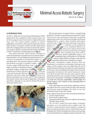
  • 1. Minimal Access Robotic Surgery INTRODUCTION On July 11, 2000, the Food and Drug Administration (FDA) approved the first completely robotic surgery device, the da Vinci surgical system from Intuitive Surgical (Mountain View, CA). The system enables surgeons to remove gallbladders and perform other general surgical procedures while seated at a computer console and three-dimensional (3D) video imaging system across the room from the patient. The surgeons operate controls with their hands and fingers to direct a robotically controlled laparoscope (Fig. 1). This system and other robotic devices developed or under development by companies such as Computer Motion (Santa Barbara, CA) and Integrated Surgical Systems (Davis, CA) have the potential to revolutionize surgery and the operating room. They provide surgeons with the precision and dexterity necessary to perform complex, minimally invasive surgery (MIS) procedures, such as beating-heart single-ordouble-vesselbypassandneurological,orthopedic, and plastic surgery, among many other future applications. Manufacturers believe that their products will broaden the scope and increase the effectiveness of MIS; improve patient outcomes; and create a safer, more efficient, and more cost- effective operating room. It is the vision of these companies that robotic systems will one day be applicable to all surgical specialties, although it is too early to tell the full extent to which they will be used. The first generation of surgical robots is already being installed in a number of operating rooms around the world. These are not true autonomous robots that can perform surgicaltasksontheirown,buttheyarelendingamechanical helping hand to surgeons. These machines still require a human surgeon to operate them and input instructions. Remote control and voice activation are the methods by which these surgical robots are controlled. Robotics are being introduced to minimal access surgery because they allow for unprecedented control and precision of surgical instruments. So far, these machines have been used to position an endoscope, perform gallbladder surgery, and correct gastroesophageal reflux and heartburn. The ultimate goal of the robotic surgery field is to design a robot that can be used to perform closed-chest, beating-heart surgery. Recently, transatlantic surgery between USA and Strasbourg is a revolution in transatlantic minimal access surgery. In this surgery, there was slight delay (66 ms) in transfer of data but in future, this delay can easily minimized. In the future, remote handling technology will overcome the manipulative restriction in the current instruments. There is no doubt 10 years from now that some surgeons will be operating exclusively via a computer interface controlling a master-slave manipulation. If computer-controlled machinery can mimic the awareness, adaptability, and knowledge of a human surgeon, such a takeover in the operating theater is actually realistic. In the operating room of the future, physicians will use tiny high-tech tools to travel inside the body with dexterity and precision beyond imaging. The future of any new technology depends upon the training. Three surgical robots that have been recently developed are: 1. da Vinci surgical system 2. ZEUS robotic surgical system 3. Automated Endoscopic System for Optimal Positioning (AESOP) robotic system. The “da Vinci” system has seven degree of freedom movement, so it can perform more complex task. The “da Vinci” system has been used to perform a number of general surgical procedures such as cholecystectomy and fundoplication.Roboticfundoplicationallowseasierpassage around and behind the esophagus during its dissection and Fig. 1: Robotic surgery port placement for nephrectomy. Prof. Dr. R. K. Mishra
  • 2. 592 SECTION 6: Miscellaneous easier mobilization of the curvature of the stomach. Suturing the wrap and the crural approximation are also easier with the help of these robots. The $1 million da Vinci system consists of two primary components: 1. A viewing and control console 2. A surgical arm unit (Figs. 2A to C). In using da Vinci for gallbladder surgery, three incisions, no larger than the diameter of a pencil, are made in the patient’s abdomen, which allows for three stainless-steel rods to be inserted. The rods are held in place by three robotic arms. One of the rods is equipped with a camera, while the other two are fitted with surgical instruments that are able to dissect and suture the tissue of the gallbladder. Unlike in conventional surgery, these instruments are not directly touched by the doctor’s hands. Effect or tips of the da Vinci surgical system incorporate miniature wrists that allow them to mimic any movement made by the surgeon at the control console. Sitting at the control console, a few feet from the operating table, the surgeon looks into a viewfinder to examine the 3D images being sent by the camera inside the patient. The images show the surgical site and the two surgical instruments mounted on the tips of two of the rods. Joystick-like controls, located just underneath the screen, are used by the surgeon to manipulate the surgical instruments. Each time one of the joysticks is moved, a computer sends an electronic signal to one of the instruments, which moves in synchronization with the movements of the surgeon’s hands. Another robotic system is the ZEUS system, made by Computer Motion, which is already available in Europe. However, both the da Vinci and ZEUS systems must receive governmental approval for each procedure that a surgeon plans to use it for. The $750,000 ZEUS has a similar setup to that of the da Vinci. It has a computer workstation, a video display, and hand controls that are used to move the table- mounted surgical instruments. While the ZEUS system has not yet been cleared for American use beyond clinical trials, German doctors have already used the system to perform coronary bypass surgery. The ZEUS system employs the assistance of the AESOP robotic system. Released by Computer Motion in 1994, AESOP was the first robot to be cleared by the FDA for assisting surgery in the operating room. AESOP is much simpler than the da Vinci and ZEUS systems. It is basically just one mechanical arm, used by the physician to position the laparoscope. Foot pedals or voice-activated software allows the surgeon to position the camera, leaving his or her hands free to continue operating on the patient (Fig. 3). The use of a computer console to perform operations from a distance opens up the idea of telesurgery, which would involve a doctor performing delicate surgery thousands of Figs. 2A to C: (A and B) Robotic arms of da Vinci surgical system; (C) Effector tips of the da Vinci surgical system. A B C
  • 3. 593 CHAPTER 47: Minimal Access Robotic Surgery Fig. 3: Robotic surgery. (1) Surgeon console; (2) Image processing equipment; (3) EndoWrist instruments; (4) Surgical arm cart; and (5) High- resolution three-dimensional (3D) endoscope. miles away from the patient. If it were possible to use the computer console to move the robotic arms in real time, then it would be possible for a doctor in New Delhi to operate on a patient in New York. A major obstacle in telesurgery has been the time delay between the doctor moving his or her hands to the robotic arms responding to those movements. Currently, the doctor must be in the room with the patient for robotic systems to react instantly to the doctor’s hand movements. Having fewer personnel in the operating room and allowing doctors the ability to operate on a patient, long distance could lower the cost of health care. In addition to cost-efficiency, robotic surgery has several other advantages over conventional surgery, including enhanced precision and reduced trauma to the patient. For instance, heart bypass surgery now requires that the patient’s chest 30.48 cm long incision. However, with the da Vinci or ZEUS systems, it is possible to operate on the heart by making three small incisions in the chest, each only 10 mm in diameter. The patient would experience less pain and less bleeding, which means a faster recovery. Robotics also decreases the fatigue that doctors experi- ence during surgeries that can last several hours. Surgeons can become exhausted during those long surgeries and can experience hand tremors as a result. Even the steadiest of human hands cannot match those of a surgical robot. The da Vinci system has been programmed to compensate for tremors, so if the doctor’s handshakes, the computer ignores it and keeps the mechanical arm steady. While surgical robots offer some advantages over the human hand, we are still a long way from the day when autonomous robots will operate on people without human interaction. But, with advances in computer power and artificial intelligence, it could be that in this century a robot will be designed that can locate abnormalities in the human body, analyze them, and operate to correct those abnormalities without any human guidance. Fig. 4: Robotic surgery via master-slave manipulator. The fallopian tube reconnection procedure, referred to as tubal reanastomosis, was performed by Dr Tommaso Falcone, who is Head of the Reproductive Endocrinology and Infertility Section at the Cleveland Clinic. Dr Falcone used Computer Motion’s ZEUS robotic surgical system as part of a clinical trial, approved by the United States FDA. The patient, a 38-year-old woman, and her healthy 10-day-old son are both in excellent condition and have returned to their Cleveland-area home. The mother had originally undergone a tubal ligation sterilization operation in her 20s. She and her partner later wished to have a child together and began preparing for a reversal operation. The patient saw an advertisement for the ZEUS study and consulted with trial leader Dr Falcone. The lady was informed in detail of the investigational protocol and agreed to have the robotically-assisted procedure. In addition to the ZEUS System, Computer Motion markets the AESOP 3000, a voice-controlled endoscope positioning system, and the HERMES Control Center, a centralized system, which enables the surgeon to voice control a network of “smart” medical devices. Currently, the ZEUS system is under an FDA-approved investigational device exemption and is also CE marked for commercial sale in Europe. Robotics is rapidly developing in surgery, although the word is slightly misused in this connection. None of the systems under development involves a machine acting autonomously. Instead, the machine acts as a remote extension of the surgeon. The correct term for such a system is a “master-slave manipulator,” although it seems unlikely that this term will gain general currency. Minimal invasive surgery is itself a form of telemani- pulation because the surgeon is physically separated from the workspace. Telerobotics is an obvious tool to extend the surgeons capabilities. The goal is to restore the tactile cues and intuitive dexterity of the surgeon, which are diminished by MIS. A slave manipulator, controlled through a spatially consistent and intuitive master with a force feedback system, could replace the tactile sensibilities and restore dexterity (Fig. 4).
  • 4. 594 SECTION 6: Miscellaneous Fig. 5: Master-slave manipulator. Fig. 6: Robotic console. Fig. 7: Robotic arm. Fig. 8: Ports in cardiac minimal access surgery. Althoughthepotentialofroboticsurgeryisjustbeginning but progress may come quickly. Laparoscopic gallbladder surgery was first done in 1987, but it became standard within 5 years. Just think about a surgeon! He picks up this black box and waves it over your body and you are fixed. How is that going to happen? One day, a surgeon may use robotic devices to enter the body through its own orifices. They could carry medical instruments inside the body, where they would be manipulated by simple computer commands (Figs. 5 to 7). The robotic arm after addition of wrists permits the sur- geon to mimic his own movements, rather than experience limitations of the rigid long cylindrical laparoscopic instru- ment and has obvious advantages in terms of dexterity and complexity of instrument (Fig. 8). INSTRUMENTS OF ROBOTIC SURGERY da Vinci Surgical System (Figs. 9 to 20) Using the most advanced technology available today, the da Vinci surgical system enables surgeons to perform delicate and complex operations through a few tiny incisions with increased vision, precision, dexterity, and control. The da Vinci surgical system consists of several key components, including: an ergonomically designed console where the surgeon sits while operating, a patient-side cart where the patient lays during surgery, four interactive robotic arms, a high-definition 3D vision system, and proprietary EndoWrist instruments.
  • 5. 595 CHAPTER 47: Minimal Access Robotic Surgery Fig. 9: da Vinci high-definition surgical robot. Fig. 10: da Vinci surgeon console. Fig. 11: Patient cart. Fig. 12: Seven-degree movement is possible. Fig. 13: da Vinci vision cart.
  • 6. 596 SECTION 6: Miscellaneous Fig. 14: Robotic EndoWrist instruments. Figs. 15A and B: Robotic scissors and bipolar dissector. Figs. 16A and B: Robotic tenaculum and grasper. Figs. 17A and B: Robotic needle holder. Figs. 18A and B: Robotic atraumatic grasper. Figs. 19A and B: Robotic clip applicator. Figs. 20A and B: Robotic scissor and Maryland. da Vinci is powered by state-of-the-art robotic techno- logy that allows the surgeon’s hand movements to be scaled, filtered, and translated into precise movements of the EndoWrist instruments working inside the patient’s body. Core Technology ■ System components ■ Three-dimensional high-definition (HD) vision. A B A B A B A B A B A B
  • 7. 597 CHAPTER 47: Minimal Access Robotic Surgery Instrumentation Using the da Vinci surgical system, the surgeon operates seated comfortably at a console while viewing an HD, 3D image inside the patient’s body. ■ The surgeon’s fingers grasp the master controls below the display with hands and wrists naturally positioned relative to his or her eyes ■ The system seamlessly translates the surgeon’s hand, wrist, and finger movements into precise, real-time movements of surgical instruments ■ The patient-side cart is where the patient is positioned during surgery. It includes either three or four robotic arms that carryout the surgeon’s commands ■ The robotic arms move around fixed pivot points, which reduce trauma to the patient, improve the cosmetic outcome, and increase overall precision ■ The system requires that every surgical maneuver be under the direct control of the surgeon. Repeated safety checks prevent any independent movement of the instruments or robotic arms ■ A full range of EndoWrist instruments is available to the surgeon while operating ■ The instruments are designed with seven degrees of motion—a range of motion even greater than the human wrist ■ Each instrument has a specific surgical mission such as clamping, suturing, and tissue manipulation ■ Quick-release levers speed instrument changes during surgery ■ The vision system is equipped with an HD, 3D endoscope (flexible tube with a camera and light at the tip) and image processing equipment that provides true-to-life images of the patient’s anatomy ■ A view of the operating field is available to the entire operative room (OR) team on a large viewing monitor (vision cart). This widescreen view provides the surgical assistant at the patient’s side with a broader perspective and visualization of the procedure. INSTRUMENTS AND ACCESSORIES Intuitive surgical’s exclusive EndoWrist instruments are designed to provide surgeons with natural dexterity and full range of motion for precise operation through tiny incisions. Modeled after the human wrist, EndoWrist instruments can offer an even greater range of motion than the human hand. They truly allow the da Vinci® system to take surgical precision and technique beyond the limits of the human hand. Similar to human tendons, an EndoWrist instrument’s internal cables provide maximum responsiveness, allowing rapid and precise suturing, dissection, and tissue manipulation. EndoWrist instruments provide enhanced dexterity, precision, and control: ■ 7° of freedom ■ 90° of articulation ■ Intuitive motion and finger-tip control ■ Motion scaling and tremor reduction. The wrist-like movement, responsiveness, and robotic control afforded by the da Vinci system and its exclusive EndoWristinstrumentsprovidesurgeonsfluidambidexterity and unparalleled precision. The EndoWrist instruments are available in a wide selection of specialized tip designs to enable a broad range of da Vinci procedures. As da Vinci surgery is adopted in new specialties, our engineers work side-by-side with surgeons to develop new EndoWrist instruments to address new clinical needs. The following is a list of instrument categories for which intuitive offers specialized tip designs. Energy instruments are used by the da Vinci surgeon to provide coagulation, cutting, and dissection of tissues. These include monopolar and bipolar cautery instruments (electrical energy), the Harmonic™ ACE (mechanical energy), the PK™ dissecting forceps (advanced bipolar), and laser. Grasping instruments allow for manipulation of a wide range of tissues, from fine, thin tissues such as peritoneum to dense, fibrous tissues such as uterus. Needle drivers provide the ability to suture with the finest of needles used in cardiovascular surgery as well as the thickest needles used in repair of uterine defects. Suture Cut™ needle drivers include an integral cutting blade for efficient cutting of suture after knot tying and increased surgeon autonomy and efficiency. Retracting instruments allow the surgeon to dynamically provide exposure of the surgical field. This minimizes dependency on the patient-side assistant, providing the da Vinci surgeon with full control of the operative field. Clip appliers, probe graspers, and cardiac stabilizers are available to allow the da Vinci surgeon to perform specialized procedures such as vessel clipping, cryoablation, and beating-heart surgery. A selection of 5 mm EndoWrist instruments provides the surgeon with the ability to use smaller access ports. Some surgeons prefer smaller access ports when performing pediatric, thoracic, bariatric, gynecologic, and general surgery. OPERATING ROOM CONFIGURATION, PORT PLACEMENT, AND DOCKING Before any procedure, the robot has to be prepared for surgery. It includes the connection of all necessary parts such as sterile drapes and connectors needed for surgery and the calibration process. These steps are conducted by a core team of scrub nurses specifically trained in handling the robot while the patient is in preparation for surgery. Currently, the only available system for laparoscopic surgery is the da Vinci surgical system, developed by Intuitive Surgical (Sunnyvale, CA, USA). Anyroboticprocedureisperformedbyateamofsurgeons and nurses. It includes the console surgeon, patient-side
  • 8. 598 SECTION 6: Miscellaneous assistant surgeon, and the scrub nurse. Although console surgeon is the leader of the team, a trained assistant surgeon is of paramount importance as he/she is the person responsible for robot docking, instrument change, manipulation of laparoscopic instruments, application of hemostatic instruments/clips, lavage and aspiration, specimen extraction, drainage, and closure of abdominal wall. A trained scrub nurse is also important in draping the robotic arms, attaching the optics, instrument changes, and undocking of the robot. Operating Room Configuration This topic includes the optimum positioning of the following components in the operating room (OR) so as to allow for maximum functionality: ■ Surgeon console positioning ■ Patient cart positioning ■ Vision cart positioning. Surgeon Console Positioning The surgeon console is placed outside of the sterile field. It should be oriented in such a position so that the surgeon has a clear view of the operative field, vision cart, and able to communicate directly with the assistant surgeon and the scrub nurse. For moving or positioning the surgeon console, only the handle (Fig. 21) on the back of the console is used. It should never be pushed or pulled by the console body or armrest to maneuver the console into place. Wheel locks located on the rear wheels of the surgeon console should be locked after positioning of console for the surgery. Patient Cart Positioning The patient cart is placed in the sterile field. It should be draped in a separate area in the room prior to moving it into place for surgery. This should be an area of the room where it will not easily come into contact with nonsterile objects or impedetraffic.Oncethepatientcartisdrapedandthepatient is positioned, prepared, and draped, ports are placed. Then, use the patient cart motor drive to help move the cart into the sterile field. The patient cart brakes are designed to automatically engage when the motor drive is not in use. Motor drive operation: The motor drive interface consists of the following components (Fig. 22): ■ Throttle ■ Throttle enable switch ■ Shift switches. To operate the motor drive: ■ Ensure that the patient cart is powered on ■ Ensure the shift switches are in the drive position (Figs. 23A and B) ■ Throttle enable switch is to be held and throttle rotated away from the operator or toward him/her depending on the intended direction of movement z The cart power light-emitting diode (LED) will flash green whenever the throttle enable switch is activated z The drive speed of the cart can be controlled by rotating the throttle to different extent in each direction. The motor drive will not engage whenever cannulas or instruments are installed on the system. A yellow LED on motor drive interface labeled “cannula installed: cart drive disabled” will indicate when cannulas or instruments are installed and motor drive is nonoperational. This has been done for safety purpose. Shift switches: The patient cart can be moved without the use of the motor drive (for example, during a power loss) by rotating the shift switches (Fig. 23B) to the neutral (N) position. The cart can then be moved manually. When the cart has been moved, shift switches should be placed in the drive (D) position to set the patient cart brakes. Fig. 21: Surgeon console handle. Fig. 22: Location of throttle, throttle enable switch, and shift switches.
  • 9. 599 CHAPTER 47: Minimal Access Robotic Surgery Figs. 23A and B: (A) Drive motor engaged; (B) Drive motor disengaged. (D: drive; N: neutral) For patient safety, the shift switches must be kept in the drive (D) position so that the motor drive remains engaged during surgery (Fig. 23A). Vision Cart Positioning The vision cart is placed adjacent to the patient cart, just outside of the sterile field, to allow the patient cart operator to see the component displays (Fig. 24). ■ The vision cart should be close enough to the patient cart to allow unrestricted camera cable movement during surgery ■ Wheel locks are located on the rear wheels of the vision cart. These should be locked after the cart is positioned for surgery. Steps of docking: ■ Position patient and OR table, including table tilt ■ Position patient cart over patient ■ Set patient cart brakes ■ Docking the camera arm ■ Docking the instrument arms ■ Check system setup. Fig. 24: Position of vision cart. Fig. 25: Camera port positioning. The patient table should be positioned according to surgeon preference (depending on the contemplated procedure) before docking the robotic arms. Once the arms are docked to the ports and instruments are placed, patient position should not be changed. The pneumoperitoneum is created and the ports are inserted by either the lead surgeon or the patient-side assistant surgeon. Port Placement The port positions vary from patient-to-patient, procedure- to-procedure, and surgeon-to-surgeon. It is very difficult to form guidelines-specific position of ports, but broad guidelines are framed to maximize endoscopic view, instrument reach, and to minimize external arm clashing. Camera port is inserted keeping following principles in mind (Fig. 25): ■ Should be in line with the target anatomy (TA) ■ Should beat 10–20 cm distance from the TA A B
  • 10. 600 SECTION 6: Miscellaneous Figs. 26A and B: (A) Disposable 12 mm trocar cannula assembly for camera port; (B) Camera port being mounted on the camera arm. Figs. 27A and B: Port placement. ■ Should be in line with the center column of the patient cart. A diagnostic laparoscopy is done after insertion of the camera port to look for safe entry, any adhesions, and surgical feasibility. A disposable 12 mm trocar cannula assembly is used as the camera port (Figs. 26A and B). There are specialized camera arm cannula mounts (on the patient cart camera arm) corresponding to each validated third-party cannula. Working Ports An 8 mm da Vinci ports are inserted for robotic arms that are inserted keeping in mind the following principles: ■ >8 cm distance between the da Vinci ports (Fig. 27A) ■ 10–20 cm distance should be maintained between the da Vinci ports and TA (Fig. 27B). A 10 cm distance from TA is good but 20 cm distance is better. Ports placed closed to the TA impede the view of the surgical site and make the operation technically challenging. Conversely, ports placed >20 cm from TA make it difficult to see or reach with robotic instruments. Assistant port, if needed, is inserted 5–10 cm away from the da Vinci ports in the desirable position. 5 or 10 mm ports can be used according to the intended function of the port. The da Vinci provides 8 mm reusable cannulas with disposable seals for the robotic arms. They come with bladeless obturator for insertion. These come in two lengths (Fig. 28): 1. Short (11 cm cannula) 2. Long (16 cm cannula) for high body mass index (BMI) patients. Remote Center Technology Remote center is the fixed point in the space around which surgical arm and cannula move. It helps in maneuvering A B A B
  • 11. 601 CHAPTER 47: Minimal Access Robotic Surgery instruments/endoscopes in surgical site while exerting minimal force on the abdominal or thoracic wall (Figs. 29A and B). It is marked on the da Vinci cannulas at a point to minimizestresstothepatient.Remotecentercanbeadjusted on patient side at the patient cart using the clutch button. Positioning the Patient Cart Once the patient is positioned and the ports are inserted, it is time to attach patient cart instrument arms to the patient in a process called docking. Before moving patient cart into position over the patient, it is important to align the OR table and the patient. Then, push the patient cart over the patient using the motor drive on the cart. Motor drive can be used in integrated mode when patient cart is connected to the rest of the system or in standalone mode as well. The shift switches on the base of the patient cart need to be set in drive (D) position (Fig. 30). Then, the Figs. 29A and B: Remote center technology. cart can be moved by pressing the throttle enable switch and throttling either forward or backward (Fig. 31). It can be moved manually if the shift switch is set in neutral (N) position. Two people should be used to move the cart, one pushes or pulls the cart and the second person verbally directs regarding the direction of movement. When the cart is in position, shift switch should be set to drive position for locking the cart. Installing the camera or instrument arm locks the patient cart automatically for patient safety. Care should be taken to align the camera port, TA, and the center column (Fig. 32). Docking the Camera Arm Camera arm should be docked first after positioning the patient cart. Align the camera port, TA, and the center column of the patient cart. Use clutch button to change the angle of camera arm to match the angle of cannula so that it points to TA (Fig. 33). Stabilize the cannula at the port site with one hand pointing it toward the TA. Bring cannula into the cannula mount on the camera arm and clip both wings shut to hold the cannula in place (Fig. 34). Camera arm setup joint #2 is placed opposite the instrument arm 3 (Fig. 35). Setup joints are numbered starting from the joint closest to the center column. Setting the system in this position allows maximum range of motion for all instrument arms. Thereisathickbluelineandabluearrowonsetupjoint#2 on the camera arm indicating the sweet spot (Fig. 36). Sweet spot should be aligned by lining up the blue arrow within the boundaries of blue line. Setting sweet spot gives patient cart arms maximum range of motion ensuring instrument and endoscope reach of all parts of TA. Fig. 28: The da Vinci cannula. (BMI: body mass index) A B
  • 12. 602 SECTION 6: Miscellaneous Fig. 30: Shift switch. Fig. 31: Throttle with throttle enable switch. Fig. 32: Alignment of camera arm, target anatomy, and center column of patient cart. Fig. 33: Docking the camera arm. Fig. 34: Mounting cannula to camera arm. Fig. 35: Camera arm setup joint #2 facing opposite instrument arm 3 (arrow). Remember that overextending or not extending the cameraarmenoughwilllimittheinstrumentrangeofmotion. Align the camera arm clutch button, third setup joint, and the center column (Fig. 37). Strive to maintain the sweet spot and alignment of the camera arm throughout the docking process. Docking of Instrument Arms After docking the camera arm, instrument arms are positioned in place so as to allow maximum range of motion of the arms. It is done in following steps: ■ Position the instrument arm with the arm number and sterile adaptor facing forward (Fig. 38)
  • 13. 603 CHAPTER 47: Minimal Access Robotic Surgery Fig. 36: Sweet spot and its alignment. Fig. 37: Alignment of camera arm clutch button, third setup joint, and the center column. Fig. 38: Positioning of instrument arm. Fig. 39: Alignment of instrument arms with respect to camera arm and each other. ■ Allow approximately 45° angle between each arm (Fig. 39). Note that the position of instrument arm 3 can vary according to patient body habitus and the procedure. After positioning, ensure that the arms will not collide with the patient or interfere with each other ■ Dock the instrument arms by bringing them to the can- nula using the port clutch button. Bring the instrument arm to the cannula and lock the wings of the quick click cannula mount on the arm to clip the arm to the cannula (Fig. 40). Confirm that the remote center of the port is present at the desired place in the abdominal wall. Remember to stabilize the cannula with one hand at the port site while docking the instrument arm. Check System Setup After docking the instrument arms, check the arm setup. Start by confirming that the sweet spot of the camera arm is in right position (i.e., arrow is pointing toward the thick blue line). If needed, move the arm back into position taking care to stabilize the cannula at the port site. Fig. 40: Example of mounting instrument arm onto the cannula. Next, check the alignment of the camera port, TA, and the center column of the patient cart. Now check the instrument arm setup. Separate the instrument arms to maximize the range of motion (Fig. 41). Check the setup joint angles to minimize potential collisions.
  • 14. 604 SECTION 6: Miscellaneous Fig. 41: Relative positions of the instrument arms. Fig. 42: Right angle at setup joint #2. Fig. 43: Depiction of the correct method of docking of patient cart. The angle at the setup joint #2 should be approximately 90° (Fig. 42). Figure 43 depicts the correct method of docking the patient cart. Endoscope Insertion and Removal First, insert the endoscope into the cannula keeping the intuitive logo on the camera head facing the camera arm. Place the body of the endoscope into the camera arm sterile adaptor making sure that the body of endoscope is fully connected (Fig. 44). Give it a gentle turn to ensure it is locked in place. Next, secure the camera cable using the camera cable clip and drape the cable across the instrument arm (Fig. 45). Push the endoscope into the cannula by pressing the camera arm clutch button till it is past the cannula tip toward the TA. Press the clutch button again to lock the camera assembly into place. To remove the endoscope, remove the camera cables fromthecliponthecameraarm.Then,unlocktheendoscope Fig. 44: Endoscope insertion into camera arm sterile adaptor of the camera arm. by opening the two latches on the camera arm sterile adaptor. Then, remove the endoscope.
  • 15. 605 CHAPTER 47: Minimal Access Robotic Surgery Fig. 45: Camera cable clip highlighted. Fig. 46: Inserting the instrument into the cannula. Fig. 47: Sliding the instrument into sterile adaptor on the instrument arm. Fig. 48: Light-emitting diode (LED) turning blue indicating proper insertion. Instrument Insertion Begin by straightening the instrument tip. Insert the instrument into the cannula (Fig. 46). Slide the instrument housing into the sterile adaptor (Fig. 47) sandwiching the instrument housing and the instrument arm between both hands. Press the arm clutch button and push the instrument into the surgical field, keeping the tip under endoscopic vision. If there is any resistance while inserting the instrument, one should stop and check for the reason. When the LED lights turn blue (Fig. 48), the surgeon can take control of the instrument and start operating. Instrument Removal and Guided Tool Change For removing the instruments, surgeon should straighten the instrument tip and open the jaws of the instrument to ensure that it does not hold any tissue (Fig. 49). Then, press the release levers on the instrument housing and simply pull off the instrument (Fig. 50). Fig. 49: Straightening the instrument tip and opening of jaws of the instrument before removal. Removal of the instrument should be done with utmost care and with complete knowledge of the operating surgeon, so as to prevent any inadvertent injury to the tissues.
  • 16. 606 SECTION 6: Miscellaneous Fig. 50: Pressing the levers to remove the instrument. Fig. 51: Light-emitting diode (LED) blinking alternately white and green indicating activation of guided tool change. Fig. 52: Pushing the instrument with one finger. Guided Tool Change This feature helps in aligning a new instrument in the same position as the previous one removed. It adapts the replaced instrument tip just short of the position of previously placed instrument tip. Whenthisfeatureisactivated,theLEDsontheinstrument arm alternately blink white and green (Fig. 51). Just push the instrument with one finger (Fig. 52) and guided tool change (GTC) will guide it into correct position. Stop as soon as you encounter any resistance. This function is disabled when there is a change in the position of instrument arm during changing the instrument. This is because the position memory of the instrument arm is reset when the position is changed. ROBOTIC CHOLECYSTECTOMY Introduction In general surgery, advanced robotics will likely find its place in the most complex laparoscopic procedures where the enhanceddexterityandsuperiorvisualizationwillextendthe feasibility of the minimally invasive approach, particularly in patients requiring advanced suturing and precise tissue dissection. Robot-assisted laparoscopic cholecystectomy is a safe and effective bridge to advanced robotics in general surgery. Laparoscopic cholecystectomy is a prime operation with which to begin robot applications for several reasons. First, gallstone disease is the most common of all abdominal diseases for which patients undergo a laparoscopic procedure. Moreover, it is an operation with a very standardized approach to prevent complications. This standardized approach has an added advantage of having aspects that may be more broadly applied to other more complex minimally invasive operations. For example, the dissection of the Calot’s triangle is analogous to dissection and isolation of vasculature, the cystic duct and artery can be tied instead of using clips, and removal of the gallbladder from the gallbladder fossa requires a vascular dissection and the appreciation of tissue planes. Therefore, robotic cholecystectomy may allow general surgeons to acquire clinical da Vinci experience in a familiar setting. Segments of Robotic Cholecystectomy Segment Operative Tasks ■ Skin incision, port placement, exploration, adhesiolysis, patient positioning, and robotic arm draping ■ Positioning of da Vinci, docking of robot arms, and camera ■ Initial dissection of gallbladder until placement into endoscopic bag ■ Gallbladder extraction, performance of additional procedures, and incision closure. Preoperative Preparation Indications and preoperative preparation are similar to conventional laparoscopic procedure. An informed consent is to be taken from the patient explaining the new technology
  • 17. 607 CHAPTER 47: Minimal Access Robotic Surgery and its possible complications. A prophylactic dose of intravenous antibiotic is given just prior to commencement of surgery. An orogastric or nasogastric tube is placed prior to creation of pneumoperitoneum to decompress stomach. Sequential compression devices are placed on legs for deep vein thrombosis (DVT) prophylaxis. Footboard is placed to keep patient from sliding off table. A seat belt is tied at midthigh region. Patient Position Patient is placed in a supine position and right arm is tucked by the side of the patient. Standard surgical preparation is done from nipple line to thigh. A standard protection bar over the patient’s head may easily block the robotic arms, giving protection to the head. Convex protection shields or bars that protect the patient’s nose and the ventilation tube are good protection for robotic-assisted surgical interventions. Pneumoperitoneum is created with closed (Veress needle) or open technique. A 12-mm camera port is inserted at the umbilicus, which should be around 20 cm away from the TA (cystic pedicle). A diagnostic laparoscopy is done. Then, a mild left tilt of the table is done with reverse Trendelenburg position. Port Position (Fig. 53) Three 8 mm da Vinci ports are inserted at least 10 cm apart for the robotic arm placement. The remote centers of the ports are placed at the level of abdominal wall. ■ Robotic arm 1: Left midclavicular line, below the subcostal margin ■ Robotic arm 2: Right midclavicular line, 5–10 cm below the subcostal margin ■ Robotic arm 3: 10 cm below and lateral to robotic arm 2 (optional, some surgeons use a 5/12 mm assistant port rather than a robotic arm as the third port). Docking and Operating Room Setup The arms are draped with disposable sterile drapes. The patient cart is brought over the patient’s right shoulder (Fig. 54). This implies that the robot is located behind the operating field and that the robotic arms cross this area and seem to work in a backward direction. The center column of the robotic cart acts as the vertical central axis of the robot. This column should be positioned at the far end of an axis running from the umbilicus through the hilar region of the liver. The patient table should be tilted according to the surgeon’s choice before docking. Align the “sweet spot” for proper camera arm positioning. Align the clutch button, third setup joint, and the center column. Allow 45° angle between each arm. Dock the camera arm and other arms to the respective ports. Camera and the instruments are placed in the ports. The operating room setup is shown in Figure 55. Operative Technique Step 1 (Diagnostic Laparoscopy and Port Position) Patient is kept in supine position. Parts cleaned and draped from nipple line to midthigh. Pneumoperitoneum is created with Veress needle at umbilical position. A 12-mm camera port is inserted at the umbilicus using Optiview cannula (Fig. 56). Fig. 53: Port positioning. (TA: target anatomy) Fig. 54: Position of patient cart and ports. (MCL: midclavicular line; SUL: spinoumbilical line)
  • 18. 608 SECTION 6: Miscellaneous Fig. 55: Operating room setup for cholecystectomy. Fig. 56: Camera port insertion under vision with 0° endoscope and Optiview cannula. Fig. 57: Diagnostic laparoscopy. Fig. 58: da Vinci port placement under vision. Fig. 59: Final port position for robotic cholecystectomy. A diagnostic laparoscopy is done and gallbladder is visualized (Fig. 57). Patient is positioned in reverse Trendelenburg position with left tilt. Three da Vinci 8 mm ports are inserted under vision as per the guiding principles described above (Figs. 58 and 59). Any adhesiolysis, if required, is done at this particular time with standard laparoscopic instruments. Step 2 (Docking of Robotic Cart) Patient cart is brought over the right shoulder of the patient. Docking of camera arm is done first followed by instrument arms. The principles of docking are kept in mind and system checkup is done after docking is complete. Instruments are inserted under endoscopic vision such as R1: Maryland dissector and R2 and R3: Prograsp forceps (Figs. 60 to 62).
  • 19. 609 CHAPTER 47: Minimal Access Robotic Surgery Step 3 Fundus is grasped with prograsp forceps (R3) and pushed toward the right shoulder of the patient (Figs. 63 and 64). This traction is constant once the arm is locked unlike in laparoscopy where it depends on the expertise of the holding assistant. Care should be taken to avoid injury to diaphragm because of inadvertent slippage of fundus from the grasper. Due to lack of haptic feedback to the main surgeon, it can go unnoticed and cause trauma to diaphragm. Step 4 Hartmann’s is grasped with prograsp forceps (R2) and Calot’s triangle is dissected with Maryland EndoWrist instruments (R1). Seven degrees of freedom in the EndoWrist instruments give a distinct advantage in this dissection (Figs. 65 to 69). Step 5 Cystic duct and artery are dissected completely. Clips are applied with EndoWrist hemolock clip applicator (R1) (Figs. 70 to 73). Fig. 60: Docking the camera arm. Fig. 61: Docking the instrument cannula to the instrument arm. Fig. 62: Insert the instruments under endoscopic vision. Fig. 63: Holding the fundus with R1 Maryland dissector so as to enable R3 grasping forceps to retract it. Fig. 64: Pushing the fundus toward the right shoulder. Step 6 The cystic duct and artery are divided with the EndoWrist scissors (R1) in between the clips (Figs. 74 to 78). The EndoWrist movements enable the surgeon to cut precisely in the direction required.
  • 20. 610 SECTION 6: Miscellaneous Fig. 65: Hartmann’s is grasped by R2 and Calot’s triangle being dissected by Maryland in R1. Fig. 66: Posterior peritoneum being dissected off the Calot’s triangle. Fig. 67: Posterior window creation. Fig. 68: Cystic duct being dissected using the degrees of freedom of the Maryland dissector. Fig. 69: Window made posterior to cystic duct. Fig. 70: Clip being applied by robotic clip applicator. The degrees of freedom can be used for optimum clip application. Step 7 The gallbladder is dissected off the gallbladder fossa (Figs. 79 and 80) with the help of hook electrode with monopolar cautery (R1). The EndoWrist function of the hook provides a great deal of versatile movements, which aid in this step of surgery.
  • 21. 611 CHAPTER 47: Minimal Access Robotic Surgery Fig. 71: Clip applied to cystic duct. Fig. 72: Second clip being applied. Fig. 73: Third clip being applied. Fig. 74: Cystic duct being divided with scissors. Fig. 75: Cystic duct divided. Fig. 76: Cystic artery dissected. Step 8 Just before taking off the gallbladder from the fossa, it is retracted cephalad and the gallbladder fossa is inspected for any bleed or bile leak (Fig. 81). Step 9 The R3 arm is undocked and Endobag is inserted by the patient-side assistant surgeon using standard laparoscopic grasper. Any suction/irrigation can also be done by the
  • 22. 613 CHAPTER 47: Minimal Access Robotic Surgery Fig. 83: Gallbladder (GB) being extracted out. while advantages of such tools are hard to prove for this specific procedure. There are two situations in which robotics can be applied to this particular procedure: 1. Complicated gallstone disease such as acute cho- lecystitis, common bile duct (CBD) exploration, or choledochoenterostomy 2. Ideal learning and teaching environment for gastrointes- tinal robot-assisted surgery. Advantages Robotic instruments clearly provide superiority in some aspects of the surgery. The increased degrees of freedom (wrist action) allow the surgeon to easily reach behind structures and negotiate difficult surgical angles. The ergonomics of the operating surgeon can be significantly improved by operating in the sitting position and facing directly forward toward the 3D operative image. These can be of value in cases of acute and chronic cholecystitis, where vision is impaired by inflammation, where there is an increased tendency for diffuse bleeding in the dissection field, and edema and fibrosis make the dissection difficult. By adjusting movement scaling, surgeon tremor can be significantly reduced; thereby, increasing operative precision. The translation of the surgeon’s hand and wrist movements is reliable and can be scaled down to improve precision and increase steadiness. Limitations There is a need for more variety in type and size of robotic trocars. The robotic arms are bulky, taking up significant space and limiting the capability of the surgical assistant. Furthermore, the da Vinci system is not attached to the operating table, so the position of the table cannot be changed without undocking the instrument and camera. The robotic arms lack haptic feedback, so the surgeon must rely on visual cues to avoid stretching and damaging tissue or suture. Financially, surgical robotic systems remain quite expensive and are associated with significant operating cost. Current robotic systems and instrumentation are still considered to be first generation and should improve as the technology evolves. Conclusion Robotic cholecystectomy is feasible and safe. There is a significant learning curve (around 20–30 cases) to gain experience for setting up the robotic instrumentation, which appears to be much less steep for the actual use of the machine. Itisunlikelythatroboticcholecystectomywillberoutinely performed in the near future. Further studies are needed to identify the benefits to the patient and compare it to the additional cost of robotic cholecystectomy before routine application of this technique can be justified. However, robotic cholecystectomy may prove its value in cases of complex gallstone disease and is an excellent procedure for teaching the basics of robotic surgery. ROBOTIC CORONARY BYPASS SURGERY Four ports are used and robotic arm is introduced. One of the arms contains telescope, which sends 12–15 times magnified clear image on the monitor. Another arm is for a stabilizer, which is used to hold the diseased coronary artery in place while bypass is performed. The other two remaining right and left instrument is used to perform microvascular anastomosis. Currently, robotic coronary bypass surgery should be considered only for the patient who have single vessel disease but in near future, we have hope to perform double or even triple bypass surgery (Fig. 84). The attached instruments are controlled by the surgeon, who sits at an adjacent console. Several passive mechanical devices, primarily used to hold the telescope, have been developed as assistants for general laparoscopic surgery. They successfully reduce the stress on the surgeon by eliminatingtheinadvertentmovementsofahumanassistant, which can be distracting and disorienting. More and more surgeries from prostate to heart are being performed by doctors remotely guiding robotic arms. In such procedures, the surgeon’s hands never enter the patient. After the initial incisions are made, robotic arms wielding a tiny camera and surgical tools make the snips, stanch the blood flow, and sew up inside when all is done. The surgeon sits at a console usually in the operating room, although the technology would allow a doctor to operate on a patient on the other side of the world peering into binocular-like lenses at views provided by the camera inside the patient. The doctor guides the robot’s work by twisting his wrists in stirrup-like handles, moving his thumb and forefinger in scissor-like loops, or tapping foot pedals to focus the camera or move a robotic arm.
  • 23. 614 SECTION 6: Miscellaneous Fig. 84: Instruments used by the da Vinci surgical system. Fig. 85: Comparison between open and da Vinci prostatectomy. ROBOTIC PROSTATECTOMY The most common robotic surgery is radical prostatectomy. Its use has grown from just 1,500 procedures in 2000 to an estimated 20,000 last year. More than 8,000 prostate glands were removed robotically last year, up from 36 in 2000. The procedure accounted for >10% of the 75,000 prostatectomies done in 2004. Busy professionals like the fact that they can be out of the hospital in a day, versus two or three for open surgery, and resume normal activities in 1 week rather than 6 weeks. Cutting nerves around the prostate can lead to incontinence or impotence, so precision is important. Between 25 and 60% of conventional prostatectomy, patients sufferfrompostoperativeimpotence.Smallstudiesofrobotic surgery have shown at least a short-term benefit in terms of impotence and incontinence. Not every patient is a candidate. Complicated cases when the patient is very sick, needs multiple procedures, or has had previous chest surgery are not suited to robotics. Nor can a heart transplant or artificial heart implant bed one this way. If the complication arises, the patient may windup being opened. So, patients who want robotics smaller scars and quicker recovery times will need to ask hard questions. Find out how many times, the procedure you need has been done robotically and what the advantages and disadvantages are. Just as important, ask how many robotic surgeries, the doctor who will do yours has performed. In real life, you want the force of experience to be with you. For qualified candidates, the robotic prostatectomy offers numerous potential benefits over the traditional open prostatectomy, including (Fig. 85): ■ Shorter hospital stay ■ Less pain ■ Less risk of infection ■ Less blood loss and transfusions ■ Less scarring ■ Faster recovery ■ Quicker return to normal activities. While still a technology that is in its infancy, the use of robots to assist in surgery is becoming more and more widespread. There are now large case series reported in the literature that show possible benefits to the patient in terms of recovery from such surgeries as prostate and cardiac procedures. Additionally, as these systems continue to develop, improved technology and software provide the surgeon with “assistance” that improves precision and accuracy (Figs. 86A to D). Robotic technology requires a tremendous financial investment, so you might not see it at every community hospital in the near future. However, as with all technology, the price will likely fall quickly as the applications are expanded, as more widespread adoption occurs and as the field of robotics experiences additional breakthroughs. For now, many major academic institutions are beginning to purchase and deploy these systems. Technology allows amazing things to be accomplished. Robotic surgery is a new and expensive tool that is beginning to see adoption. MIS with a robot and without bypass is the next logical step in the development of cardiac surgery. It is beginning to show clear benefits for patients and this means that it will likely become more and more popular with time and as the price falls. ROBOTIC VERSUS LAPAROSCOPIC PROSTATECTOMY In laparoscopy, the surgeon uses long instruments through small openings and maneuvers them with direct hand contact. Robotic systems use even more delicate instruments that possess two additional degrees of movement excursion (for a total of six, as with a human hand). Comfortably seated at the robot console, the surgeon can maneuver the instruments via a computer interface. Laparoscopic radical prostatectomy is associated with a steep learning curve. Even in the hands of expert surgeons, laparoscopic radical prostatectomy requires extensive
  • 24. 615 CHAPTER 47: Minimal Access Robotic Surgery Figs. 86A to D: Robotic-assisted nephrectomy. learning; approximately 40 cases are needed to master this technique. In contrast, the learning of robotic prostatectomy seems to be more intuitive and less demanding. Robotic prostatectomy is a safe, effective, and reproducible technique for removing the prostate. In most patients, it can be performed within 1.5–2 hours with minimal blood loss and few complications. The procedure incorporates principles of both laparoscopic and open radical prostatectomy. The patients enjoy benefits of surgical treatment in the setting of less invasion, minimal pain, limited blood loss, and early functional and overall recovery. NEW SURGICAL ROBOTS Senhance Surgical System Robotic-assisted digital laparoscopy (Senhance Surgical System) provides a digitized interface between the surgeon and the patient designed to increase surgeon control and reduce surgical variability. The physical open architecture of the system is composed of independent robotic-assisted manipulator arms that are compatible with conventional trocars and familiar laparoscopic instruments and, thus, taking advantage of the existing operating room and surgical suite ecosystem (Fig. 87). Avatera Surgical System The Avatera robotic surgical system gained CE mark last month for use in keyhole surgery, with its initial focus on gynecology and urology. It consists of the main surgical robot and its control unit and various endoscopes and instruments (Fig. 88). Fig. 87: Senhance surgical robot system. A C B D
  • 25. 616 SECTION 6: Miscellaneous Fig. 88: The Avatera robotic surgical system. Fig. 89: SSI MANTRA is made in India surgical robot. Indian Robotic Surgical System The SSI MANTRA is made in India. Surgical robot is the brainchild of cardiac surgeon Dr Sudhir Srivastava and was developed in an Atal Incubation Centre. It has conducted 18 successful surgeries and the cost is likely to be one-fourth of leading global competitor, da Vinci robot (Fig. 89). Artificial intelligence is a buzzword on everyone’s lips today. Johnson & Johnson has partnered with Google to form VerbSurgical,whichisdevelopinga“digitalsurgeryprogram” . While it is still quite early in development, it is expected to include advancements in data analytics, machine learning, and connectivity. A recent study from this device reported on completely autonomous suturing using the robot in an animal model. Artificial intelligence could also potentially play a significant role in presurgical planning, intraoperative 3D imaging, and navigation allowing for even more precise and subtle operations. People expect that ultimately robots will replace surgeons and actually perform the surgery. Nevertheless, it is believed that this still remains well in the future. However, given the speed at which artificial intelligence research is progressing, you never know when surgeons’ jobs could be under complete hand of surgical robot. BIBLIOGRAPHY 1. Berguer R. Surgery and ergonomics. Arch Surg. 1999;134:1011-6. 2. Berguer R, Rab GT, Abu-Ghaida H, Alarcon A, Chung J. A comparison of surgeons’ posture during laparoscopic and open surgical procedures. Surg Endosc. 1997;11:21-39. 3. BeurgerR,RemberM,BeckleyD.Laparoscopicinstrumentscause increased forearm fatigue: a subjective and objective comparison of open and laparoscopic techniques. Minim Invasive Ther Allied Tech. 1997;6:36-40. 4. BuessGF,SchurrMO,FischerSC.Roboticsandalliedtechnologies in endoscopic surgery. Arch Surg. 2000;132:229-35. 5. Cadière GB, Himpens J, Germany O, Izizaw R, Degueldre M, Vandromme J, et al. Feasibility of robotic laparoscopic surgery: 146 cases. World J Surg. 2001;25:1467-77. 6. Cadière GB, Himpens J, Bruyns J. How to avoid esophageal perforation while performing laparoscopic dissection of the hiatus. Surg Endosc. 1995;9:450-2. 7. Cadière GB, Himpens J, Vertruyen M, Favretti F. The world’s first obesity surgery performed by a surgeon at a distance. Obes Surg. 1999;9:206-9. 8. Cadière GB, Houben JJ, Bruyns J, Himpens J, Panzer JM, Gelin M. Laparoscopic Nissen fundoplication technique and preliminary results. Br J Surg. 1994;81:400-3. 9. Chan A, Chung S, Yim A, Lau J, Ng E, Li A. Comparison of two-dimensional vs three-dimensional camera systems in laparoscopic surgery. Surg Endosc. 1997;11:438-40. 10. Cheah WK, Lenzi JE, So J, Dong F, Kum CK, Goh P. Evaluation of a head-mounted display (HMD) in the performance of a simulated laparoscopic task. Surg Endosc. 2001;15:990-1. 11. Davies B. A review of robotics in surgery. Proc Inst Mech Eng. 2000;214:129-40. 12. Dion YM, Gaillard F. Visual integration of data and basic motor skills under laparoscopy. Influence of 2-D and 3-D video-camera systems. Surg Endosc. 1997;11:995-1000. 13. Dosis A, Bello F, Rockall T, Munz Y, Moorthy K, Martin S, et al. ROVIMAS: a Software Package for Assessing Surgical Skills Using the da Vinci Telemanipulator System. Birmingham: IEEE Publisher; 2003. 14. Felger JE, Nifong LW, Chitwood WR. The evolution of and early experience with robot-assisted mitral valve surgery. Surg Laparosc Endosc Percutan Tech. 2002;12:58-63. 15. Gallagher AG, Satava RM. Virtual reality as a metric for the assessment of laparoscopic psychomotor skills. Learning curves and reliability measures. Surg Endosc. 2002;16:1746-52. 16. Garcia-Ruiz A, Gagner M, Miller JH, Steiner CP, Hahn JF. Manual vs robotically assisted laparoscopic surgery in the performance of basic manipulation and suturing tasks. Arch Surg. 1998;133:957-61. 17. Garcia-Ruiz A, Smedira NG, Loop FD, Hahn JF, Miller JH, Steiner CP, et al. Robotic surgical instruments for dexterity enhancement in thoracoscopic coronary artery bypass graft. J Laparoendosc Adv Surg Tech A. 1997;7:277-83. 18. Hanna GB, Cuschieri A. Influence of two-dimensional and three- dimensional imaging on endoscopic bowel suturing. World J Surg. 2000;24:444-9. 19. Hernandez JD, Bann SD, Munz Y, Moorthy K, Datta V, Martin S, et al. The learning curve of a simulated surgical task using the da Vinci telemanipulator system. Br J Surg. 2002;89:17-8. 20. Himpens J, Leman G, Cadière GB. Telesurgical laparoscopic cholecystectomy. Surg Endosc. 1998;12:1091. 21. Johns DB, Brewer JD, Soper NJ. The influence of three- dimensional video systems on laparoscopic task performance. Surg Laparosc Endosc. 1996;6:191-7. 22. Kim VB, Chapman WHH, Albrecht RJ, Bailey BM, Young JA, Nifong LW, et al. Early experience with telemanipulative robot- assisted laparoscopic cholecystectomy using da Vinci. Surg Laparosc Endosc Percutan Tech. 2002;12:33-40. 23. McDougall EM, Soble JJ, Wolf JS, Nakada SY, Elashry OM, Clayman RV. Comparison of three-dimensional and two- dimensional laparoscopic video systems. J Endoural. 1996;10: 371-4.
  • 26. 617 CHAPTER 47: Minimal Access Robotic Surgery 24. Mentges B, Buess G, Schäfer D, Manncke K, Becker HD. Local therapy of rectal tumors. Dis Colon Rectum. 1996;39:886-92. 25. Mueller MD, Camartin C, Dreher E, Hanggi W. Three- dimensional laparoscopy: gadget or progress? A randomized trial on the efficacy of three-dimensional laparoscopy. Surg Endosc. 1999;13:469-72. 26. Munz Y, Hernandez H, Bann S, Bello F, Dosis A, Martin S, et al. The advantages of 3D visualization in surgical performance with the da Vinci telemanipulation system. J Soc Laparosc Surg. 2002;6:264-94. 27. Satava RM. Emerging technologies for surgery in the 21st century. Arch Surg. 1999;134:1197-202. 28. Shea JA, Healey MJ, Berlin JA, Clarke JR, Malet PF, Staroscik RN, et al. Mortality and complications associated with laparoscopic cholecystectomy: a meta-analysis. Ann Surg. 1996;224:609-20. 29. Taffinder N, Smith S, Mair J, Russell R, Darzi A. Can a computer measure surgical precision? Reliability, validity and feasibility of the ICSAD. Surg Endosc. 1999;13:81. 30. Voorhorst FA, Overbeeke CJ, Smets GJ. Spatial perception during laparoscopy: implementing action-perception coupling. Stud Health Technol Inform. 1997;39:379-86. 31. Wappler M. Medical manipulators: a realistic concept? Minim Invasive Ther. 1995;4:261-6. 32. Williams LF, Chapman WC, Bonau RA, McGee EC, Boyd RW, Jacobs JK. Comparison of laparoscopic cholecystectomy with open cholecystectomy in a single center. Am J Surg. 1993;165:459-65. 33. Yohannes P, Rotariu P, Pinto P, Smith AD, Lee BR. Comparison of robotic versus laparoscopic skills: is there a difference in the learning curve? Urology. 2002;60:39-45.