Download to read offline

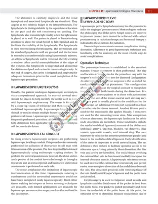
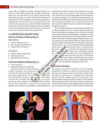


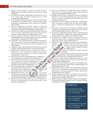

Laparoscopic urological surgery encompasses diagnostic, extirpative, and reconstructive procedures, with laparoscopic applications expanding particularly in pediatric urology and surgeries like adrenalectomy and varicocele ligation. The techniques involve various methods for identifying and preserving anatomical structures, with discussions around best practices, such as the necessity of preserving the testicular artery during varicocelectomy. Challenges include increased operative time and concerns regarding tumor spillage during nephrectomy, highlighting the need for continual advancements in laparoscopic technologies.


