Laparoscopic Hysterectomy Guide
•
0 likes•695 views

Laparoscopic hysterectomy is a minimally invasive surgical procedure to remove the uterus through small incisions in the abdomen using laparoscopic instruments and visualization. There are several types of laparoscopic hysterectomy depending on the extent of the procedure and whether it is assisted vaginally. Key advantages over traditional abdominal hysterectomy include less postoperative pain, shorter hospital stay, and faster recovery time. Important anatomical structures like the ureters must be carefully identified and protected during the procedure.

Recommended
More Related Content
What's hot
What's hot (20)
Similar to Laparoscopic Hysterectomy Guide
Similar to Laparoscopic Hysterectomy Guide (20)
More from World Laparoscopy Hospital
More from World Laparoscopy Hospital (20)
Recently uploaded
Recently uploaded (20)
Laparoscopic Hysterectomy Guide
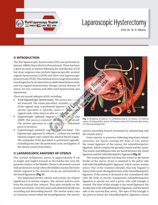
- 1. Laparoscopic Hysterectomy INTRODUCTION The first laparoscopic hysterectomy (LH) was performed in January 1988 by Harry Reich in Pennsylvania. There has been a great increase in interest following the introduction of LH but most surgeons now perform laparoscopically-assisted vaginal hysterectomy (LAVH) and then total laparoscopic hysterectomy (TLH). This minimal access surgical procedure was designed to be an alternative to abdominal hysterectomy and not vaginal hysterectomy. Benign uterine diseases of uterus are very common and often need hysterectomy and laparotomy. There are several subtypes of LH, including: ■ Total laparoscopic hysterectomy: The uterus and cervix are removed. The entire procedure, including suturing of the vaginal vault, is performed laparoscopically. The uterine specimen is typically removed through the vaginal vault, either intact or after morcellation. ■ Laparoscopic subtotal (supracervical) hysterectomy (LSH): The uterus is removed; the cervix is conserved. The uterine specimen is extracted via the abdominal ports or incisions. ■ Laparoscopic-assisted vaginal hysterectomy: The laparoscopic approach is utilized to perform any needed adnexal surgery and control the adnexal blood supply. The remainder of the procedure is performed vaginally, including entry into the peritoneal cavity and ligation of the uterine vessels from below. LAPAROSCOPIC ANATOMY OF UTERUS The normal nulliparous uterus is approximately 8 cm in length and angled forward so the fundus lies over the posterior surface of the bladder. Uterus is all around covered with peritoneum except where the bladder touches the lower uterine segment at the anterior cul-de-sac and laterally at the broad ligaments (Fig. 1). Two important arteries, uterine and ovarian, are of great significance in uterine surgery. The uterine arteries arise from the internal iliac arteries. They pass medially on the levator ani muscle, cross the ureter and ultimately divide into ascending and descending branch. The uterine artery runs in a tortuous course within the broad ligaments. The uterine arteries ascending branch terminates by anatomizing with the ovarian artery. From anterior to posterior, following important tubular structures are found crossing the brim of true pelvis: the round ligament of the uterus, the infundibulopelvic ligament, which contains the gonadal vessels and the ureter. The ovaries and fallopian tube are found between the round ligament and the infundibulopelvic ligament (Fig. 2). The ovarian ligaments run from the ovaries to the lateral border of the uterus. Ovary is attached to the pelvic side wall with infundibulopelvic ligament, which carries ovarian artery. One of the common mistakes that may happen is injuryoftheureterduringdissectionoftheinfundibulopelvic ligament. If the uterus is deviated to the contralateral side with the help of uterine manipulator infundibulopelvic ligament is spread out and a pelvic side wall triangle is created. The base of this triangle is the round ligament, the medial side is the infundibulopelvic ligament, and the lateral side is the external iliac artery. The apex of this triangle is the point at which the infundibulopelvic ligament crosses Fig. 1: Anatomy of uterus. (1) Umbilical artery; (2) Ureter; (3) Uterine artery; (4) Internal iliac artery; (5) Ovarian artery; (6) Common iliac artery; (7) Uterosacral ligament. Prof. Dr. R. K. Mishra
- 2. 431 CHAPTER 34: Laparoscopic Hysterectomy TABLE 1: Postoperative pain levels. Day LAVH (n = 19) TAH (n = 19) p 1 6.6 6.4 NS 3 4.4 4.3 NS 7 2.8 3.6 S 14 1.6 2.4 S 21 1.46 1.8 S Week 6 1.35 1.4 NS Wilcoxon’s signed-rank test. Ten-point activity scale: 1 = no pain; 10 = unbearable pain. S = significant at p < 0.005; NS = not significant at p < 0.01 Fig. 2: Position of uterus. (1) Uterus; (2) Round ligament; (3) Utero-ovarian ligament (proper ovarian ligament); (4) Uterosacral ligament; (5) Ovary; (6) Suspensory ligament of the ovary; (7) Ureter. the external iliac artery. The ureter always enters medial to this triangle into the pelvis. It is visible under the peritoneum overlying the external iliac artery. The ureters enter the pelvis in close proximity to the female pelvic organ and are at risk for injury during laparoscopic surgery of these organs. As the ureters course medially over the bifurcation of the iliac vessels, they pass obliquely under the ovarian vessels and then run in close proximity to the uterine artery. Laparoscopy hysterectomy needs careful identification of ureter with some dissection of retroperitoneum. An incision is made in the peritoneum overlying the pelvic side wall triangle between the fallopian tube and iliac vessel. Pelvic lymph node dissection is also necessary if gynecologist plans to perform radical LH. Node dissection as far distal as Cloquet’s node in the femoral triangle may be included and proximally dissection may be necessary up to para-aortic lymph nodes. If a vaginal hysterectomy can be performed in the first place, there would be no point in adding the costs and complications of laparoscopy. Its greatest benefit is the potential to convert what would have been an abdominal hysterectomy into a vaginal hysterectomy. An abdominal hysterectomy requires both a vaginal incision and a 4–6 inch long incision in the abdomen, which is associated with greater postoperative discomfort and a longer recovery period than for a vaginal procedure. Another advantage of the LH may be the removal of the tubes and ovaries which on occasion may not be easily removed with a vaginal hysterectomy. The most common medical reasons for performing hysterectomies include uterine fibroids (30%), abnormal uterine bleeding (20%), endometriosis (20%), genital prolapse (15%), and chronic pelvic pain (10%). For most of theseconditions,othertreatmentsshouldfirstbeconsidered, and hysterectomy should be reserved as a last resort. Laparoscopic hysterectomy results in a significantly shorter hospital stay, and a much more rapid return to normal activities than in total abdominal hysterectomy (TAH). The drug requirement to control pain and the level of pain patients experienced were also significantly less. Blood loss was not different for the two procedures (Tables 1 and 2). Postoperative recovery times and pain levels were assessed in 37 patients with a primary complaint of pelvic pain and diagnoses of fibroid uterus, adenomyosis, and severe endometriosis who underwent LH. Women reported an activity level of 8.7 on a scale of 1–10 (10 no limits on activity) by postoperative day 14. In another study, those undergoing abdominal hysterectomy had a mean uterine weight of 418 g compared with 150 g for those undergoing LAVH. The mean hospital stay after abdominal hysterectomy was4.5daysandafterLH2.5days.Animportantpublicpolicy issue now confronts us. As it is currently performed, LH is more expensive than TAH. The issue is whether the benefits of shorter convalescence and faster return to the work force, shorter hospitalization, and less need for narcotics for postoperative pain outweigh the disadvantage of the higher cost. If total healthcare system costs are evaluated, the short- term disability costs of 2 weeks of recovery after LH should TABLE 2: Postoperative activity levels. Day LAVH (n = 19) TAH (n = 19) p 1 3.4 3.3 NS 3 5.4 4.4 NS 7 7.8 5.8 S 14 9.2 6.4 S 21 9.6 7.9 S Week 6 9.95 8.5 S Wilcoxon’s signed-rank test. Ten-point activity scale: 1 = extremely limited activity, 10 = no limits on activity S = significant at p < 0.005; NS = not significant at p < 0.01
- 3. 432 SECTION 3: Laparoscopic Gynecological Procedures be compared with disability costs of 6–8 weeks of recovery after abdominal hysterectomy. For LH to be economically viable compared with TAH, savings in disability costs and the increased contribution to the gross domestic product must offset the increased healthcarecosts.Inthecurrentsystem,insurancecompanies and hospitals do not have share in these benefits, only consider the costs. The economic impact of laparoscopic surgery must take into account both the cost to the hospital and insurance payers and these productivity and social issues. Insurance is based on a risk pool whereby the cost of a premium is based on the cost of treatment, not the ability of the subscriber to return to work. An economic and social cost–benefit analysis must be performed before decisions are made to modify or judge a procedure that provides substantial benefits to the patient. Since its introduction in 1989, no one could have imagined that with continued improvement of techniques will progress so rapidly that LH can be performed on daycare basis for many women, and will result in shorter recovery time. Thus, the increased operating room time of approximately 46 minutes is significantly outweighed by the benefits available with widespread application of this procedure. CLASSIFICATION Garry and Reich Classification of Laparoscopic Hysterectomy ■ Type 1: Diagnostic lap + VH ■ Type 2: Lap vault suspension after VH ■ Type 3: LAVH ■ Type 4: LH (lap ligation of uterine artery) ■ Type 5: TLH ■ Type 6: LSH (lap supracervical hysterectomy) ■ Type 7: LHL (lap hysterectomy with lymphadenectomy) ■ Type 8: LHL + O (as above + omentectomy) ■ Type 9: RLH (radical lap hysterectomy) AAGL Classification of Laparoscopic Hysterectomy American Association of Gynecologic Laparoscopists (AAGL) classification of laparoscopic hysterectomy is shown in Table 3. TABLE 3: Laparoscopic hysterectomy classification according to American Association of Gynecologic Laparoscopists. Type 0 Laparoscopic-directed preparation for vaginal hysterectomy Type I* Dissection up to but not including uterine arteries Type IΑ Ovarian artery pedicle(s) only Type IΒ¶ IΑ + anterior structures Type IC IΑ + posterior culdotomy Type ID¶ IΑ + anterior structures and posterior culdotomy Type II* Type I + uterine artery occlusion and division, unilateral or bilateral Type IIΑ Ovarian artery(ies) and uterine artery(ies) occlusion and division only Type IIΒ¶ IIΑ + anterior structures Type IIC IIΑ + posterior culdotomy Type IID¶ IIΑ + anterior structures and posterior culdotomy Type III* Type II + portion of cardinal-uterosacral ligament complex only, unilateral or bilateral Type IIIΑ Uterine and ovarian artery pedicles with portion of the cardinal-uterosacral complex only, unilateral or bilateral Type IIIΒ¶ IIIΑ + anterior structures Type IIIC IIIΑ + posterior culdotomy Type IIID¶ IIIΑ + anterior structures and posterior culdotomy Type IV* Type II + total cardinal-uterosacral ligament complex, unilateral or bilateral Type IVΑ Uterine and ovarian artery pedicles with complete detachment of the total cardinal-uterosacral ligament complex only, unilateral or bilateral Type IVΒ¶ IVΑ + anterior structures Type IVC IVΑ + posterior culdotomy Type IVD¶ IVΑ + anterior structures and posterior culdotomy Type IVE Laparoscopically directed removal of entire uterus (AAGL: American Association of Gynecologic Laparoscopists) The system describes the portion of the procedure completed laparoscopically. *A suffix "o" may be added if unilateral or bilateral oophorectomy is performed concomitantly (e.g., type IoA). ¶ The B and D subgroups may be further subclassified according to the degree of dissection involving the bladder and whether an anterior culdotomy is created: (1) incision of vesicouterine peritoneum only, (2) dissection of any portion of bladder from cervix, and (3) creation of an anterior culdotomy.
- 4. 433 CHAPTER 34: Laparoscopic Hysterectomy Figs. 3A and B: Pervaginal examination should be routine. LAPAROSCOPIC-ASSISTED VAGINAL HYSTERECTOMY Laparoscopically-assisted vaginal hysterectomy is one of the most frequently performed gynecologic operations, and numerous authors have demonstrated its safety and feasibility. A laparoscopic-assisted vaginal hysterectomy is a type 3 Garry and Reich hysterectomy in which the adnexal pedicles of the round ligament, fallopian tube, and ovarian ligament are released abdominally through laparoscopic approach while the uterine artery and vein are secured throughthevaginalapproach.Furtherdissectionofthebroad ligament anteriorly to free the bladder from the uterovesical fold is done laparoscopically. A 2 cm posterior colpotomy in between the uterosacral ligaments at the base of the pouch of Douglas is also done laparoscopically. This dissection allows completion of the surgery vaginally with ease. Preoperative Measures Patients are evaluated same way as that of any major surgery. Routine preoperative tests include a complete blood count with differential, serum electrolyte, bleeding time, and urinalysis. More comprehensive blood studies include thrombin time, partial thrombin time, ECG, chest X-ray, and endometrial biopsy. Mechanical and antibiotic bowel preparation is advised. Peglec powder 1 sachet with water a night prior to surgery is advised. Patient Position Patient should be in steep Trendelenburg and lithotomy position. One assistant should remain between the legs of patient to do uterine manipulation whenever required (Figs. 3A and B). Position of Surgical Team (Fig. 4) Surgeon stand left to the patient, camera assistant should be left to the surgeon. Second assistant should be the opposite side of the body of patient. One more assistant is required between the legs to handle uterine manipulator. The patient should ideally get general anesthesia with endotracheal intubation. A Foley catheter should be inserted to provide bladder drainage throughout the operation. Port Position A 10-mm umbilical port for camera should be along the inferior crease. Two 5-mm ports should be placed at 5 cm away from umbilicus on either side. Sometime, accessory port at right or left iliac region may be required according to need. Port position should be in accordance with baseball diamond concept. If the left side of tube has to be operated, one port should be in right iliac fossa and another below left iliac fossa (Fig. 5). Operative Technique It is important throughout the procedure to be able to manipulate the uterus for optimal observation. Fig. 4: Surgical team position in laparoscopically-assisted vaginal hysterectomy. Fig. 5: Port position for laparoscopically-assisted vaginal hysterectomy. A B
- 5. 434 SECTION 3: Laparoscopic Gynecological Procedures Figs. 6A and B: Laparoscopically-assisted vaginal hysterectomy using bipolar. A B Figs. 7A and B: Successive desiccation and dissection. A B Different types of uterine manipulators are available. Depending on the laparoscopic procedure, digital examination, probes, and sponge stick applicators are used in the cul-de-sac for identification of structures during laparoscopy. The direction and location of both ureters should be identified as much as possible (Figs. 6A and B). With the patient in lithotomy position, after the pneumoperitoneum insufflated to a pressure of 12–14 mm Hg, three ports should be introduced. The 10 mm optical umbilical trocar, 5 mm in lateral border of the right rectus abdominis in right iliac fossa, 5 mm in the same position on the left side for the Ligasure forceps. The ureters were visualized transperitoneally. If adnexectomy is planned, following electrodesiccation and cutting of the round ligaments 2–3 cm from the uterus, then infundibulopelvic ligament is desiccated and cut, taking progressive bites of tissue starting at pelvic brim and moving towards the round ligament. It is important to avoid the coagulation of the round ligament near the uterus because of higher bleeding (Figs. 6A and B). In order to preserve the adnexa, the coagulation and section is performed proximal to the fallopian tubes and the utero-ovarian ligament. The dissection continues posteriorly on the broad ligament, taking care not to cut the uterine pedicle’s vessels (Figs. 7A and B). The anterior leaf of the broad ligament is opened towards the vesicouterine fold and bladder flap is developed. The anterior leaf of the broad ligament is grasped with forceps, elevated and dissected from the anterior lower uterine segment. The utero-ovarian ligament, proximal tube, and mesosalpinx are progressively dissected and cut and posterior leaf of the broad ligament is opened. Similarly, the round ligament, fallopian tube, and utero-ovarian ligament are grasped closed to their insertion into the uterus then secured with the bipolar, Ligasure, or harmonic scalpel and cut. The distal end of the energized instruments must be kept free of the bladder and ureter (Fig. 8). The uterovesical junction is identified, grasped, and elevated with forceps while being cut with scissors. The bladder pillars are identified desiccated and cut. The bladder can be completely freed from the uterus by pushing downward with the tip of a blunt probe along the vesicocervical plane until the anterior cul-de-sac is exposed completely. In patients with severe anterior cul-de-sac
- 6. 435 CHAPTER 34: Laparoscopic Hysterectomy Fig. 8: Dissection of bladder peritoneum. endometriosis, previous CS or adhesions, sharp dissection can be performed. Injecting 5 mL of indigo carmine in the patient’s bladder helps to detect bladder trauma (Figs. 9 and 10). Colpotomy A folded gauze in sponge forceps is used to mark the fornix. The vaginal wall is tented and transacted horizontally with hook electrode (Figs. 11A to C). Once the dissection is extended to the lower uterine segment or to the level of cardinal ligament, laparoscopic procedure is temporarily terminated and vaginal part of LAVH started. VAGINAL PART OF LAPAROSCOPIC- ASSISTED VAGINAL HYSTERECTOMY The sponge forceps occluding the colpotomy is withdrawn together with the uterine manipulator. Three vaginal specula are used to get proper access for vaginal part of LAVH (Figs. 12A to D). With aid of appropriate-sized speculum and adequate lateral and anterior retraction by the assistant, the anterior and posterior lips of the cervix are held by tenaculum. Using monopolar energy in circumcision of the skin at 2.5 cm above the external os towards the vaginal vault is done. Further dissection with gloved finger or dissecting scissors creates a flap in which the superior Sims speculum or narrow angled retractor is placed to lift the bladder away. Gradual release of the tissues results in entry to the abdominal cavity (Figs. 13A to H). Using appropriate-sized caucus clamp, the uterosacral pedicles are identified bilaterally clamped, secured, cut, and tied with the tail of the thread left as mark or coagulated with Ligasure, harmonic scalpel, or bipolar instrument specially made for vaginal hysterectomy (Figs. 12A to D). With release of the uterosacral, the uterus descends into the vaginal cavity. Similarly, the cardinal ligaments are bilaterally clamped, secured, cut, and tied with a mark or coagulated and dissected. Further descent of the uterus is gained, and the uterine pedicles are clamped, secured, cut and tied. The uterus is then delivered vaginally. Once the uterus is removed, the vaginal vault is closed to ensure support of the vault; the vaginal angles are attached to the uterosacral and cardinal ligaments with 2-0 vicryl. Any coexisting cystocele or rectocele is repaired. A very large fibroid uterus should be debulked by morcellation for removal vaginally. It can be combined with laparoscopic adnexal surgery, e.g., ovariectomy or adhesiolysis. Once the vaginal surgery is completed again laparoscopic inspection of the pelvis is done. Total Laparoscopic Hysterectomy Preparation and Positioning of Patient Preoperative check of consent form and patient past medical history should be taken followed by proper preanesthetic checkup (PAC). Patients is placed in a dorsal lithotomy position. The arms are tucked at the sides and a foam mattress is situated directly under the patient to prevent sliding during steep Trendelenburg. Gynecologist should keep the table in a low position and have a monitor directly facing each surgeon to promote an ergonomic working environment (Fig. 14). Abdominal Entry and Trocar Placement A 10-mm supraumbilical skin incision is made using a #15 blade and insert a Veress needle is introduced into the peritoneal cavity. Once intraperitoneal pressure has reached 15 mm Hg, insert trocar through the supraumbilical incision, followed by a complete survey of the abdomen to rule out any visceral injury at the time of entry. The lower quadrant trocar sleeves are placed under direct vision. These trocars are placed lateral to the rectus abdominis muscles, 2 cm above and 2 cm medial to the anterior superior iliac spine. These secondary trocar position varies according to the size of uterus. Usually, a 5-mm trocar is placed on the right and a 5-mm trocar on the left. For TLH port position can be ipsilateral or contralateral. Two ports on the left greatly facilitate suturing and help to maintain an ergonomic position for the surgeon throughout the procedure (Figs. 15A and B). Insertion of a Uterine Manipulator Different types of uterine manipulators are available. Generally, most of the gynecologists use the RUMI® or Mangeshkar Uterine Manipulator; however, in patients with a very narrow introitus the VCare® Uterine Manipulator is useful because this is easier to insert (Fig. 16).
- 7. 436 SECTION 3: Laparoscopic Gynecological Procedures Fig. 9: Opening of anterior and posterior leaf broad ligament. Fig. 10: Separation of bladder. Figs. 11A to C: Steps of colpotomy. Figs. 12A to D: Anterior and posterior colpotomy. A B C A C B D
- 8. 437 CHAPTER 34: Laparoscopic Hysterectomy Figs. 13A to H: Successive clamping and desiccation of uterine pedicle through the vaginal route using bipolar. A C E G B D F H
- 9. 438 SECTION 3: Laparoscopic Gynecological Procedures Fig. 14: Position of the patient for total laparoscopic hysterectomy. Dissection of Infundibulopelvic Ligament The infundibulopelvic (IP) ligament or the utero-ovarian ligament is initially desiccated with a reusable bipolar grasper, Ligasure, or harmonic scalpel. It is important to stay close to the ovary as this helps to avoid the pelvic sidewall during ovarian removal and the ascending uterine vessel during ovarian conservation. The gynecologists should take special care to desiccate the parametrial veins that run between the ovary and the round ligament as these can be quite tortuous and tend to bleed if left unattended. The IP ligament or utero-ovarian ligament is then transacted close to the ovary. During this step of the procedure, the uterine manipulator is being pushed upward and to the contralateral side to provide maximal visualization (Fig. 17). Mobilization of the Bladder Transect the round ligament and separate the anterior and posterior leaves of the broad ligament with the harmonic scalpel or monopolar scissors. It is important to find the correct plane; this is where the peritoneum separates easily with gentle manipulation. After peritoneal separation, identify the vesicouterine peritoneal fold and continue the dissection anteriorly, thereby mobilizing the bladder Figs. 15A and B: Abdominal entry and trocar placement. off the lower uterine segment. It is important to stay in the loose areolar tissue if at all possible. In patients who have had a prior cesarean section, this area may be scarred and it is important to stay relatively high on the uterus during the dissection. In case of adhesion a pledget dissection combined with sharp dissection with cold scissors is helpful. A reevaluation of the route of dissection is advised if fat is encountered because the fat belongs to the bladder; this may indicate that the dissection is moving too close to the bladder (Fig. 18). Securing the Uterine Vessels Uterine artery course is diverse and due to a wide variety in anatomy and in the course of the uterine vessels, it is helpful to initially skeletonize them with the harmonic scalpel. Then desiccate the ascending uterine vessels with the bipolar grasper or Ligasure at the level of internal cervical os. Note that pushing cephalad with the uterine manipulator helps to move the uterine vessels away from the ureter. Complete desiccation of the vessels can be assessed visually by observing the bubbles coming and going during this process; when the bubbles stop forming and tissue color turns brown the vessel is desiccated and safe to transect with the harmonic scalpel or Ligasure. It is important to take the uterine vessels high and then dissect medially to the uterine vessels down to the cup of colpotomizer. This averts ureteral injury and provides a healthy vascular pedicle that can be safely desiccated further in the event of bleeding (Figs. 19A and B). Colpotomy After dissection of uterine pedicle the next step is to identify the vaginal fornices while pushing cephalad with the uterine manipulator. Gynecologist will either see the indentation of the KOH colpotomizer or be able to palpate it with a laparoscopic instrument. The harmonic scalpel is then used to cut circumferentially around the cup. Take care not to direct the harmonic scalpel directly into the metal because this may result in failure of the device and may even break A B
- 10. 439 CHAPTER 34: Laparoscopic Hysterectomy Fig. 16: Insertion of a uterine manipulator. Fig. 17: Dissection of upper pedicle, conserving ovary. Fig. 18: Mobilization of bladder. the active blade or harmonic (Figs. 20A to D). For colpotomy hook can also be used if harmonic is not available. Removal of the Uterus Once complete separation of uterus with vaginal cuff is achieved pull the uterus into the vagina if it fits (Figs. 21A and B). The uterus can remain there to maintain pneumoperitoneum during suturing. Alternatively, the uterus is removed and a glove with a pair of 4 × 4 sponges is placedintothevaginatomaintainpneumoperitoneum.Ifthe uterus is too large to fit through the vagina, it can be carefully morcellated transvaginally by using a 10-blade scalpel and triple hooks for retraction. In patients with limited vaginal access, the uterus can be morcellated laparoscopically using an electronic morcellator. It is important to keep the tip of the morcellator in clear view at all times. Vaginal Cuff Closure There are various ways of vaginal cuff closure (Figs. 22A to D). One can use barbed suture, extracorporeal square knot, intracorporeal continuous suture, or interrupted suture. Most commonly used technique of vault closure is intracorporeal continuous suturing. Closure begins at the distal angle of the vaginal cuff and proceeds in a running fashion, making sure to include the vaginal mucosa and the pubocervical and rectovaginal fascia. Many surgeons do not tie a knot at the end of vault if they use barbed suture. Extracorporeal knot with Clark knot pusher is also very good to close the vault especially in deep pelvis. The pelvis can now be irrigated and hemostasis at all sites is assured. Port Site Closure The fascia at the 10 mm incision is closed using 0 vicryl sutures with a fascia closure device. The skin is closed with 4-0 monocryl suture in a continuous subcutaneous fashion or skin stapler. The 5-mm incisions are closed with Dermabond. Many gynecologists then inject 20 cc of 0.5% Marcaine at all incision sites to reduce immediate postoperative pain. It is not necessary to perform cystoscopy routinely, but in selected cases cystoscopy is performed after vaginal closure to check ureteral patency and for any signs of bladder injury. The patient is given 5 cc of Indigo carmine intravenously 5 minutes prior to closure. Please note that a normal cystoscopy does not exclude a delayed thermal injury to either the ureters or the bladder. Subtotal Hysterectomy Supracervical hysterectomy is performed to preserve libido ofpatient.Theprocedureisperformedfullylaparoscopically. After desiccating and cutting the uterine vessels at the level of cardinal ligaments above the uterosacral ligament, uterus is retracted, and its lower segment is amputated with the scissors and unipolar cutting current. After transecting the uterus from the cervix, uterine manipulator is removed vaginally, the cervical stump is irrigated, and hemostasis is achieved. The endocervical epithelium, lining the cervical canal, is vaporized or coagulated with laser or electrosurgery. The rest of the endocervical canal is ablated vaginally to reduce the risk of intraepithelial cervical neoplasia. The cervical stump is closed with interrupted absorbable sutures and covered with peritoneum, which is stitched transversely with interrupted sutures. The dissected uterus is morcellated and removes through a 10-mm cannula. Mini-laparotomy or posterior colpotomy can also be performed to remove the uterus in case of subtotal hysterectomy. These patients are advised for annual examination for Pap smear.
- 11. 440 SECTION 3: Laparoscopic Gynecological Procedures Figs. 19A and B: Securing uterine artery. Figs. 20A to D: Colpotomy by harmonic scalpel. Figs. 21A and B: Removal of uterus through the vaginal route. A B A B C D A B
- 12. 441 CHAPTER 34: Laparoscopic Hysterectomy Figs. 22A to D: Vault closure with by continuous suturing. Ending the Procedure One of the benefits of LAVH or TLH over nondescent vaginal hysterectomy (NDVH) is inspection of pedicles at the end of surgery. The vaginal cuff can be closed from below or above but after that pneumoperitoneum is again restored to see the pelvic and abdominal cavity. Irrigation and suction should be performed. In case of any residual bleeding it is controlled laparoscopically. At the end pelvis is filled with 300 mL Ringer’s lactate and it should be seen for any change in color. Once inspection is satisfactory, the fluid is sucked and instrument and cannula are removed after deflating the abdominal cavity. It has been demonstrated that TLH and LAVH are associated with a shorter hospital stay and patients require less pain medication compared to TAH. LAVH or TLH can replace most of the abdominal hysterectomy for the benign disease of uterus and with the technology available today, it has definite benefit over nondescended vaginal hysterectomy. DISCUSSION Vaginal hysterectomy is a part of repertoire of every trained gynecologist. It is considered as a feasible option to abdominal hysterectomy and many studies have shown that vaginal hysterectomy has fewer complications, short recovery, and smaller hospital stay than laparotomy. LH requires greater surgical expertise and has a steep learning curve. Randomized trials have shown advantages of laparoscopy vs. laparotomy, including reduced postoperative pain, shorter hospitalization, rapid recovery, and substantial financial benefits to society. The objective of performing hysterectomy laparoscopically can be achieved but the question is, does this offer any advantage over vaginal route? Every mode of hysterectomy has advantages and disadvantages but the indications for each remain controversial. Good surgical practice is when the indication for hysterectomy is considered as the primary criterion for selecting the route of hysterectomy and not factors such as surgeon’s choice and experience. A major determinant of the route of hysterectomy is not the clinical situation but the attitude of the surgeon. There is no need for extra training and special skills or complicated equipment for vaginal hysterectomy. Laparoscopic hysterectomy took a long time to perform in all studies. However, with increasing weight of the uterus, there was a linear increase in operating time and blood loss in hysterectomy performed vaginally which was not observed in laparoscopic-assisted vaginal hysterectomy. Thereisnostatisticallysignificantdifferenceinpostoperative analgesia requirement, hospital stay, recovery milestones, or complication rates. The biggest drawback of laparoscopic A B C D
- 13. 442 SECTION 3: Laparoscopic Gynecological Procedures route over vaginal one is its cost due to expensive disposable instruments, prolonged operating and anesthesia time, and the need for a trained senior gynecologist. For laparoscopic- assisted vaginal hysterectomy to be cost effective expensive disposable instruments must be eliminated. Laparoscopic surgeons argue that subtotal hysterectomy can be performed laparoscopically but most randomized trials have failed to demonstrate any benefit of subtotal hysterectomy over total hysterectomy. In women who wish to retain their cervix vaginal subtotal hysterectomy described by Döderlein-Krönig technique can be performed. The disadvantage of vaginal approach is vault hematomas. The abdominal approach to hysterectomy does ensure good hemostasis under direct vision, while during the vaginal operation, the vault is closed and subsequent bleeding from the vagina between the mucosa and the peritoneum can give rise to problems, especially if a vasoconstrictor has been given that subsequently wears off. Laparoscopic approach can help check hemostasis and reduce the incidence of vault hematomas. However; this aspect needs to be evaluated in studies. Lack of uterine descent and nulliparity, fibroid uterus, need for oophorectomy, and previous pelvic surgery are no more considered as contraindications to the vaginal route. With adequate vaginal access and technical skill, and good uterine mobility, vaginal hysterectomy can easily be achieved. Multiparity, lax tissues due to poor involution following multiple deliveries and lesser tissue tensile strength afford a lot of comfort to vaginal surgeon even in the presence of significant uterine enlargement. None of the evidence supports the use of LH rather than VH if latter can be performed safely. No outcomes are significantly worse for vaginalhysterectomyascomparedtoLAVH.Thereareclinical situations where vaginal surgeries are not appropriate such as dense pelvic adhesions, severe endometriosis, adnexal disease, when vaginal access is reduced wherein LH is indicated as it has advantages over the abdominal approach. Laparoscopic approach may be helpful postoperatively to rule out hemorrhage in some cases. Laparoscopic assistance should not be used to supplant inadequate skills of vaginal hysterectomy. Lack of training in laparoscopic surgery is not a reason for not removing uteri vaginally. The learning curve of VH is noticeably short as compared to laparoscopic surgery, however, the current scenario in residency programs is not providing a level of surgical competency in performing difficult vaginal hysterectomies. There is a need to improve this training. In order to compare the complication rates of different types of hysterectomies, considering an incidence of 4–5% of serious complications of hysterectomies, at least 1,460 women would be required in each arm of the study to detect 50% increase in the complication rate. Therefore, larger randomized controlled trials are required to compare different types of hysterectomies. When the size of the uterus is greater than 16 weeks gestation, there is an increase in the operative time and blood loss in VH compared to LAVH which is statistically significant. Laparoscopically-assisted vaginal hysterectomy is a useful adjunct to transvaginal hysterectomy for lysis of extensive adhesions and sometimes for certain concomitant adnexal surgery. Besides, LAVH can also secure almost all the main blood supplies to the uterus, i.e., the uterine vessels and the adnexal collaterals. Although, a skilled surgeon can do transvaginal hysterectomy with a larger uterus by employing volume-reducing techniques, Kohler reported that laparoscopic coagulation hemostasis of the uterine vessels was associated with less blood loss. It may take time to achieve these goals, but they may make subsequent extirpation or volume-reducing procedures easier and safer to perform. Therefore, the average operative time and estimated blood loss for LAVH remained almost constant regardless of increasing uterine weight. Generally, the average operative time for LAVH is longer than that for transvaginal hysterectomy. It takes time to secure the uterine blood supply before extirpation and volume-reducing procedures, but it also makes LAVH superior to transvaginal hysterectomy when dealing with a larger uterus. In our opinion, LAVH might be considered for a larger uterus in view of the relatively shorter operative time and less blood loss, whereas transvaginal hysterectomy is preferable for a small uterus, not only for shorter operative time and minimal wound, but also for much lower costs. BIBLIOGRAPHY 1. Carley ME, McIntire D, Carley JM, Schaffer J. Incidence, risk factors and morbidity of unintended bladder or ureter injury during hysterectomy. Int Urogynecol J Pelvic Floor Dysfunct. 2002;13:18-21. 2. Chapron C, Dubuisson JB, Aubert V. Total laparoscopic hysterectomy: preliminary results. Hum Reprod. 1994;9: 2084-9. 3. Chapron C, Dubuisson JB. Laparoscopic hysterectomy. Lancet. 1995;345:593. 4. Chapron C, Fauconnier A, Goffinet F, Bréart G, Dubuisson JB. Laparoscopic surgery is not inherently dangerous for patients presenting with benign gynaecologic pathology. Results of a meta-analysis. Hum Reprod. 2002;17:1334-42. 5. Chapron C, Laforest L, Ansquer Y, Fauconnier A, Fernandez B, Breart G, et al. Hysterectomy techniques used for benign disorders: results of a French multicentre study. Hum Reprod. 1999;14:2464-70. 6. Chauveaud A, de Tayrac R, Gervaise A, Anquetil C, Fernandez H. Total hysterectomy for a nonprolapsed, benign uterus in women without vaginal deliveries. J Reprod Med. 2002;47:4-8. 7. Cosson M, Querleu D, Crepin G (Eds). Hystérectomies pour pathologies bénignes. Masson: Paris; Williams et Wilkins; 1997. pp. 160. 8. Councell RB, Thorp JM Jr, Sandridge DA, Hill ST. Assessments of laparoscopic-assisted vaginal hysterectomy. J Am Assoc Gynecol Laparosc. 1994;2:49-56.
- 14. 443 CHAPTER 34: Laparoscopic Hysterectomy 9. Dandolu V, Mathai E, Chatwani A, Harmanli O, Pontari M, Hernandez E. Accuracy of cystoscopy in the diagnosis of ureteral injury in benign gynecologic surgery. Int Urogynecol J Pelvic Floor Dysfunct. 2003;14:427-31. 10. Daraï E, Soriano D, Kimata P, Laplace C, Lecuru F. Vaginal hysterectomy for enlarged uteri, with or without laparoscopy assistance: randomized study. Obstet Gynecol. 2001;97:712-6. 11. Davies A, Vizza E, Bournas N, O’Connor H, Magos A. How to increase the proportion of hysterectomies performed vaginally. Am J Obstet Gynecol. 1998;179:1008-12. 12. Dicker RC, Greenspan JR, Strauss LT, Cowart MR, Scally MJ, Peterson HB, et al. Complications of abdominal and vaginal hysterectomy among women of reproductive age in the United States. The Collaborative Review of Sterilization. Am J Obstet Gynecol. 1982;144:841-8. 13. Dorairajan G, Rani PR, Habeebullah S, Dorairajan LN. Urological injuries during hysterectomies: a 6-year review. J Obstet Gynaecol Res. 2004;30:430-5. 14. Dorsey JH, Steinberg EP, Holtz PM. Clinical indications for hysterectomy route: patient characteristics or physician preference? Am J Obstet Gynecol. 1995;173:1452-60. 15. Dwyer PL, Carey MP, Rosamilia A. Suture injury to the urinary tract in urethral suspension procedures for stress incontinence. Int Urogynecol J Pelvic Floor Dysfunct. 1999;10:15-21. 16. Farquhar CM, Steiner CA. Hysterectomy rates in the United States 1990–1997. Obstet Gynecol. 2002;99:229-34. 17. Garry R, Fountain J, Brown J, Manca A, Mason S, Sculpher M, et al. Evaluate hysterectomy trial. A multicentre randomised trial comparing abdominal, vaginal and laparoscopy methods of hysterectomy. Health Technol Assess. 2004a;8:1-154. 18. Garry R, Fountain J, Mason S, Hawe J, Napp V, Abbott J, et al. (2004b) The evaluate study: two parallel randomised trials, one comparing laparoscopy with abdominal hysterectomy, the other comparing laparoscopy with vaginal hysterectomy. BMJ. 2004;328:129. Erratum in BMJ. 2004;328:494. 19. GilmourDT,DasS,FlowerdewG.Ratesofurinarytractinjuryfrom gynecologic surgery and the role of intraoperative cystoscopy. Obstet Gynecol. 2006;107:1366-72. 20. Gilmour DT, Dwyer PL, Carey MP. Lower urinary tract injury during gynecologic surgery and its detection by intraoperative cystoscopy. Obstet Gynecol. 1999;94:883-9. 21. Gimbel H, Settnes A, Tabor A. Hysterectomy on benign indication in Denmark 1988–1998. Acta Obstet Gynecol Scand. 2001;80:267-72. 22. Härkki-Siren P, Kurpi T, Sjöbery J, Tiitinen A. Safety aspects of laparoscopic hysterectomy. Acta Obstet Gynecol Scand. 2001;80:383-91. 23. Härkki-Siren P, Sjöberg J, Mäkinen J, Heinonen PK, Kaudo M, Tomas E, et al. Finnish national register of laparoscopic hysterectomies: A review and complications of 1165 operations. Am J Obstet Gynecol. 1997;176:118-122. 24. Härkki-Siren P, Sjöberg J, Tiitinen A. Urinary tract injury after hysterectomy. Obstet Gynecol. 1998;92:113-8. 25. Harris MB, Olive DL. Changing hysterectomy patterns after introduction. Gynecol. 1994;171:340-3. 26. Hurd WW, Bude RO, De Lancey JO, Pearl ML. The relationship of the umbilicus to aortic bifurcation: implications for laparoscopic technique. Obstet Gynecol. 1992;80:48-51. 27. Hwang JL, Seow KM, Tsai YL, Huang LW, Hsieh BC, Lee C. Comparative study of vaginal, laparoscopically-assisted vaginal and abdominal hysterectomies for uterine myoma larger than 6 cm in diameter or uterus weighing at least 450 g: a prospective randomized study. Acta Obstet Gynecol Scand. 2002;81:1132-8. 28. Johns DA, Carrera B, Jones J, DeLeon F, Vincent R, Safely C. The medical and economic impact of laparoscopically-assisted vaginal hysterectomy in a large, metropolitan, not-for-profit hospital. Am J Obstet Gynecol. 1995;172:1709-15. 29. Johnson N, Barlow D, Lethaby A, Tavender E, Curr E, Garry R. Surgical approach to hysterectomy for benign gynaecological disease. Cochran Database Syst Rev. 2006;(2):CD003677. 30. Johnson N, Barlow D, Lethaby A, Tavender E, Curr L, Garry R. Methods of hysterectomy: systematic review and meta-analysis of randomised controlled trials. Br Med J. 2005;330:1478. 31. KadarN.Dissectingthepelvicretroperitoneumandidentifyingthe ureters. A laparoscopic technique. J Reprod Med. 1995;40:116-22. 32. Kreiker G, Bertoldi A, Sad Larcher J, Ruiz Orrico G, Chapron C. Prospective evaluation of the learning curve of total laparoscopic hysterectomy in a university hospital. J Am Assoc Gynecol Laparosc. 2004;11:229-35. 33. Leonard F, Chopin N, Borghese B, Fotso A, Foulot H, Coste J, et al. Total laparoscopic hysterectomy: preoperative risk factors for conversion to laparotomy. J Minim Invasive Gynecol. 2005;12:312-17. 34. Leonard F, Chopin N, Borghese B, Fotso A, Foulot H, Coste J, et al. Total laparoscopy hysterectomy: preoperative risk factor for conversion to laparotomy. J Minim Invasive Gynecol. 2005;12:312-7. 35. LiuCY,ReichH.Complicationsoftotallaparoscopichysterectomy in 518 cases. Gynecol Endoscopy. 1994;3:203-8. 36. Mäkinen J, Johansson J, Tomas C, Tomas E, Heinonen PK, Laatikainen T, et al. Morbidity of 10110 hysterectomy by type approach. Hum Reprod. 2001;16:1473-8. 37. McMaster-Fay RA, Jones RA. Laparoscopic hysterectomy and ureteric injury: a comparison of the initial 275 cases and the last 1,000 cases using staples. Gynecol Surg. 2006;3:118-21. 38. Meikle SF, Nugent EW, Orleans M. Complications and recovery from laparoscopy-assisted vaginal hysterectomy compared with abdominal and vaginal hysterectomy. Obstet Gynecol. 1997;89:304-11. 39. Mteta KA, Mbwambo J, Mvungi M. Iatrogenic ureteric and bladder injuries in obstetric and gynaecologic surgeries. East Afr Med J. 2006;83:79-85. 40. National Centre for Disease Control and Prevention 1997. Hysterectomy surveillance United States 1980–1993. CDC surveillance summaries, August. Mabille de Poncheville L (1998) Coeliochirurgie gynécologique en France, instantanée 1996. Résultats d’une enquête nationale. Thèse de médecine, Tours, France. Moller C, Kehlet H and Ottesen BS. Hospita- lization and convalescence after hysterectomy. Open or laparoscopy surgery? Ugeskr Laeger. 1999;161:4620-4. 41. NezhatF,NezhatC,AdmonD,GordonS,NezhatC.Complications and results of 361 hysterectomies performed at laparoscopy. J Am Coll Surg. 1995;180:307-16. 42. O’SheaRT,PetruccoO,GordonS,SemanE.Adelaidelaparoscopic hysterectomy audit (1991–1998): realistic complications rates. Gynaecol Endoscopy. 2000;9:369-72. 43. Oh BR, Kwon DD, Park KS, Ryu SB, Park YI, Presti JC Jr. Late presentation of ureteral injury after laparoscopic surgery. Obstet Gynecol. 2000;95:337-39. 44. Ou CS, Beadle E, Presthus J, Smith M. A multicenter review of 839 laparoscopic-assisted vaginal hysterectomies. J Am Assoc Gynecol Laparosc. 1994;1:417-22. 45. Paulson JD. Laparoscopically-assisted vaginal hysterectomy. A protocol for reducing urinary tract complications. J Reprod Med. 1996;41:623-8. 46. Phipps JH, Tyrrell NJ. Transilluminating ureteric stents for preventing operative ureteric damage. Br J Obstet Gynaecol. 1992;99:81. 47. Reich H, De Caprio J, McGlynn F. Laparoscopic hysterectomy. J Gynecol Surg. 1989;5:213-6. 48. Ribeiro S, Reich H, Rosenberg J, Guglielminetti E, Vidali A. The value of intraoperative cystoscopy at the time of laparoscopic hysterectomy. Hum Reprod. 1999;14:1727-9.
- 15. 444 SECTION 3: Laparoscopic Gynecological Procedures 49. Ribeiro SC, Ribeiro RM, Santos NC, Pinotti JA. A randomized study of total abdominal, vaginal and laparoscopy hysterectomy. Int J Gynaecol Obstet. 2003;83:37-43. 50. Rutkow IM. Obstetric and gynecologic operations in the United States, 1979 to 1984. Obstet Gynecol. 1986;67:755-9. 51. Saidi MH, Sadler RK, Vancaillie TG, Akright BD, Farhart SA, White AJ. Diagnosis and management of serious urinary complications after major operative laparoscopy. Obstet Gynecol. 1996;87:272-6. 52. Shen CC, Wu MP, Kung FT, Huang FJ, Hsieh CH, Lan KC, et al. Major complications associated with laparoscopic-assisted vaginal hysterectomy: ten-year experience. J Am Assoc Gynecol Laparosc. 2003;10:147-53. 53. Soriano D, Goldstein A, Lecuru F, Daraï E. Recovery from vaginal hysterectomy compared with laparoscopy-assisted vaginal hysterectomy: a prospective, randomized, multicenter study. Acta Obstet Gynecol Scand. 2001;80:337-41. 54. University of York (UK) Centre for Health Economics. The management of menorrhagia. Effective Healthcare. 1991;1(9). 55. Vakili B, Chesson RR, Kyle BL, Shobeiri SA, Echols KT, Gist R, et al. The incidence of urinary tract injury during hysterectomy: a prospective analysis based on universal cystoscopy. Am J Obstet Gynecol. 2005;192:1599-1604. 56. VesseyMP,Villard-MackintoshL,McPhersonK,CoulterA,YeatesD. The epidemiology of hysterectomy: findings in a large cohort study. Br J Obstet Gynaecol. 1992;99:402-7. 57. Visco AG, Taber KH, Weidner AC, Barber MD, Myers ER. Cost- effectiveness of universal cystoscopy to identify ureteral injury at hysterectomy. Obstet Gynecol. 2001;97:685-92. 58. Wattiez A, Soriano D, Cohen SB, Nervo P, Canis M, Botchorisvili R, et al. The learning curve of total laparoscopic hysterectomy: comparative analysis of 1647 cases. J Am Assoc Gynecol Laparosc. 2002;9:339-45. 59. Wood EC, Maher P, Pelosi MA. Routine use of ureteric catheters at laparoscopic hysterectomy may cause unnecessary complications. J Am Assoc Gynecol Laparosc. 1996;3:393-7. 60. Wu SM, Chao Yu YM, Yang CF, Che HL. Decision making tree for women considering hysterectomy. J Adv Nurs. 2005;51:361-8.