
Recommended
More Related Content
Similar to MEDICAL ELECTRONICS.pdf
Similar to MEDICAL ELECTRONICS.pdf (20)
Recently uploaded
Recently uploaded (20)
MEDICAL ELECTRONICS.pdf
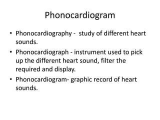
- 1. Phonocardiogram • Phonocardiography - study of different heart sounds. • Phonocardiograph - instrument used to pick up the different heart sound, filter the required and display. • Phonocardiogram- graphic record of heart sounds.
- 5. Two categories • Heart sound: • Transient characteristics with short duration occurs due to opening and closing of the heart valves. • In abnormal heart additional sounds are heard between the normal heart sound. • Murmurs: • additional sounds • Noisy characteristics with long duration occurs due to turbulent blood flow in heart. • Caused by improper opening of the valves
- 6. Heart Sound • Mechanical working processes of the heart produce sound - health status. • Relationship between blood volumes, pressures and flows within the heart determines the opening and closing of the heart valves. • Normal heart sounds- lub and dub • Valvular theory - heart sounds originate from a point sources located near the valves. • Cardiohemic theory -the heart and the blood represent an interdependent system that vibrates as a whole and propagates sound as waves of alternate pressure.
- 7. CLASSIFICATION OF HEART SOUND • It is divided into four types: • Valve closure sound • Ventricular filling sound • Valve opening sound • Extra cardiac sound
- 8. Types of Heart Sound • Valve closure sound - occurs at the beginning of systole and at the beginning of diastole. • Ventricular filling sound - occurred at the time of filling of the ventricles. • Valve opening sound - occurs at the time of opening of atrio- ventricular valves and semi lunar valves. • Extra cardiac sound - occur in mid systole or late systole or early diastole
- 9. • Systole: • Contraction of the heart muscle • Systolic pressure: 120 mm • Diastole: • Relaxation of the heart muscle • Diastolic pressure is 80 mm
- 11. First Heart Sound Initial vibrations occur when first contraction of ventricle move blood towards atria, closing AV valves Abrupt tension of closed AV valves, decelerating the blood Oscillation of blood between root of aorta and ventricular walls Vibrations caused by turbulence in ejected blood flowing into aorta
- 13. Second Sound S2 • The second sound (S2) signals the end of systole and the beginning of diastole • Heard at the time of the closing of the aortic and pulmonary valves • S2 - the result of oscillations in the cardiohemic system caused by deceleration and reversal of flow into the aorta and the pulmonary artery
- 14. S3 and S4 Third heart sound (S3) • connected with the diastolic filling period. The rapid filling phase starts with the opening of the semilunar valves. • attributes energy released with the sudden deceleration of blood that enters the ventricle throughout this period Fourth heart sound (S4) • connected with the late diastolic filling period • occur during atrial systole where blood is forced into the ventricles.
- 15. Basic Heart Sounds in a Phonocardiogram Recording
- 16. PCG Signal Characteristics • 1 st Heart sound: • Closure of mitral and tricuspid valves. • Frequency- 30 to 100Hz and duration 50 to 100ms. • 2 nd Heart sound : • Closure of aortic and pulmonary valves. • Frequency- 30 to 100Hz and duration 25 to 50ms.
- 17. PCG Signal Characteristics • 3 rd heart sound: • Blood rapid movement into relaxed ventricular chambers • Frequency – 10 to 100 Hz and duration 0.04 to 0.08 S. • 4 th heart sound : • Atrial contraction. • Frequency 10 to 50HZ and duration 0.03 to 0.06S
- 18. Heart Murmers • Murmurs are extra heart sounds that are produced as a result of turbulent blood flow which is sufficient to produce audible noise. • Innocent murmurs - present in normal hearts without any heart disease. • Pathologic Murmurs - result of various problems, such as narrowing or leaking of valves, or the presence of abnormal passages through which blood flows in or near the heart. • Graded by intensity from I to VI. • Grade I - very faint and heard only with special effort • Grade VI - extremely loud
- 19. Factors involved in production of Murmers Heart Murmurs High rate of flow through valves Flow through constricted valves (stenosis) Backward flow through incompetent valve Septal defects Decreased viscosity, which causes increased turbulence
- 20. Block Diagram of PCG recording
- 21. PCG – Recording set up (i) Microphone : • used to convert heart sound into the electrical signals. • Certain positions are recommended to pick up the heart sound by using microphone. (ii) Amplifier: • Electrical signal picked up by the microphone is amplified by the amplifier block. • The amplified output is given to filter block.
- 22. PCG – Recording set up (iii) Filter: • Permit selection of suitable frequency bands • Avoid aliasing • Separate louder low frequency signals from lower intensity, much informative high frequency murmurs. • ECG electrode system and ECG amplifiers are used for reference for PCG. • ECG and PCG outputs are connected to FM tape recorder and output display unit.
- 23. Sensors • Sensors used when recording sound: Microphones Accelerometers • Microphone - air coupled sensor that measure pressure waves induced by chest-wall movements • Accelerometers - contact sensors which directly measures chest-wall movements • For recording of body sounds, condenser microphones piezoelectric accelerometers
- 24. Acquisition of Phonocardiographic Signals • Microphones picks up (i). Heart sounds (ii). Heart murmurs (iii). Extraneous noise in the immediate vicinity of the patient • Group 1- (i) . Contact microphone (ii). Air coupled microphone • Group 2- (i) Crystal microphone (ii) Dynamic microphone
- 25. Contact Microphone • also known as a pickup or a piezo microphone • made of a thin piezoelectric ceramic round • designed to transmit audio vibrations through solid objects. • contact mics act as transducers which pick up vibrations and convert them into a voltage which can then be made audible
- 26. Air coupled Microphone • shows a low-pass frequency response because of its air-chamber compliance. • Movement of chest is transferred through the air cushion. • It provides low mechanical impedance to the chest. • The sound pressure, or normal stress exerted on the chamber should be constant to keep a flat response.
- 27. Crystal and Dynamic Microphones • Crystal microphone : • contains wafer of piezo-electric material • which generates potentials when subjected to mechanical stresses due to heart sound. • Smaller in size, high sensitivity. • Dynamic microphone : • consists of a moving coil having a fixed magnetic core inside it. • coil moves with the heart sound and produce a voltage because of interaction with the magnetic flux.
- 28. Writing methods for phonocardiography • Requires a writing system capable of responding to 2000 Hz. • Types of writing methods: (i). Mechanical Recorders (ii). Optical Galvanometric Recorders (iii). Envelope detection (iv). Direct recording using Ink Jet Recorders (v). Electrostatic Recorder (vi). Thermal Recorder
- 29. Introduction • Electromyogram (EMG) - technique for evaluating and recording the activation signal of muscles. • EMG is performed by an electromyograph, which records an electromyogram. • Electromyograph detects the electrical potential generated by muscle cells when these cells contract and relax. • Electrical signals associated with the contraction of a muscular is called an electromyogram (EMG). • The study of EMG’s is called electromyography
- 30. EMG Signal Factors, which can influence the EMG signal: • Velocity of shortening or lengthening of the muscle • Fatigue - extreme tiredness resulting from mental or physical exertion or illness. • Reflex activity - involuntary and immediate movement in response to a stimulus
- 31. Electrodes used for EMG (i)Surface electrodes - used when signals are recorded on the surface and it cannot detect deep potential from within the cell. (ii)Needle electrodes - inserted deep into the tissue, which records the muscle potential. (iii)Microelectrode - used to record action potential from single nerve.
- 34. carpal tunnel • The carpal tunnel is a narrow passageway surrounded by bones and ligaments on the palm side of your hand. • When the median nerve is compressed, the symptoms can include insensible, pricking and weakness in the hand and arm • causes: Repetitive motions, like typing, or any wrist movements
- 36. Electrical Characteristics • The electrical source is the muscle membrane potential of about -70mV. • Measured EMG potentials range between < 50 μV up to 20 to 30 mV, depending on the muscle under observation. • Typical repetition rate of muscle- 7-20 Hz. • Repetition rate - rate at which recurrent signals are produced or transmitted.
- 39. Electrodes • Electrodes record EMG potentials from the tissue. • Disc shaped surface electrodes made of Ag/AgCl are used to pick the signals. • skin surface is cleaned and electrode gel is applied on the skin surface. • Elastic bands - to attach the electrodes to the skin. • When electrodes are affixed tight on the skin, it reduces the skin contact impedance to below 10kΩ.
- 40. Amplitude of EMG signal Depends on various factors like : • Type of electrode used • Placement of electrode on the muscle and • The degree of muscular exertions. • When surface electrodes are used, it picks signals • from nearby spikes and produces average voltage value from the muscles and motor units. • If needle electrodes- pick signal from single nerve fiber and it produces one voltage value.
- 41. Differential Amplifier • EMG potentials are taken from the tissue by using electrodes. • potentials are given to differential amplifier. • High gain amplifier and frequency range is 10 Hz to 10 KHz. • Bandwidth of EMG is large. • CMRR (Common mode Rejection Ratio) of this differential amplifier is 80 to 100 db. • Input Impedance of this amplifier is 10 MΩ.
- 42. Power Amplifier and CRO • Before giving the output of differential amplifier to loudspeaker, it is given to power amplifier. • Power amplifier - amplifies the signal that is received by loudspeaker. • The amplified signal from the output of the differential amplifier is displayed by using CRO. • Storage oscilloscope - Output cab be displayed and the same can be stored in CRO • signal from the differential amplifier is recorded by using tape recorder.
- 44. Cntd.. • In the motor nerves, to indicate the location and type of nerve lesions, conduction velocity is measured. • An electric shock of duration 0.2 – 0.5 ms is stimulated on the nerves. • Conduction velocity along the peripheral nerve is measured to calculate the latency. • Latency - difference in time between stimulating impulse and action potential of the muscle.
- 45. Steps involved in measurement of conduction velocity • Stimulate is applied at point A • Electrical activity of muscle is measured at point B • Space between A and - l1 meters. • Latency - Time delay between applying stimulus and receiving action potential • Time delay - t1 second. • Now change the position of A into C. Now the space is reduced. • It is noted as l2meters. • Time delay - t2 second.
- 56. Electrodes • Small metal discs usually made of stainless steel, tin, gold or silver covered with a silver chloride coating . • Placed on the scalp in special position • position of electrodes is specified by international 10/20 system. • Each electrode site is labeled with a letter and a number • Each letter refer to the area of brain underlying the electrode . • Even numbers denote the right side of the head and odd numbers denote the left side of the head .
- 57. Electrodes • Electrodes are attached to channel selector in groups of eight called a montage of electrodes. • The right ear electrode acts as reference electrode for the right brain electrodes • left ear electrode acts as reference electrode for the left brain electrodes.
- 60. Amplifier • An electronic device that increases the power of a signal. • It takes energy from a power supply and controlling the output to match the input signal shape but with a larger amplitude. • Human brain wave activity is too subtle to read unless the signal is amplified. • These units available now usually connect through a USB port and transmit signals to the therapist computer.
- 61. Filters • Use of filters in recording and displaying EEG data is an indispensable tool in producing interpretable EEG tracings. • Without filters, many segments of EEG would be essentially unreadable • The main benefit of filters is that they can appear to “clean up” the EEG tracing • More pleasing to the eye
- 63. Writing unit • Final link between the patient and a legible EEG tracing is the writer. • A pen-ink-paper system is employed • The speed of the paper mechanism should include 30 mm/s with at least the additional speeds of 15 mm • 60 mm/s selectable during operation. • Writing unit may be replaced by a digital screen in modern EEG devices
- 64. Mechanism Of EEG • Billions of nerve cells in brain produce very small electrical signals that form patterns called brain waves. • During an EEG, small electrodes and wires are attached to your head. • Electrodes detect your brain waves and the EEG machine amplifies the signals and records them in a wave pattern on graph paper or a computer screen. • EEG waveforms are generally classified according to their frequency, amplitude, and shape, as well as the sites on the scalp at which they are recorded. • Classification of EEG waveform- alpha, beta, theta, and delta
- 67. Artifact correction • Artifacts are signals recorded by EEG but not generated by brain. • Physiologic artifacts originate from the patient • Non-physiologic artifacts originate from the environment of the patient • Independent component analysis techniques have been used to correct or remove EEG contaminants • This would result in clean EEG by nullifying (zeroing) the weight of unwanted components • Surface Laplacian has been shown to be effective in eliminating muscle artifact
- 68. Why is an EEG performed ? • An EEG is used to detect problems in the electrical activity of the brain that may be associated with certain brain disorders. • Measurements given by an EEG are used to confirm or rule out various conditions, including: • Disorders (such as epilepsy) • Head injury • Brain tumor • Encephalopathy (disease that causes brain dysfunction) • Memory problems • Sleep disorders • When someone is in a coma, an EEG may be performed to determine the level of brain activity.
- 69. Risks of EEG (Disadvantages) • EEG can cause seizures in a person with a seizure disorder. • This is due to the flashing lights or the deep breathing that may be involved during the test. • seizure - a sudden, uncontrolled electrical disturbance in the brain. • It can cause changes in your behavior, movements or feelings, and in levels of consciousness • Certain factors or conditions may interfere with the reading of an EEG test. These include: • A. caused Low blood sugar • B. Body or eye movement during the tests. • C. Lights, especially bright or flashing ones • D. Drinks containing caffeine, such as coffee and cola • E. Oily hair or the presence of hair spray
- 70. Preparation for EEG: what a person should do before the test: • patient will be asked to sign a consent form that gives him/her permission to do the procedure. • The patient must wash his/her hair with shampoo, but conditioner must not be used the night before the test. • The patient must Tell his/her healthcare provider of all medicines and herbal supplements that they are taking. • The patient must Discontinue using medicines that may interfere with the test if the healthcare provider has directed him/her to do so. • The patient must Avoid consuming any food or drinks containing caffeine for 8 to 12 hours before the test. • G- The patient must Avoid fasting the night before or the day of the procedure. Low blood sugar may influence the results.
- 71. During the EEG procedure: • A standard EEG takes about 1 hour. The patient will be positioned on a padded bed or table. • To measure the electrical activity in various parts of the brain, a nurse or EEG technician will attach 16 to 20 electrodes to the scalp. • The brain generates electrical impulses that these electrodes will pick up. • To improve the conduction of these impulses to the electrodes, a gel will be applied to them. • The electrodes only gather the impulses given off by the brain and do not transmit any stimulus to the brain. • The technician may tell the patient to breathe slowly or quickly and may use visual stimuli such as flashing lights to see what happens in the brain when the patient sees these things. • The brain's electrical activity is recorded continuously throughout the exam on special EEG paper.
- 72. After EEG procedure: • After the test is complete, the technician will remove the electrodes. • The patient will be instructed when to resume any medications. • The patient generally will be ready to go home immediately following the test. No recovery time is required. • The patient should avoid activities that may harm them if a seizure occurs, until they have resumed their seizure medication for an adequate length of time. • The doctor or technician will tell the patient when and how they will learn the results of their EEG.
- 73. EEG results • When the EEG is finished, the results are interpreted by a neurologist • EEG records the brain waves from various locations in the brain. • Each area produces a different brain wave strip for the neurologist to interpret. • When examining the recordings, the neurologist looks for certain patterns that represent problems in a particular area of the brain. • For example, certain types of seizures have specific brain wave patterns that the trained neurologist recognizes. • Likewise, a normal brain has a specific brain wave pattern that the trained neurologist recognizes • The neurologist must look at all recorded tracings, decide what is normal and what is not, and determine what the abnormal tracings represent.
- 74. Uses of EEG • Clinical Use • Distinguish epileptic seizures from non- epileptic seizures • To serve as an adjunct test of brain death • Monitor human and animal brain development • Test epilepsy drug effects • Investigate sleep disorder and physiology • Control anesthesia depth
- 79. ELECTROENCEPHALOGRAPH (EEG) • Electroencephalography: • This is the technique by which the electrical activities of the brain are studied. • Electroencephalograph: • This is the instrument by which the electrical activities of the brain are recorded. • Electroencephalogram: • This is the record or graphical registration of electrical activities of the brain.
- 80. EEG • EEG is a medical device for analyzing the electrical • activity of the brain. • it can detect Epilepsy or Alzheimer’s diseases. . • used in humans by Hans Berger in 1924 • the general mechanism is picking up the charge of electrical potentials • the neurons are negative (-) when they are at rest and become positive (+) when they synapse
- 81. EEG Waves • It records the signals as wavy lines on to a computer screen or paper in order of microvolt. • EEG waves: • frequency range = 0.1 to 100 • amplitude = 2 to 200 micro volt.
- 82. Significance of EEG: • EEG is useful in the diagnosis of neurological disorders and sleep disorders. • Helps to detect and localize cerebral brain lesions. • Brain lesions a region in an organ or tissue which has suffered damage through injury or disease, such as a wound, ulcer, abscess, or tumors. • An EEG can identify areas of the brain that are not working properly • EEGs are also used to determine the level of brain function in people who are in a coma
- 83. Types of EEG • Routine EEG: Around 20 electrodes are stuck to the scalp using a special paste and EEG signals are recorded • Sleep EEG: The EEG tracing will be recorded along with the heart rate,airflow, respiration, oxygen saturation and limb movement • Ambulatory EEG: It involves recording the brain activity throughout the day and night A small portable EEG recorder is clipped on to the clothing
- 84. EEG Electrodes • Transform ionic currents from cerebral tissue into electrical current used in EEG preamplifier. • 5 types of electrodes are used. 1. Scalp: silver pads, discs or cups, stainless steel rods, chlorided silver wires. 2. Sphenoidal: Alternating insulated silver and bare wire and chlorided tip inserted through muscle tissue by a needle. 3. Nasopharyngeal: Silver rod with silver ball at the tip inserted through the nostril. 4. Electrocorticographic: Cotton threads soaked in saline solution that rests on brain surface. 5. Intracerebral: sheaves of Teflon coated gold or platinum wires used to stimulate the brain.
- 85. TYPES OF LOBES (i) Frontal lobes: (ii) Parietal lobes: (iii)Temporal lobes: (iv)Occipital lobes:
- 86. Frontal Lobe • Most anterior portion of the cerebrum(under forehead) • Controls motor function, personality and speech • Like center of reasoning, planning, emotions,problem soving • Also called as motor cortex
- 87. Parietal Lobes • Most superior portion of the cerebrum(top of head) • Receives and interprets nerve impulses from sensory receptors and interprets language • Receives sensory input from the skin(touch, temperature, pain) • Also called as sensory cortex
- 88. Occipital Lobe • Most posterior portion of the cerebrum(back of the head) • Receives input from the eyes and control vision • Also called as visual cortex
- 89. Temporal lobe • Left and right lateral portion of the cerebrum(on the sides of your head above ears) • Controls hearing and smell • Also called as auditory cortex
- 90. EEG Waves • Electrical recordings from the surface of the brain • Recordings from the outer surface of the head demonstrate continuous electrical activity in the brain. • Intensities of the brain waves on the surface of the scalp range from 0-300uV and their frequencies range from 0.5 Hz to 100Hz. • Brain waves are irregular and no general pattern can be discerned in the EEG. • However at other times distinct patterns will appear. • In normal persons it is classified as alpha, beta, theta and delta waves.
- 91. Alpha Waves • Alpha waves are found in normal persons (resting state), when they are awake . • They occur in occipital region . • Frequency : 8 - 13 HZ. • Alpha waves are present, they indicate a calm & relaxed state • During sleep they disappear.
- 92. Beta Waves • Beta waves are recorded from parietal & frontal region of scalp. • Divided into 2 types: • Beta - 1 which is inhibited by cerebral activity. • Beta - 2 excited by mental activity like tension. • Frequency: 13 to 30 Hz
- 93. Theta waves • Theta waves are recorded from temporal region of scalp from children. • They occur stress & frustration. • Frequency : 4 - 8 HZ. • Theta waves observed when you are day dreaming & drowsiness.
- 94. Delta waves • Delta waves are recorded from cortex region. • They occur deep sleep in premature babies & incases of brain diseases. • Frequency: 0.5 - 4 HZ. • Delta waves observed when you are day dreaming & drowsiness. • occur only once in every 2 or 3 seconds during deep sleep
- 96. Placement of Electrodes • In EEG, electrodes are placed in standard positions on the skull • Arrangement 10-20 system - placement scheme devised by the International Federation of societies of EEG.
- 97. Placement of Electrodes • Draw a line on the skull from the nasion,the root of the nose, to the inion • Draw a similar line from the left preauricular (ear) point to the right preauricular point. • Mark the intersection of these two lines as Cz which is the mid point of the distance between the nasion and inion
- 98. Placement of Electrodes • Points Fpz,Fz, Cz, Pz and Oz: Mark points at 10, 20, 20, 20, 20 and 10% of the total nasion - inion distance. • T3, C3, Cz, C4 and T4 : Mark points at 10, 20, 20, 20, 20 and 10% of the total distance between the preauricular points • In these odd numbered points T3 and C3 are on the left • Even numbered points C4 and T4 are on the right.
- 99. Placement of Electrodes • Measure the distance between Fpz and Oz along the great circle passing through T3 and mark points at 5, 10, 10, 10, 10 and 5% of this distance. These are the positions of Fp1, F7, T3, T5 and O1. • Repeat this procedure on the right side and mark the positions of Fp2, F8, T4, T6 and O2.
- 100. Placement of Electrodes • Measure the distance between Fp1 and O1 along the circle passing through C3 and mark points at 25 and 20%intervals. These points give the positions of F3, C3 and P3. • Repeat this procedure on the right side and mark the positions of F 4, C4 and P4.
- 101. Placement of Electrodes • Check that F7, F3, Fz, F4 and F8 are equidistant along the transverse circle passing through F7, Fz and F8 and check that T5, P3, Pz, P4 and T6 are equidistant along the transverse circle passing through T5, Pz and T6.
- 102. Placement of Electrodes • Before placing the electrodes, the scalp is cleaned, lightly abraded and electrode paste is applied between the electrode and the skin. • By means of this application of electrode paste, the contact impedance is less than 10KΩ. • Generally disc like surface electrodes are used. In some cases, needle electrodes are inserted in the scalp to pick up EEG.
- 103. Biological Amplifiers • Generally, biological/bioelectric signals have low amplitude and low frequency. • To increase the amplitude level of biosignals amplifiers are designed. • The outputs from these amplifiers are used for further analysis and they appear as ECG, EMG, or any bioelectric waveforms.. • Need to amplify biopotential which are generated in the body • Bioamplifiers are required to increase the signal strength while maintaining fidelity
- 104. Basic Requirements for Biological Amplifiers • The biological amplifier should have a high input impedance value. The range of value lies between 2 MΩ and 10 MΩ depending on the applications. Higher impedance value reduces distortion of the signal. • When electrodes pick up biopotential from the human body, the input circuit should be protected. Every bio-amplifier should consist of isolation and protection circuits, to prevent the patients from electrical shocks. • Since the output of a bioelectric signal is in mill volts or microvolt range, the voltage gain value of the amplifier should be higher than 100dB. • Throughout the entire bandwidth range, a constant gain should be maintained. • A bio-amplifier should have a small output impedance. • A good bio-amplifier should be free from noise.
- 105. ECG - Electrocardiograph • Recording of electrical activity of heart on a graph • Graphical representation of electrical activity of heart • Machine which is used to record the electrical activity of heart: • ECG machine and power lab • Graph on which electrical activity recorded - Electrocardiogram
- 106. Significance of ECG • Gives rate and rhythm of heart • Does not provide any information about the mechanical activity • Its diagnostic tool for measuring various heart conditions • It is a tool to determine the presence and severity of acute myocardial issues(heart attack)
- 107. ECG Wave • Electrocardiogram is a graphical representation of the bio-electrical currents generated by myocardial cells. • By placing a pair of electrodes on the body, an ECG can be measured and recorded. • Graphical representation (ECG) can be either printed on a paper or displayed on a monitor. • The device which displays the ECG on a screen is called monitor or cardiac monitor.
- 108. Depolarization and Repolarization • Depolarization - is the loss of resting membrane potential due to the alteration of the polarization of cell membrane • Repolarization - is the restoration of the resting membrane potential after each depolarization event. • Depolarization in the right and left atria, causing contraction, which corresponds to the P wave on an electrocardiogram. • It is the restoring of the resting state. In the ECG, repolarization includes the J point, ST segment, and T and U waves
- 109. Chambers of Heart • The heart has four chambers: two atria and two ventricles. • Right atrium receives oxygen- poor blood from the body and pumps it to the right ventricle. • Right ventricle pumps the oxygen-poor blood to the lungs. • Left atrium receives oxygen- rich blood from the lungs and pumps it to the left ventricle.
- 110. Einthoven - ECG • Inventor of the first device capable of printing the ECG on paper. • Einthoven named the waves using five capital letters from the alphabet: P, Q, R, S, and T. • width of a wave on the horizontal axis represents a measure of time. • Height and depth of a wave represent a measure of voltage • upward deflection of a wave - positive deflection • Downward deflection of a wave - negative deflection.
- 112. ECG Waveforms • Description of 5 ECG waveforms : • P wave : represents the depolarization impulse across the atria • Q, R and S waves : all these three waves represent the ventricular depolarization • the electrical impulse spreads through the ventricles and indicates ventricular depolarization • (the downward stroke followed by and upward stroke is called Q wave, the upward stroke is called R wave and any downward stroke preceded by an upward stroke is called S wave)
- 113. • T wave : represents the repolarization of the ventricles (resting state) • J-point elevation represents a family of ECG findings. • The U wave is a small (0.5 mm) deflection immediately following the T wave. U wave is usually in the same direction as the T wave • Time from beginning of P wave to the beginning of QRS wave – PR interval(0.12 to 0.20 secs) • QRS complex includes the Q wave, R wave, and S wave.
- 114. Waveform specifications of ECG
- 115. ECG lead Configuration • Leads are electrodes which record the electric potential of heart. • The 12 standard ECG leads are divided in to i) Bipolar limb leads or Standard leads or Einthoven lead system • Lead I • Lead II • Lead III ii) Augmented unipolar limb leador Wilson Lead System • aVR • aVL • aVF iii) Unipolar chest leads
- 116. Bipolar Leads • Measure the difference in electric potential between 2 different points in a body • In this lead system, the potentials are tapped from four locations of our body. They are • i) Right arm ii) Left arm iii) Right Leg iv) Left Leg • The Right Leg (RL) electrode acts as the reference electrode.
- 117. Lead I • It is a lead obtained between a negative electrode placed on the right arm and a positive electrode placed on the left arm. • It gives voltage VI, the voltage drop from the left arm(LA) to the right arm(RA).
- 118. Lead II • It is a lead obtained between a negative electrode placed on the right arm and a positive electrode placed on the left foot. • It gives voltage VII, the voltage drop from the left leg (LL) to the right arm (RA).
- 119. Lead III • It is a lead obtained between a negative electrode placed on the left arm and a positive electrode placed on the left foot. • It gives voltage VIII, the voltage drop from the left leg (LL) to the left arm (LA).
- 120. Einthoven Triangle • The closed path RA to LA to LL and back to RA is called as Einthoven Triangle. • The vector sum of the projections on all the three sides is equal to zero. • Applying KVL, the R wave amplitude of lead II is equal to the sum of the R wave amplitudes of Lead I and Lead III.
- 121. Augmented Unipolar Limb Leads • Introduced by Wilson. • The electrocardiogram is recorded between a single electrode and the central terminal which has a potential corresponding to the • center of the body. • Two equal and large resistors are connected to a pair of limb electrodes and the center of this resistive network. • Remaining limb electrode acts as exploratory single electrode. • By means of augmented ECG lead connections, a small increase in the ECG voltage can be realized. The • augmented voltage Right arm (aVR) • augmented voltage Left arm (aVL) • augmented voltage Foot arm (aVF)
- 122. Contd….. • aVR: lead obtained between the average signal obtained from three negative electrodes (left arm, left leg and right foot) and the signal obtained from a positive electrode placed on the right arm • aVL: (right arm, left foot and right foot) and the signal obtained from a positive electrode placed on the left arm • aVF: left arm, right arm and and right foot) and the signal obtained from a positive electrode placed on the left foot
- 124. unipolar chest leads • In unipolar chest leads, the exploratory electrode is obtained from one of the chest electrodes. • Exploratory electrode - an electrode placed on or near an excitable tissue • The chest electrodes are placed at six different points on the chest close to the heart. • By connecting 3 equal large resistors to RA, RL, LL, a central terminal is obtained. • This lead system is known as Wilson lead system.
- 126. Lead positions • V1: is a lead obtained between the reference negative electrode and a positive electrode placed on the chest in the V1 position • V2: is a lead obtained between the reference negative electrode and a positive electrode placed on the chest in the V2 position • V3: is a lead obtained between the reference negative electrode and a positive electrode placed on the chest in the V3 position • V4: is a lead obtained between the reference negative electrode and a positive electrode placed on the chest in the V4 position • V5: is a lead obtained between the reference negative electrode and a positive electrode placed on the chest in the V5 position • V6: is a lead obtained between the reference negative electrode and a positive electrode placed on the chest in the V6 position.
- 128. ECG Recorder (i)Electrode system: • Metal plate electrodes made of Ag/AgCl are placed at desired limb positions. • Good contact between electrodes & skin is ensured with the help of gel and belts. (ii)Lead fault detect: • To detect the improper connection of the electrodes on to the skin by continually measuring the contact resistance • warn the operator of this by using an audible tone or a visual indication. (iii)Amplifier protection circuitry: • To protect the remaining part of the circuit from large electrical discharges resulting from defibrillation process.
- 129. ECG Recorder (iv)Lead selector: • Function of this block is to select a desired lead system from 12 possible lead systems. • carried out either manually by an operator or • automatically by microprocessor or microcontroller or microcomputer. (v)Preamplifier: • Function of this block is to eliminate noise • Noises - biopotential and various electromagnetic interferences resulting from nearby communication links etc. • Differential amplifier with high input impedance and CMRR is used for this purpose. (vi)Calibration signal: • Function of this block is to calibrate the display or the recorder for predetermined amplitude. • A sine wave of 1 mV is generally used for this purpose.
- 130. ECG Recorder Baseline restoration: • Function of this block is to restore any baseline shift resulting from the low operating frequency of the amplifier. Right leg driven system: • The function of this block is to provide a reference point on the patient generally at ground potential. Isolation circuitry: • The function of this block is to provide electrical isolation between the high power section that is generally driven by 230 V 50 Hz ac mains and the low power patient section that is generally driven by a low power battery. • This is required to protect the patient from any electrical hazards resulting from leakage currents
- 131. ECG Recorder Driver amplifier: • To amplify the ECG signal sufficiently to level required for the display or the recorder. ADC & memory: • The ECG signal can be digitized and stored for future analysis. Microcomputer: • A microcomputer along with a user-friendly software package developed on a high-level language such as VC++ can be used (i) to control the entire process of acquiring the ECG and (ii) to analyze it automatically for various parameters such as heart rate, PR interval, QRS interval etc using sophisticated digital signal processing techniques. Recorder-printer/display: • A heat sensitive paper can be used to get a hard copy of the ECG signal obtained • CRO can be used to display the ECG signal obtained for visual
- 132. Holter ECG • Continuous recording of ECG at a stretch up to 24 hour and playing it in as minimum as 12 minutes • used to diagnose certain arrhythmias • Arrhythmia – irregular heart beats may be too fast or too slow or rhythm may be irregular • which occur under certain physiological conditions such as emotional stress.
- 133. Problems frequently encountered during ECG recording (1) Frequency distortion: • High frequency distortion rounds off sharp corners of ECG waveforms and reduces the amplitude of QRS complex. • Low frequency distortion shifts the base line causing monophasic waves in ECG to be biphasic. (2) Saturation or cutoff distortion: • High offset voltages at the electrodes or amplifier produce saturation or cutoff distortion – peaks of QRS complex are cut off due to this. (3) Ground loop: • When two or more equipments are grounded via different outlets, there may exist a potential difference among these grounds. • This leads to a current from one ground through the patient to another ground as shown below.
- 134. Open lead wires: • Due to rough handling or bad wiring, one or more lead wire may become disconnected from the electrodes. • This leads to invalid signals. Artifacts: • Due to large electrical discharges or patient’s large movements, serious artifacts are produced in the recorded ECG signal. • Example: Internal electrical artifacts can be caused by muscle shivering, hiccups • personal adornment such as buttons, jewelry and clothing.
- 136. BIO ELECTRODES
- 137. Bio Electrodes: Bio electrodes function as an interface between biological structures and electronic systems. Electrical activity within the biological structure is either sensed or stimulated. The electrical systems are either passively sensing (measuring) or actively stimulating (inducing) electrical potentials within the biological structure or unit. Bioelectric potentials generated in our body are ionic potentials and it is necessary to convert these ionic potentials into electronic potentials before they can be measured by conventional methods. Devices that convert ionic potential into electronic potential are called electrodes. A transducer that converts the body ionic current in the body into the traditional electronic current flowing in the electrode is a Bio Electrode. 16
- 138. Bio Electrodes: Able to conduct small current across the interface between the body and the electronic measuring circuit. Oxidation is dominant when the current flow is from electrode to electrolyte, and reduction dominate when the current flow is in the opposite. The net current that crosses the interface, passing from the electrode to electrolyte consist of Electrons moving in a direction opposite to that of current in the electrode. Cations moving in the same direction. Anions moving in direction opposite to that of current in electrolyte. 17
- 139. Electrode – Electrolyte Interface •A conductor through which electricity enters or leaves an object, substance, or region •use of electrodes : to generate electrical current and pass it through nonmetal objects to basically alter them in several ways. •Electrodes are also used to measure conductivity •Electrolyte : • A substance that dissociates into ions in solution and acquires the capacity to conduct electricity. •Examples: Sodium, potassium, chloride, calcium, and phosphate •An electrolyte is a chemical compound that dissociates into ions and hence is capable of transporting electric charge - i.e. •an electrolyte is an electric conductor; unlike metals the flow of charge is not a flow of electrons, but is a movement of ions
- 140. •When a metal is partly immersed in an electrolyte, a potential is set up across the two phases, i.e., at the electrode/electrolyte interface. •Phases- solids,liquids,gases •potential difference being set up across the interface of two phases •charge transfer occurs across the interface. •During this process, a charge separation will occur because of electron transfer across the interface
- 141. OXIDATION AND REDUCTION •Oxidation is loss of electrons •That means that an oxidizing agent takes electrons from that other substance. •So an oxidizing agent must gain electrons. •oxidizing agents are oxygen, hydrogen peroxide •Reduction is gain of electrons •A reducing agent is a substance that causes another substance to reduce •Ex:earth metals, formic acid, oxalic acid
- 142. Properties of Bio Electrodes: Good conductors. Low impedance. Should not polarize(restrict) when a current flows through them. Should establish a good contact with the body and not cause motion. Should not cause itching swelling or discomfort to the patient. Metal should not be toxic. Mechanically rugged – capable of withstanding rough handling Easy to clean. 19
- 143. Electrode – Skin/Tissue Interface Interface between body and electronic measuring device. Conducts current across the interface. Ions carry current in the body. Electrodes are capable of changing ionic current into electronic current. Termed as Electrode – Electrolyte or Electrode – Tissue Interface. 20
- 144. Electrode – Skin/Tissue Interface 21
- 145. HALF CELL POTENTIAL • Each half-cell is represented by an electrode in a solution (electrolyte) and both half-cells are connected together. •Since one of the electrodes has a higher tendency to corrode compared to the other, that electrode (anode) will oxidize and in turn will donate electrons. •To keep in equilibrium and balance the charges in the electrolytes, there will be an exchange of ions through the salt bridge. • voltmeter will measure the potential difference (voltage) between both electrodes, which indicates the rate of dissolution of the anode.
- 146. Half Cell Potential Half-Cell potential is determined by Metal involved Concentration of its ion in solution Temperature 22
- 147. Nernst Equation in context of Half Cell Potential Nernst Equation governs the half-cell potential. When two ionic solutions of different concentration are separated by semipermeable membrane, an electric potential exists across the membrane. a1 and a2 are the ionic activity of the ions on each side of the membrane.(redox) •R is the ideal gas constant (joules per kelvin per mole), T is the temperature in kelvins, F is Faraday's constant (coulombs per mole). •N- number of electrons involved in the process Ionic activity is the availability of an ionic species in solution to enter into a reaction. 23
- 148. Polarization Normally Standard Half Cell Potential (E0) is an equilibrium value and assumes zero-current across the interface. When current flows, the half-cell potential, E 0 ,changes. Overpotential ( V p ): Difference between non-zero current and zero-current half-cell potentials; also called the polarization potential. Ohmic Overpotential ( V r ) : Due to the resistance of the electrolyte (voltage drop along the path of ionic flow). Concentration Overpotential ( V c ): Due to a redistribution of the ions in the vicinity of the electrode-electrolyte interface (concentration changes). Activation Overpotential ( V a ): Due to metal ions going into solution (must overcome an energy barrier, the activation energy) or due to metal plating out of solution onto the electrode (a second activation energy). 24
- 149. Mechanism Contributed to Overpotential Ohmic overpotential: Voltage drop along the path of the current, and current changes resistance of electrolyte and thus, a voltage drop does not follow ohm’s law. Concentration overpotential: Current changes the distribution of ions at the electrode-electrolyte interface Activation overpotential: Current changes the rate of oxidation and reduction. Since the activation energy barriers for oxidation and reduction are different, the net activation energy depends on the direction of current and this difference appear as voltage. 25
- 150. Polarizable Electrodes Perfectly Polarizable Electrodes Electrodes in which no actual charge crosses the electrode-electrolyte interface when a current is applied. The current across the interface is a displacement current and the electrode behaves like a capacitor. Overpotential is due concentration. Example: Platinum electrode Perfectly Non-Polarizable Electrode Electrodes in which current passes freely across the electrode-electrolyte interface, requiring no energy to make the transition. These electrodes see no overpotentials. Example: Ag/AgCl Electrode 26
- 151. Motion Artefact: Blurring of a radiographic image, produced by respiratory, muscular, or other movement of the patient. When polarizable electrode is in contact with an electrolyte, a double layer of charge forms at the interface. Movement of the electrode will disturb the distribution of the charge and results in a momentary change in the half cell potential until equilibrium is reached again. Motion artifact is less minimum for non-polarizableelectrodes. Signal due to motion has low frequency so it can be filtered out when measuring a biological signal of high frequency component such as EMG or axon action potential. However, for ECG, EEG and EOG whose frequencies are low it is recommended to use non-polarizable electrode to avoid signals due to motion artifact. 27
- 152. Electrode Types Surface Electrode o Metal Plate o Floating Electrodes o Flexible Electrodes Microelectrodes: Internal Electrode: Needle Electrode 28
- 153. Electrode Types - Surface Primarily used in ECG, EEG and EMG With conductive path between metal and skin being electrolyte paste or jelly. Sub types are Metal Suction Floating Flexible 29
- 154. Electrode Types – Surface - Metal Metal-plate electrode used for application to limbs. Metal-disk electrode applied with surgical tape. E lectrodes were separated from subject’s skin by cotton pads socked in a strong saline solution. • Have generally smaller contact area and they do not totally seal on the patient. • Electrode slippage and displacement of plates were the major difficulties faced by these type of electrodes because they have a tendency to lose their adhesive ability as a result of contact with fluids on or near the patient. • Very sensitive, it led to measurement errors 30
- 155. Electrode Types – Surface - Suction •To measure ECG from various positions on the chest, Suction cup electrodes are used. •It suits well to attach electrodes on flat surface of the body and on soft tissue regions.. •No need for strap or adhesive and can be used frequently. • Higher source impedance since the contact area is small. 31
- 156. Electrode Types – Surface - Floating Eliminate the movement errors (called artifacts) which is a main problem with plate electrodes. • This is done by avoiding any direct contact of the metal with the skin. • Advantage of floating electrodes is mechanical reliability. • conductive path between the metal and the skin is the electrolyte paste or jelly. 32
- 157. Electrode Types – Surface - Flexible • A flexible electrode is fabricated depositing a metallic film, for example, Ag–AgCl, on a flexible substrate (polymeric); • usually the substrate side receives a layer of conductive adhesive to fix the electrode to the skin, and the metallic face, which is connected to a lead wire, receives a layer of insulating material. •Electrode must be thin, flexible, and easy to adjust • used primarily for monitoring children and premature newborns because they are light, thin, and adapt to body contours, avoiding motion artifact. 33
- 158. Microelectrodes •Microelectrodes are biopotential electrodes with an ultrafine tapered tip that can be inserted into individual biological cells. •Important role in recording action potentials from single cells and are commonly used in neurophysiological studies. •The tip of these electrodes must be small - to avoid cell damage and at the same time sufficiently strong to penetrate the cell wall.
- 159. Microelectrodes: It is an electrode of very small size, used in electrophysiology for either recording of neural signals or electrical stimulation of nervous tissue. MEAs are Circuit less chips Sufficiently small to be placed into cell. Sufficiently strong to penetrate cell membranes. Tip diameter: 0.05 – 10 microns. Useful to access the behavior of electrogenic cells. 34
- 160. Glass micropipettes, metal microelectrodes, solid-state microprobes.
- 161. Needle Electrodes It penetrates the skin to record the potentials. It reduces interface impedance. Single wire inside the needle which acts as the unipolar electrode measuring potential at the point of contact. Types are concentric, bipolar and monopolar. 37
- 162. Applications of Bio Electrodes Cardiac Monitoring Infant Cardiopulmonary Monitoring Sleep Encephalography Diagnostic MuscleActivity Cardiac Electrogram Implanted Telemetry of Biopotentials Eye Movement 38
- 163. Action Potential: change in electrical potential that occurs between inside and outside of a nerve or muscle fiber when it is stimulated , resulting to transmit nerve signals. The maximum value of action potential is generally 30 mV. The action potential is an explosion of electrical activity that is created by a depolarizing current. This means that some event (a stimulus) causes the resting potential to move toward 0 mV. When the depolarization reaches about -55 mV a neuron will fire an action potential. This is the threshold. 6
- 164. Action Potential: 7
- 165. Propagation of Action Potential: The action potential generated at the axon hillock propagates as a wave along the axon. The currents flowing inwards at a point on the axon during an action potential spread out along the axon, and depolarize the adjacent sections of its membrane. The currents flowing inwards at a point on the axon during an action potential spread out along the axon, and depolarize the adjacent sections of its membrane. The absolute refractory period keeps the direction of propagation unidirectional. 8
- 166. Propagation of Action Potential: Neurons send messages electrochemically. This means that chemicals produce electrical signals. The important ions in the nervous system are sodium and potassium (both have 1 positive charge, +), calcium (has 2 positive charges, ++) and chloride (has a negative charge, -). As sodium ions are more on the outside, and the inside of the neuron is negative relative to the outside, sodium ions rush into the neuron. When the potassium channel opens, potassium rushes out of the cell, reversing the depolarization. 9
- 167. Propagation of Action Potential: A cell in the resting state is called polarized. The process of changing from the resting state to the action potential is called depolarization and the process of returning back to the resting state is called repolarization. During the process of repolarization, sodium pump pushes three sodium ions quickly out of the cell for every two potassium ions it puts in. Following the generation of action potential, there is a small gap within which the cell cannot respond to any new stimulus and this period is called the absolute refractory period which lasts for about 1ms. Beyond this point is the relative refractory period when cells do respond but the stimulus needed is much stronger and this may last for several milliseconds. Measurement methods which are based on bio potential are ECG, EEG, EMG, EOG, VCG and several others. 10
- 168. Propagation of Action Potential: 11
- 169. Absolute refractory period: During a short period after the generation of an action potential, the cell does not respond to any stimulus at all.. Relative refractory period: It is the time period between the instant when the membrane potential becomes negative again and the instant when the membrane potential returns to RMP. During this period, the cell responds to a stimulus but less strongly than usual
- 170. Different action potential graph of many cell
- 171. Phases of action potential • : A. Depolarization phase. B. Repolarization phase. • Another phase present in cardiac muscle and others have additional phase called the Plateau phase. • Also we can divide an action potential into 5 phases: 1. The resting potential. 2. Threshold. 3. The rising phase. 4. The falling phase. 5. The recovery phase.
- 173. Depolarization Phase • Definition: Is change from negative direction toward the positive direction. • Causes: Na enter inside the cell = (+). • From: example – 70 mV upto + 35 mV. • The graph will be:
- 174. Repolarization phase • Definition: Is change from positive direction toward the negative direction. • Causes: more K exist outside the cell = (-). • From: example +35 mV down to – 70 mV. • The graph will be:
- 175. Plateau Phase • Definition: Is maintaining the positive charge inside the cell. • Causes: more Ca enter inside the cell = (+). • From: example maintaining toward +35 mV. • The graph will be:
- 176. BIOELECTRIC POTENTIALS • An electric potential that is measured between points in living cells, tissues and organisms and which accompanies all biochemical processes. • Describes the transfer of information between and within cells
- 177. Origin of Bioelectric Potentials • Bioelectric potentials - ionic voltages produced as a result of electrochemical activity of certain special types of cells such as nerve cell or muscle cells • ECG (Electrocardiogram), EMG (Electromyogram), EEG (Electroencephalogram), ENG (Electroneurogram), EOG (Electro- oculogram), ERG (Electroretinogram), etc. are some examples of biopotentials
- 178. To understand the origin of biopotentials we need to focus on: Bioelectric phenomena at the cellular level Volume conductor fields of simple bioelectric sources Volume conductor fields of complex bioelectric sources need to focus on: Bioelectric phenomena at the cellular level Volume conductor fields of simple bioelectric sources
- 179. Introduction- Cell • Cell consists of a plasma membrane, a nucleus and cytoplasm. • Plasma membrane: It is selectively permeable to (various ions such as) Na+, K+ and intracellular anions. • The fluid inside the plasma membrane called the intracellular fluid • The fluid outside the plasma membrane is called the extracellular fluid • The plasma membrane separates the cell’s contents from its surroundings. • Nucleus: It is the largest single organized cellular component. It is a distinct spherical or oval structure located near the center of the cell. It is covered by a double-layered membranous structure. • Cytoplasm: It is a gel-like mass with membrane-bound structures suspending in it.
- 180. Introduction: • All excitable tissue mainly (nerve & muscle tissue) have membrane potential. • Membrane potential: cell that exhibit the polar (charged) electricity. • Resting membrane potential or polarized cell: is a excitable cell that have positive charge outside and negative charge inside during resting state. • Two factors determine the polarity of the cell: 1. Ion Channels. 2. Electrolyte (Na, K, Ca) movement in/out of the cell.
- 181. Electrical activity of excitable cells • Cells like nerve and muscle cells in the body are encased in semi- permeable membrane that permits some substance to pass through the membrane while others are kept out. • Cells are surrounded by fluid. • Fluid contains ions such as sodium, potassium, chloride etc. • Fluid outside the cell membrane - Extracellular fluid (ECF) and it is rich in Na+ , Cl− • Fluid inside the cell membrane - Intracellular fluid (ICF) and it is rich in K+ , Mg++ , phosphates.
- 182. Contd… • Normal condition when the semi-permeable membranes are in polarized state - Sodium (Na+) ions will be outside the membrane. • Since the size of Na+ ions is more than the size of holes in semi-permeable membrane, they cannot enter inside • Other ions like potassium (K+) and Chloride (Cl−) can enter the membranes and exhibits resting potential. • The sodium ions can enter the membrane when the holes of it are increased by stimulation (excitation) • Depolarization - After stimulation of membrane, all sodium ions can enter inside by its increased diameter of pores or holes. It constitutes depolarisation and gives action potential.
- 183. Cntd….
- 184. RESTING POTENTIAL • Electric potential of a neuron or other excitable cell relative to its surroundings when not stimulated(at rest) • RMP – product of distribution of charged particles • RMP of a neuron is -70mv.which means potential inside neuron is 70mv less than outside. • At rest – more sodium ions outside neuron and more potassium ions inside the neuron
- 185. Resting potential
- 186. • Fluids surrounding the cells of the body are conducting and these conductive solutions contain atoms known as ions. • Principal ions present are: Sodium-Na+, Potassium-K+ and Chlorlde-Cl−. • Membrane of excitable cells - permits entry of K+ and Cl-, but effectively blocks Na+ Ions. • According to concentration and electric charge, various Ions seek a balance between inside and outside of cell.
- 187. Inability of Na+ ions • Due to inability of Na+, to penetrate the membrane results two conditions: • The Na+ ions inside the cell become much lower than in the Extracellular fluid outside. (Sodium ions are +ve. It tends to make outside of cell more +ve than inside). • In an attempt to balance the electric charge, additional potassium ions, which are also +ve, enter the cell causing a higher concentration of potassium on the inside than on the outside.
- 188. Contd.. • Equilibrium is reached with a potential difference across the membrane, -ve on inside and +ve on the outside. • And this membrane potential is known as resting potential of cell. • This potential is maintained until some disturbance upsets the equilibrium. • The membrane potential is made from inside the cell with respect to the body fluids - resting potential is -ve
- 189. SIGNAL function of one or several variables that carries useful information biological Signal - if it is recorded from a living system and conveys information about the state or behavior of that system. Examples: the temperature record of a patient, the voltage record by an electrode placed on the scalp, and the spatial pattern of X-ray absorption obtained from a CT scan are biological signals.
- 190. BIOMEDICAL SIGNAL Observations of physiological activities of organisms, ranging from gene and protein sequences, to neural and cardiac rhythms, to tissue and organ images. Biomedical signal processing aims at extracting significant information from biomedical signals.
- 191. Origin of Biomedical Signals Human body is made up of a number of systems e.g- respiratory, cardiovascular, nervous system, etc. Each of these systems is made up of several subsystems that carry on many physiological processes. Each physiological process is associated with certain types of signals referred as Biomedical signals that reflect their nature and activities.
- 192. SOURCES OF BIOMEDICAL SIGNALS A number of signal sources may result into a biomedical signal. bioelectric Signals, bioimpedance signals, bioacoustic signals, biomagnetic signals, biochemical signals and bio-optical signals.
- 194. BIOELECTRIC SIGNALS Bioelectric signals are specific types of biomedical signals which are obtained by electrodes that record the variations in electrical potential generated by physiological processes. Examples of bioelectric signals are: Electrocardiogram (ECG) Electroencephalogram (EEG) Electromyogram (EMG)
- 195. BIOELECTRIC SIGNALS generated by nerve cells and muscle cells. Their basic source is the cell membrane potential . The electric field generated by the action of many cell constitutes the bio-electric signal. The most common examples of bioelectric signals are the ECG (electrocardiographic) and EEG (electroencephalographic)signals
- 196. BIOACOUSTIC SIGNALS The measurement of acoustic signals created by many biomedical phenomena provides information about the underlying phenomena. The examples of such signals are; flow of blood in the heart, through the heart's valves and flow of air through the upper and lower airways and in the lungs which generate typical acoustic signal
- 197. BIOMECHANICAL SIGNALS These signals originate from some mechanical function of the biological system. They include all types of motion and displacement signals, pressure and flow signals. Example:The movement of the chest wall in accordance with the respiratory activity
- 198. BIOCHEMICAL SIGNALS The signals which are obtained as a result of chemical measurements from the living tissues or from samples analyzed in the laboratory. Examples :measurement of partial pressure of carbon dioxide (pCO2 ), partial pressure of oxygen (pO2 ) and concentration of various ions in the blood.
- 199. BIOMAGNETIC SIGNALS Extremely weak magnetic fields are produced by various organs such as the brain, heart and lungs. The measurement of these signals provides information which is not available in other types of bio signals such bioelectric signals. A typical example is that of magneto encephalograph MEG signals from the brain
- 200. BIO OPTIC SIGNALS These signals are generated as result of optical functions of the biological systems, occurring either naturally or induced by the measurement process. For example, blood oxygenation may be estimated by measuring the transmitted/back scattered light from a tissue at different wavelengths.
- 201. BIO IMPEDANCE SIGNALS The impedance of the tissue is a source of important information concerning its composition, blood distribution and volume. Example: measurement of galvanic skin resistance GSR The bio-impedance signal is also obtained by injecting sinusoidal current in the tissue and measuring the voltage drop generated by the tissue impedance.
- 202. COMMONLY USED BIOMEDICAL SIGNALS The electromyogram (EMG): It is the electrical activity of the muscle cells. The electrocardiogram (ECG): It is the electrical activity of the heart /cardiac cells. The electroencephalogram (EEG): It is the electrical activity of the brain. The electrogastogram (EGG): It is the electrical activity of the stomach. The phonocardiogram (PCG): It is the audio recording of the heart’s mechanical activity. The carotid pulse (CP): It is the pressure of the carotid artery. The electoretinogram (ERG): It is the electrical activity of the retinal cells. The electrooculogram (EOG): It is the electrical activity of the eye muscles.
- 203. IMPORTANCE OF BIOSIGNALS Diagnosis Patient Monitoring Biomedical research
- 204. SIGNAL CONDITIONING Signal conditioning is the manipulation of a signal in a way that prepares it for the next stage of processing. Many applications involve environmental or structural measurement, such as temperature and vibration, from sensors
- 206. STAGES OF BIOSIGNAL PROCESSING Transformation and reduction of signals Computation of signal parameters Interpretation or classification of signals Signal acquisition and reconstruction, Quality improvement including filtering, smoothing and digitization, Feature extraction, Signal compression, Prediction.
- 207. What is Blood Pressure Blood pressure is the force that blood exerts against blood vessel walls. The pumping action of the heart generates the flow Pressure occurs when the flow is met by resistance from blood vessel walls
- 208. Blood Pressure Heart supplies the organs and tissues of the body with blood. With every beat, it pumps blood into the large blood vessels of the circulatory system. As the blood moves around the body, it puts pressure on the walls of the vessels.
- 209. Laminar Flow Blood flows faster in the center of a blood vessel, because the blood near the sides are hitting the walls of the vessels. Is caused by the friction (resistance) between the blood and the vessel walls.
- 210. Blood Pressure Graph By taking your pulse, you can feel that blood pressure fluctuation with each heartbeat. The pulse which you feel is actually a pressure wave that travels from your heart though your arteries Systolic Dicrotic Notch Diastolic Average Pressure Pulse Pressure
- 211. Blood pressure Measured on a number of different days and when you are at rest. If several of these measurements are too high - high blood Medical term for high blood pressure is hypertension. In adults- normal blood pressure is under a systolic value of 140 mmHg under a diastolic value of 90 mmHg.
- 212. Systolic Pressure Systolic pressure is the maximum pressure exerted by the blood against the artery walls. It results when the ventricles contract. Normally, it measures 120 mm Hg. Systolic
- 213. Dicrotic Notch The Dicrotic Notch represents the interruption of blood flow due to the brief backflow of blood that closes the aortic semilunar valve when the ventricles relax. Dicrotic Notch
- 214. Diastolic Pressure Diastolic Pressure is the lowest pressure in the artery. It result when the ventricles are relaxed and is usually around 80 mm Hg. Diastolic
- 215. Blood Pressure Sounds A cuff is inflated to constrict an artery so that no blood flows. As the cuff pressure is slowly released, but the artery is still partially constricted, blood flow begins again. Sounds can be heard because the blood flows violently causing audible sounds. When the artery is fully open, the blood flows freely and the sounds disappear.
- 216. Checking Blood Pressure The first sounds that are heard indicate systolic pressure (top number). When the sounds stop, diastolic pressure has been reached (bottom number). Average blood pressure is: 120 80
- 217. How is blood pressure measured? change due to things like physical exertion, stress, pain, or extreme heat or cold. Reliable readings- while you are at rest sitting down and relaxing on a chair, and waiting about three minutes before taking a measurement circulatory system comes to rest During process - upper arm that is being used for the measurement should rest on a table, at about the same height as the heart
- 218. Digital blood pressure monitors used on the wrist, also be placed on the finger or upper arm and activated simply by pressing a button. Read the blood pressure automatically based on variations in the volume of blood in the arteries Digital meters can sometimes be inaccurate and produce unreliable readings anyway people with certain heart rhythm problems or arteries
- 219. Blood pressure Measurement Accurate measurement of the BP is important in: Assessment and management of hypotension(low blood pressure) The diagnosis and management of hypertension(high blood pressure)
- 220. POSTURAL HYPOTENSION Fall in blood pressure that occurs when changing position from lying to sitting or from sitting to standing. A fall of >20mmHg in systolic pressure on standing – postural hypotension It is also known as orthostatic hypotension.
- 221. HYPOTENSION person’s blood pressure is much lower than usual. When the blood pressure is too low, there is inadequate blood flow to the heart, brain and other vital organs. Important factor is how the BP changes from the baseline and how that change affects the person
- 222. The majority of patients have Primary (Essential) Hypertension, in other words there is no identifiable underlying cause. The remainder suffer from Secondary Hypertension whereby the raised blood pressure arises from an identifiable disease.
- 223. HYPERTENSION Bp increases when large blood vessels begin to lose their elasticity smaller vessels start to constrict, causing the heart to try to pump the same volume of blood through vessels with a smaller internal diameter. Hypertensive - if blood pressure is equal to or greater than 140mmHg systolic, or over 85mmHg diastolic.
- 224. CAUSES OF HYPERTENSION Majority of patients have Primary Hypertension, in other words there is no identifiable underlying cause. Secondary Hypertension - raised blood pressure arises from an identifiable disease. Hypertension is usually asymptomatic.
- 233. Indirect Blood Pressure Measurement - Sphygmomanometer Pressure cuff on the upper arm is first increased to a pressure well above the systolic pressure. •At this point no sound can be heard through the stethoscope, which is placed over the brachial artery Brachial Artery - major blood vessel located in the upper arm and is the main supplier of blood to the arm and hand The pressure in the cuff is then gradually reduced.
- 240. Ultrasonic Based Blood Pressure Measurement Employs a Doppler sensor that detects the motion of the blood-vessel walls in the various states of occlusion. Occlusion - blockage or closing of a blood vessel Doppler ultrasonic transducer is focused on the vessel wall and the blood. The reflected signal (shifted in frequency) is detected by the receiving crystal and decoded.
- 244. pH Measurement
- 245. What is pH? • pH is a method of measurement of hydrogen ion concentration. • The lower-case alphabet “p” in pH denotes negative common (base ten) logarithm, while the upper-case alphabet “H” denotes the element hydrogen. • pH is a negative logarithmic measurement of the number of moles of hydrogen ions (H+) per litre of solution. • In purewater, hydrogen ion concentration is 10-7 moles per litre under standard conditions (25°C), giving it a pH of 7. • pH scale ranges - 0 and 14. 3
- 246. PH Acidity describes higher concentration of hydrogen ions and reads below pH 7. •Alkali or base fully ionized in water reads pH 14.0. • Alkalinity ranges between 7 and 14, excluding 7 as it depicts pure water as a neutral solution. • As pH is measured on a logarithmic scale, one unit increase of pH corresponds to a decrease in concentration by a factor of 10. • For example, the concentration of hydrogen ions for pH 3 is 10 times greater than that of pH 4.
- 247. pH.. The pH value is expressed as 𝑝𝐻 = 1 𝑙𝑜𝑔10𝐶 ,𝐶 = 𝑐𝑜𝑛𝑐𝑒𝑛𝑡𝑟𝑎𝑡𝑖𝑜𝑛 𝑜𝑓𝐻+𝑖𝑜𝑛𝑠 Solution's pH is measured on its net concentration of hydrogen ions [H+] compared to concentration of hydroxide ions [OH-]. Acids dissociates to produce H+ ions whereas bases dissociates to produceOH- ions. The product of these two concentrations of H+ and OH – gives the dissociation constant pH + pOH = pKW. At 25°C under standard conditions, pH + pOH = 14, which is why the scale for pH usually ranges from 0 to14. 4
- 248. Nernst’s Equation This equation enables the determination of cell potential under non – standard conditions. It establish the relationship between measured cell potential and reaction quotient allowing accurate determination of equilibrium constants. This equation relates potential of electrodes to ion concentration at equilibrium. The equation is expressedas 𝑅𝑇 𝐸 = 𝐸0 + 𝑛𝐹 ln(𝑎𝐶) E = e.m.f of the half-cell; E0 = e.m.f of the half-cell under standard conditions; R = Gas constant (8.314 J/°C); T = Absolute temperature (K); n = valence ofion; F = Faraday constant (96493 C); a = activity coefficient (0 ≤ a ≤ 1) C = molar concentration of ions. The pH of any solution is measured on the basis of this equation where the potential developed across membranes reflects the concentration of H+ ions. 5
- 249. Measurement of pH When very precise and accurate pH measurement is not required, litmus papers that change colour on coming in contact with solutions of certain pH values are used. • But continuous and process measurement demands more sophisticated measurement techniques. Essentially two electrodes, measuring and reference electrodes are employed for its measurement. Both these electrodes are dipped in the solution whose pH is to bemeasured. These two electrodes form two half-cells and the total potential generated is the difference between these two electrodes separately produced in each one ofthem. The generation of this potential is dependent on the H+ ion concentration governed by Nernst equation. This potential is sensitive to the H+ ion concentration, having a sensitivity of 59.2 mv/pH at 25°C. 𝐸 = 𝐸0 − 2.303𝑅𝑇. 𝑝𝐻 𝐹 6
- 250. Measuring Electrodes Measuring electrodes of specific make (thin ion selective glass) having buffer solution of constant H+ ion concentration and silver wire inside the glass bulb is dipped in the unknown solution where a potential is generated across the glassbulb. It forms one of the two half-cells. The measurement electrode’s purpose is to generate the voltage used to measure the solution’s pH. 7
- 251. Reference Electrodes provides continuity to the electric circuit as one half-cell is unable to measure the potential generated. Commonly of two types : i) Calomel (Mercury – Mercurous Chloride) and ii) Silver – Silver Chloride electrode. The two sets of silver wires coming out from both the measuring electrode and reference electrode complete the circuit measuring the potentialgenerated. RE consists of a chemical solution of neutral (7) pH buffer solution (usually potassium chloride) allowing exchange of ions with the process solution through a porous separator. purpose is to provide the stable, zero-voltage connection to the liquid solution so that a complete circuit can be made to measure the glass electrode’s voltage and the electrical connection is maintained through a salt bridge. 8
- 252. Electrodes The resistance offered by the measuring electrode and reference electrode is substantially high while the voltage produced per scale of pH is of the order of millivolts. The common solution to this problem is to use an amplified meter with an extremely high internal resistance to measure the electrode voltage, so as to draw as little current through the circuit as possible. The other method is to use a potentiometric “null-balance” voltage measurement setup to measure this voltage without drawing any current from the testcircuit. 9
- 253. Electrodes A new method for pH value measurement is the use of an ion selective field effect transistor (ISFET). The ISFET is a transistor with power source and drain, divided by an isolator. This isolator (gate) is made of a metal oxide where hydrogen ions accumulate in the same way as an electrode. The positive charge that accumulates outside the gate is 'mirrored' inside the gate by an equal negative charge generates. Once this happens the gate begins to conduct electricity. The lower the pH value the more hydrogen ions accumulate and the more power can flow between source and drain. The ISFET sensors act according to the Nernstequation. The advantage of an ISFET is that they are very small. The disadvantage of using an ISFET for pH measurements is that they have comparatively short durability and low long-term stability. 10
- 254. Electrode Types Electrodes are designed to allow H+ ions in the solution to migrate through a selective barrier, producing a measurable potential (voltage) difference proportional to the solution’s pH. The glass is chemically doped with lithium ions, which is what makes it react electrochemically to hydrogen ions. All pH electrodes have a finite life depending greatly on the type and severity of service. The pH electrodes come in different types based on different kind of application and use. They are as given below I. Glass Electrode II. Hydrogen Electrode III. Quinhydrone Electrode Saturated Calomel Electrode and Silver Chloride IV. Reference Electrodes - Electrode V. Combination Electrode VI. Ion-Selective Electrodes 11
- 255. Glass Electrode It is one type of ion-selective electrode made of doped glass membrane and sensitive to a specific ion. The response may be to H+ ion or it may be to the other cations based on the glass composition. Construction wise, the glass electrode consists of a thin walled bulb of pH sensitive glass sealed to a stem of non pH sensitive high resistanceglass. The pH response is limited to the special glass membrane making it independent of the depth of immersion. On the inside of the membrane is a system of effectively constant pH. It is composed of Ag – AgCl or calomel electrode dippedin HCl acid. Changes in the electrical potential of the outer membrane surface are measured by means of an external reference electrode and its associated saltbridge. 12
- 256. Glass Electrode The complete pH cell is represented as follows: Glass Electrodes have two disadvantages and they are a) Measuring solutions containing particulates can damage the glassmembrane b) The glass membrane is vulnerable towards breaking 13 Internal Reference Electrode Internal Electrolyte Glass Membrane Test Solution External Reference Electrode
- 258. Hydrogen Electrode The hydrogen electrode is the primary electrode to which all electrochemical measurements are referred. The performance of all other electrodes is always evaluated in terms of the hydrogen electrode. The hydrogen electrode consists of an inert but catalytically active metal surface, most frequently platinum, over which hydrogen is bubbled to achieve electrochemical equilibrium with the hydrogen ions in the solution. 14
- 259. Hydrogen Electrode The choice of platinum for the hydrogen electrode is due to severalfactors: • Inertness of platinum (it does not corrode) • The capability of platinum to catalyse the reaction of proton reduction • A high intrinsic exchange current density for proton reduction onplatinum • Excellent reproducibility of the potential (bias of less than 10 μV when two well- made hydrogen electrodes are compared with one another) The surface of platinum is platinized (i.e. covered with platinum black)to: •Increase total surface area. This improves reaction kinetics and maximum possible current •Use a surface material that absorbs hydrogen well at its interface. This also improves reaction kinetics. 15
- 261. Quinhydrone Electrode The quinhydrone electrode is a redox electrode which is used to measure the hydrogen ion concentration (pH) of a solution in a chemical experiment. • It acts as an alternative to the common glass electrode in a pH meter. The electrode consists of an inert metal electrode (usually a platinum wire) in contact with quinhydrone crystals and a water-based solution. • Quinhydrone is slightly soluble in water, dissolving to form a mixture of two substances, quinone and hydroquinone, with the two substances present at equal concentration. • Each one of the two substances can easily be oxidised or reduced to the other. The potential at the inert electrode depends on the ratio of the activity of two substances (quinone-hydroquinone), and also the hydrogen ion concentration. The quinhydrone electrode is not reliable above pH 8. • unreliable in the presence of strong oxidising or reducing agents, which would disturb the equilibrium between hydroquinone and quinone. • subject to errors in solutions containing proteins or high concentrations of salts. 17
- 262. Reference Electrode - Calomel Reference electrode provides a stable, reproducible voltage to which the working electrode potential may be referenced. A reference electrode acts as a battery whose voltage is dependent on the reaction taking place between a solid conductor (metal salt) and the electrolyticsolution. It is necessary that the pH cell be completed by means of a stable reference electrode, whose potential remains unchanged by changes in the composition of the cellsolution. The Saturated calomel electrode (SCE) is a reference electrode based on the reaction between elemental mercury and mercury (I) chloride. The aqueous phase in contact with the mercury and the mercury (I) chloride (Hg2Cl2, "calomel") is a saturated solution of potassium chloride in water. The electrode is normally linked via a porous membrane (the salt bridge) to the solution in which the other electrode is immersed. 18
- 263. Reference Electrode - Calomel SCE consists of a metallic internal element, typically of mercury – mercurous chloride (calomel) or silver – silver chloride, immersed in an electrolyte, which is usually a saturated solution of potassium chloride. One drawback of calomel electrode is its mercury content which sometimes may create health hazard. Also one cause of malfunction is due to the trapped airbubbles. 19
- 264. Reference Electrode – Silver Chloride This type is used usually in electrochemical measurements. The electrode functions as redox electrode where reaction takes place between silver (Ag) and Silver Chloride salt. This type is constructed with glass having a porous ceramic membrane at the interface. Sodium Chloride is used as the filling solution which is in a semi – solidstate. A porous reference junction separates the filling solution in the electrode from the solution whose pH is to be measured. The filling solution’s constant chloride ion concentration generates potential at a pure silver wire with silver chloride on it. The silver wire passes a signal from the solution being measured to the electrode’s cable. This configuration of the electrode is called “Single JunctionReference”. 20
- 265. Reference Electrode – Silver Chloride The potential developed is dependent on the effective concentration of Cl- ions as established by Nernst’s equation. The salient features of this type are simple construction, stable potential, inexpensive and non – toxic components. But the issues of concern are it is very sensitive to bromide ion traces and the electrodes get easily damaged by drying. 21
- 266. Combination Electrode As stated before, here the measuring and reference electrodes are builttogether. Here the internal reference electrode and external reference electrode are identical both being Ag/AgCl type. Inner solution in both the electrodes are also same held at same temperature and protection against light is provided by ruby red glasses. The potential of a combination pH electrode is due to the difference in activities of H+ ions between the test solution and reference solution sides of the glassmembrane. The potential of the combination electrode is proportional to the pH of the testsolution. 22
- 267. Ion – Selective Electrode (ISE) As the name implies, these electrodes are sensitive to the activity of a particular ion in solution and quite insensitive to the other ions present necessitating different electrodes for different measurement. ISEs work on the basic principle of the galvanic cell. By measuring the electric potential generated across a membrane by selected ions and comparing it to a reference electrode, a net charge is determined. The strength of this charge is directly proportional to the concentration of the selected ion. An ion selective membrane is fixed at one end so that the external solution can only come into contact with the outer surface and the other end is fitted with a noise cable or gold plated pin for connection to the millivolt measuring device. Most commonly used ISE: Cations: Ammonium (NH+), Barium (Ba++), Calcium (Ca++), Cadmium (Cd++), Copper (Cu++), Lead (Pb++), Mercury (Hg++), Potassium (K+), Sodium (Na+), Silver (Ag+). Anions: Bromide (Br-), Carbonate (CO3-), Chloride (Cl-), Cyanide (CN-), Fluoride (F-), Iodide (I-), Nitrate (NO3-), Nitrite (NO2-), Perchlorate (ClO4-), Sulphide (S-), Thiocyanate (SCN-). 23
- 268. Ion – Selective Electrode (ISE) Advantages of ISE: a) Relatively inexpensive and simple to use. b) Robust and durable c) Rapid operation even in relative dilute aqueous solution viz. lakes or rivers d) Able for continuous monitoring e) Measure activity of ions directly rather than measuring the concentration f) Higher accuracy and precision g) Can measure both positive and negative ions h) Unaffected by colour or turbidity i) Has got a wide temperature range Limitations of ISE: a) Effect of interference with other ions in solution b) Effect of ionic strength of the solution, reducing activity c) Drift in electrode potential during a sequence of measurement d) Vulnerable towards contamination by organic molecules 24
- 269. Buffer Solution A buffer (more precisely, pH buffer or hydrogen ion buffer) is an aqueous solution comprising a mixture of a weak acid and its conjugate base. Adding a small amount of strong acid or base changes its pH value very slightly, therefore it is used to prevent changes in pH of solution, keeping the pH value at a nearly constant value. The consistency of buffer pH value is maintained as it maintains the equilibrium between acid HAand its conjugate baseA-. 𝐻𝐴 ⇋ 𝐻+ + 𝐴− Adding strong acid to an equilibrium mixture of weak acid and its conjugate base, the equilibrium is shifted to the left. Increase in H+ ion concentration is lesser than expected for the quantity of strong acid added. Likewise happens in case of addition of strongalkali. A buffering agent is a weak acid or base applied to maintain the acidity of a solution near a chosen value even after addition of another acid or base, thus preventing rapid changes in pH value. 25
- 270. Buffer Solution Buffer capacity, β, is a quantitative measure of the resistance of a buffer solution to pH change on addition of hydroxide ions. It can be expressedas 𝛽 = 𝑑 𝑛 𝑑(𝑝[𝐻 +]) 26 . d n = infinitesimal amount of added base; d (p[H+]) = resulting infinitesimal change in the co logarithm of the hydrogen ion concentration Buffer capacity for a 0.1 M solution of an acid with p Ka of 7
- 271. There are three regions of high buffercapacity. • At very low p [H+] the first term predominates and β increases in proportion to the hydrogen ion concentration. This is independent of the presence or absence of buffering agents and applies to all solvents. • In the region p [H+] = p Ka ± 2 the second term becomes important. Buffer capacity is proportional to the concentration of the buffering agent, CA, so dilute solutions have little buffer capacity. • At very high p [H+] the third term predominates and β increases in proportion to the hydroxide ion concentration. This is due to the self-ionization of water and is independent of the presence or absence of buffering agents.
- 272. Buffer Calibration Buffers are standard solutions formulated to preserve a known pH in spite of small amounts of impurity. Buffer calibrations use two buffer solutions, separated by 3 pH units allowing the pH analyser to evaluate a new slope and zero value to be used for deriving pH from the millivolt and temperature signals. The slope and zero value resulting from a buffer calibration provide an indication of the state of the glass electrode from the scale of its slope, while the zero value indicates reference poisoning or asymmetry potential. Buffer calibration demonstrates how well the pH sensor responds topH. 27
- 273. Process Effects on The Glass pH Electrode Temperature Effects: Fluctuating and increasing temperature accelerate the aging of electrodes. Elevated temperature affects the interior and exterior of the electrodes resulting in the shift of zero point. Sodium Error: Also termed as alkali error occurs in high pH where Na+ concentration is more than H+ concentration. Under this condition, electrodes start responding to Na+ ions resulting lower reading than actual. Li+ ion effects are even more prominent than Na+ whereas K+ ions effect is negligible. Components attacking pH Electrodes: High concentrations of hydroxyl ions shorten the life of pH electrodes. Solutions that reach a pH in excess of 14 pH (equivalent to 4% caustic soda) can destroy a pH electrode within hours. Hydrofluoric Acid: This can even dissolve pH glass decreasing the life of electrodes. HF acids attack glass but not the fluoride ion (F-). Alkaline Error: This error can result when cations other than H+ are present in solution. These cations can exchange for H+ in the gellayer. 28
- 274. Process Effects on Reference Electrodes Reference Poisoning: If instead of Ag-AgCl, other silver compounds viz. bromide, iodide and sulphide ions are used, they may cause this effect as the salts produced are less soluble than AgCl leaving behind insoluble particulates in the fill solution. • To counter this effect, multiple reference electrodes are used which slows down the effect of poisoning Plugging of the Liquid Junction: Large concentrations of an ion that forms an insoluble precipitate with silver ion (most notably sulphide ion) will precipitate within the liquid junction and plug it. • Metal ions that form insoluble salts with chloride ion will also precipitate in the liquid junction. • Multiple junction reference electrodes with the outermost fill solution containing potassium nitrate, rather than potassium chloride are used Liquid Junction Potential: Potassium chloride is chosen for the fill solution because of its ability to solubilize silver ion 29
- 275. Merits of pH Meters • Advantages are that they produce reasonably good and reproducible measurements. • Recent advances do provide measurements up to the 4th place of decimal results with digital outputs. 30
- 276. Demerits of pH • They are slow to register devices with little drifts in final values. •Temperature influences the output results greatly so definite temperature compensation is required. •Though glass electrodes are selective for H+ ions but not uniquely responsive to them only leading to response to other ions as well. •Depositions on glass electrodes affect results. •Carbon dioxide absorption influences the output and measurement becomes seemingly tough with solutions having varying pH
- 277. Application of pH Meters The measurement of pH reflects the effective concentration and activity of H+ and other ions present in solution. For chemical reactors and scrubbers, they provide indications of the solution used being acidic or basic qualitatively. These meters find major application to correct the hypochlorite concentration for an Oxidation – Reduction Potential (ORP) measurement. Water treatment plants, micro-electronics laboratories and pharmaceutical laboratories are in constant need of pH level monitoring and control for their very accurate and precise applications. 31
- 288. The methods used to detect volume (pulse) change due to blood flow are, 1. Electrical Impedance changes 2. Strain Gauge or microphone (mechanical) 3. Optical change (Changes in density
- 289. Electrical Impedance changes Measures the impedance change between 2 electrodes caused by the change in blood volume between them. change in the impedance (0.1 ohm) may be as small as compared to the total impedance (Several hundred ohms). Impedance is measured by applying an alternating current between electrodes attached to the body. An alternating current (10 – 100 KHz) is used.
- 290. Strain Gauge or microphone Mechanical method involves the use of strain gauge connected to a rubber band placed around the limb or finger. Expansion in the band due to change in blood volume causes a change in resistance of the strain gauge. A sensitive crystal microphone is placed on the skin surface to pick up the pulsation.
- 291. Optical change (Changes in density) Commonly used method to measure blood volume change is photo electric method. In this method we have 2 types of method 1. Transmittance method 2. Reflectance method
- 292. Transmittance method Light emitting diode (LED) and photoresistor are mounted in an enclosure that fits over the tip of the patient’s finger. The light is transmitted through the finger tip Resistance of photoresistor is determined by the amount of light reaching it With each contraction of the heart, blood is forced to the extremities and the amount of blood in the finger increases.
- 294. Transmittance Method It alters the optical density resulting in light transmission through the finger reduces Resistance of the photoresistor increases The photoresistor is connected as part of a voltage divider circuit Produces a voltage that varies with the amount of blood in the finger. Voltage follows the pressure pulse and its waveshape can be displayed on an oscilloscope
- 296. Reflectance method Photo resistor is placed adjacent to the exciter lamp. Part of the light rays emitted by the LED is reflected and scattered from the skin and the tissues and falls on the photoresistor. Quantity of light reflected depends upon the amount of blood filling the capillaries voltage drop across the photoresistor, connected as a voltage divider, will vary in proportion to the volume changes of the blood vessels.
- 297. Plethysmographic signal A plethysmograph is an instrument for measuring changes in volume within an organ or whole body resulting from fluctuations in the amount of blood or air it contains used to measure the functional residual capacity (FRC) of the lungs
- 298. plethysmographic pulse rate circuit consists of two parts, a LED oscillator and driver produce 300 Hz, 50 µS infrared light pulses to the finger probe attached to the patient phototransistor that picks up the attenuated light The electrical signal obtained from the phototransistor is amplified and its peak value is sampled and filtered.
- 300. An automatic gain control circuit adjusts the amplifier gain to yield a constant average pulse height at the output The ac component with a frequency in the heart rate range (0.8-5 Hz) is further amplified to output the plethysmographic pulse rate form. This signal is transmitted across the isolation barrier, demodulated, low-pass filtered and transmitted to the analog multiplexer resident on the CPU board